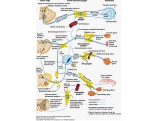
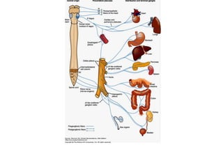
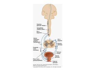
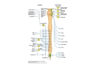
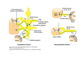
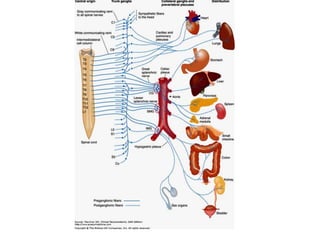
AUTONOMIC NERVOUS SYSTEM
INTRODUCTION
• Concerned with control of target tissues: the
  cardiac muscle, the smooth muscle in blood
  vessels and viscera, and the glands.
• It helps in homeostasis
• Consists of efferent pathways, afferent
  pathways, and groups of neurons in the brain
  and spinal cord that regulate the system's
  functions.
• Autonomic reflex activity in the spinal cord
  accounts for some aspects of autonomic
  regulation and homeostasis.
• Modulated by supraspinal centers such as
  brain stem nuclei and the hypothalamus-
  hierarchical organization
Divisions
•   anatomically distinct
•   largely opposing actions
•   sympathetic (thoracolumbar)
•   parasympathetic (craniosacral) division
Critical point
• Many commonly used medications (e.g.
  medications for treating high blood pressure,
  for regulating gastrointestinal function, or for
  maintaining a regular heart beat) have their
  major actions on neurons within these
  systems.
AUTONOMIC OUTFLOW/EFFERENTS

• efferent components arise from preganglionic
  cell bodies in different locations.
• organized more diffusely than the somatic
  motor system
Comparison with somatic nervous
               system
• In the SNS, LMN project directly from the
  spinal cord or brain, without an interposed
  synapse, to innervate a relatively small group
  of target cells (somatic muscle cells).
• A more slowly conducting two-neuron chain
  characterizes the autonomic outflow.
• The cell body of the primary neuron (the
  presynaptic, or preganglionic, neuron) within
  the central nervous system is located in the
  intermediolateral gray column of the spinal
  cord or in the brain stem nuclei.
• It sends its axon, which is usually a small-
  diameter, myelinated B fiber out to synapse
  with the secondary neuron (the postsynaptic,
  or postganglionic, neuron) located in one of
  the autonomic ganglia
• From there, the postganglionic axon passes to
  its terminal distribution in a target organ.
• Most postganglionic autonomic axons are
  unmyelinated C fibers.
• The autonomic outflow system projects widely
  to most target tissues and is not as highly
  focused as the somatic motor system.
• Postganglionic fibers outnumber the
  preganglionic neurons by a ratio of about
  32:1, a single preganglionic neuron may
  control the autonomic functions of a rather
  extensive terminal area.
Sympathetic Division

• Arises from preganglionic cell bodies located
  in the intermediolateral cell columns of the 12
  thoracic and upper 2 lumbar segments of the
  spinal cord
PREGANGLIONIC SYMPATHETIC
       EFFERENT FIBER SYSTEM
• Mostly myelinated
• Coursing with the ventral roots, they form the
  white communicating rami of the thoracic
  and lumbar nerves, through which they reach
  the ganglia of the sympathetic chains or
  trunks
• These trunk ganglia/paravertebral ganglia lie
  on the lateral sides of the bodies of the
  thoracic and lumbar vertebrae.
• On entering the ganglia, the fibers may
synapse with a number of ganglion cells,
 pass up or down the sympathetic trunk to
 synapse with ganglion cells at a higher or
 lower level, or
pass through the trunk/paravertebral ganglia
 and out to one of the collateral
 (intermediary)/prevertebral sympathetic
 ganglia (eg, the celiac and mesenteric
 ganglia).
Splanchnic nerves
• Arise from the lower seven thoracic segments
• pass through the paravertebral ganglia to the
  celiac and superior mesenteric ganglia.
• Synaptic connections occur with ganglion
  cells-postganglionic axons then pass to the
  abdominal viscera via the celiac plexus.
Splanchnic nerves
• arising from spinal cord segments in the
  lowest thoracic and upper lumbar region
  convey fibers to synaptic stations in the
  inferior mesenteric ganglion and to small
  ganglia associated with the hypogastric
  plexus, through which postsynaptic fibers are
  distributed to the lower abdominal and pelvic
  viscera.
THE ADRENAL GLAND

• Preganglionic sympathetic axons in the
  splanchnic nerves also project to the adrenal
  gland, where they synapse on chromaffin cells
  in the adrenal medulla.
• The adrenal chromaffin cells, which receive
  direct synaptic input from preganglionic
  sympathetic axons, are derived from neural
  crest and can be considered to be modified
  postganglionic cells that have lost their axons
POSTGANGLIONIC EFFERENT FIBER
            SYSTEM
• mostly unmyelinated postganglionic fibers
  form the gray communicating rami.
• fibers may course with the spinal nerve for
  some distance or go directly to their target
  tissues.
• Gray communicating rami join each of the
  spinal nerves and distribute the vasomotor,
  pilomotor, and sweat gland innervation
  throughout the somatic areas
• Branches of the superior cervical sympathetic
  ganglion enter into the formation of the
  sympathetic carotid plexuses around the
  internal and external carotid arteries for
  distribution of sympathetic fibers to the head
• After exiting from the carotid plexus, these
  postganglionic sympathetic axons project to
  the salivary and lacrimal glands, the muscles
  that dilate the pupil and raise the eyelid, and
  sweat glands and blood vessels of the face
  and head.
• superior cardiac nerves from three pairs of
  cervical sympathetic ganglia→ cardiac plexus
  → cardioaccelerator fibers
• Vasomotor branches from the upper five
  thoracic ganglia →thoracic aorta → posterior
  pulmonary plexus→bronchi[dilatation]
Parasympathetic Division

from preganglionic cell bodies in
A. the gray matter of the brain stem
 medial part of the oculomotor nucleus,
    Edinger–Westphal nucleus,
 superior and inferior salivatory nuclei)
B. the middle three segments of the sacral cord
  (S2–4)
• Most of the preganglionic fibers from S2, S3,
  and S4 run without interruption from their
  central origin to
• either the wall of the viscus they supply or
• the site where they synapse with terminal
  ganglion cells associated with the plexuses of
  Meissner and Auerbach in the wall of the
  intestinal tract
• the parasympathetic postganglionic neurons
  are located close to the tissues they supply-
  relatively short axons.
• parasympathetic distribution confined entirely
  to visceral structures.
Cranial nerves conveying
 preganglionic parasympathetic fibers
• CN III, VII, and IX distribute parasympathetic
  or visceral efferent fibers to the head
• axons in these nerves synapse with
  postganglionic neurons in the ciliary,
  sphenopalatine, submaxillary, and otic
  ganglia, respectively
• The vagus nerve (cranial nerve X) →thoracic
  and abdominal viscera via the prevertebral
  plexuses.
• The pelvic nerve (nervus erigentes) →most of
  the large intestine and to the pelvic viscera
  and genitals via the hypogastric plexus.
Autonomic Plexuses
• conduit for the distribution of the sympathetic
  and parasympathetic (and afferent) fibers that
  enter into their formation
Cardiac plexus
• Formed from the cardiac sympathetic nerves
  and cardiac branches of the vagus nerve
• Supplies the myocardium and the vessels
  leaving the heart.
Pulmonary plexuses
• joined with the cardiac plexus
• formed from the vagus and the upper thoracic
  sympathetic nerves
• distributed to the vessels and bronchi of the
  lung.
Celiac (solar) plexus
• located in the epigastric region over the
  abdominal aorta
• formed from vagal fibers reaching it via the
  esophageal plexus, sympathetic fibers arising
  from celiac ganglia, and sympathetic fibers
  coursing down from the thoracic aortic plexus
• projects to most of the abdominal viscera,
  which it reaches by way of numerous
  subplexuses, including phrenic, hepatic,
  splenic, superior gastric, suprarenal, renal,
  spermatic or ovarian, abdominal aortic, and
  superior and inferior mesenteric plexuses.
Hypogastric plexus
• located in front of the fifth lumbar vertebra
  and the promontory of the sacrum.
• receives sympathetic fibers from the aortic
  plexus and lumbar trunk ganglia and
  parasympathetic fibers from the pelvic nerve.
• Its two lateral portions, the pelvic plexuses, lie
  on either side of the rectum.
• Supplies pelvic viscera and genitals via
  subplexuses that extend along the visceral
  branches of the hypogastric artery.
• middle hemorrhoidal plexus, to the rectum;
• the vesical plexus, to the bladder, seminal
  vesicles, and ductus deferens;
• the prostatic plexus, to the prostate, seminal
  vesicles, and penis;
• the vaginal plexus, to the vagina and clitoris;
• uterine plexus, to the uterus and uterine
  tubes.
VISCERAL AFFERENT PATHWAYS

• Visceral afferent fibers have their cell bodies in
  sensory ganglia of some of the cranial and
  spinal nerves.
• Most are unmyelinated and have slow
  conduction velocities
Pathways to the Spinal Cord

• Via middle sacral, thoracic, and upper lumbar
  nerves.
• The sacral nerves carry sensory stimuli from
  the pelvic organs-nerve fibers are involved in
  reflexes of the sacral parasympathetic outflow
  that control various sexual responses,
  micturition, and defecation.
• Axons carrying visceral pain impulses from the
  heart, upper digestive tract, kidney, and
  gallbladder travel with the thoracic and upper
  lumbar nerves.
• Pathways are associated with sensations such
  as hunger, nausea, and poorly localized
  visceral pain
• Pain impulses from a viscus may converge
  with pain impulses arising in a particular
  region of the skin, causing referred pain.
• Examples:
• shoulder pains associated with gallstone
  attacks
• the pains of the left arm or throat associated
  with myocardial ischemia
Examples
• shoulder pains associated with gallstone
  attacks
• the pains of the left arm or throat associated
  with myocardial ischemia
Pathways to the Brain Stem

• Visceral afferent axons in CN IX and especially
  CN Xcarry a variety of sensations to the brain
  stem from the heart, great vessels, and
  respiratory and gastrointestinal tracts.
• The ganglia involved are the inferior
  glossopharyngeal and the inferior vagal
• The afferent fibers are also involved in reflexes
  that regulate blood pressure, respiratory rate
  and depth, and heart rate through specialized
  receptors or receptor areas.
• Baroreceptors- aortic arch and carotid sinus .
• Chemoreceptors -aorta and carotid bodies.
• Chemosensitive zone –medulla
• Chemoreceptor neurons -alter their firing
  patterns in response to alterations of pH and
  pCO2 within the cerebrospinal fluid.
Autonomic nervous system by DR.ARSHAD

Autonomic nervous system by DR.ARSHAD

  • 2.
  • 4.
    INTRODUCTION • Concerned withcontrol of target tissues: the cardiac muscle, the smooth muscle in blood vessels and viscera, and the glands. • It helps in homeostasis • Consists of efferent pathways, afferent pathways, and groups of neurons in the brain and spinal cord that regulate the system's functions.
  • 5.
    • Autonomic reflexactivity in the spinal cord accounts for some aspects of autonomic regulation and homeostasis. • Modulated by supraspinal centers such as brain stem nuclei and the hypothalamus- hierarchical organization
  • 6.
    Divisions • anatomically distinct • largely opposing actions • sympathetic (thoracolumbar) • parasympathetic (craniosacral) division
  • 8.
    Critical point • Manycommonly used medications (e.g. medications for treating high blood pressure, for regulating gastrointestinal function, or for maintaining a regular heart beat) have their major actions on neurons within these systems.
  • 9.
    AUTONOMIC OUTFLOW/EFFERENTS • efferentcomponents arise from preganglionic cell bodies in different locations. • organized more diffusely than the somatic motor system
  • 10.
    Comparison with somaticnervous system • In the SNS, LMN project directly from the spinal cord or brain, without an interposed synapse, to innervate a relatively small group of target cells (somatic muscle cells). • A more slowly conducting two-neuron chain characterizes the autonomic outflow.
  • 11.
    • The cellbody of the primary neuron (the presynaptic, or preganglionic, neuron) within the central nervous system is located in the intermediolateral gray column of the spinal cord or in the brain stem nuclei.
  • 12.
    • It sendsits axon, which is usually a small- diameter, myelinated B fiber out to synapse with the secondary neuron (the postsynaptic, or postganglionic, neuron) located in one of the autonomic ganglia
  • 14.
    • From there,the postganglionic axon passes to its terminal distribution in a target organ. • Most postganglionic autonomic axons are unmyelinated C fibers.
  • 15.
    • The autonomicoutflow system projects widely to most target tissues and is not as highly focused as the somatic motor system. • Postganglionic fibers outnumber the preganglionic neurons by a ratio of about 32:1, a single preganglionic neuron may control the autonomic functions of a rather extensive terminal area.
  • 16.
    Sympathetic Division • Arisesfrom preganglionic cell bodies located in the intermediolateral cell columns of the 12 thoracic and upper 2 lumbar segments of the spinal cord
  • 17.
    PREGANGLIONIC SYMPATHETIC EFFERENT FIBER SYSTEM • Mostly myelinated • Coursing with the ventral roots, they form the white communicating rami of the thoracic and lumbar nerves, through which they reach the ganglia of the sympathetic chains or trunks
  • 18.
    • These trunkganglia/paravertebral ganglia lie on the lateral sides of the bodies of the thoracic and lumbar vertebrae. • On entering the ganglia, the fibers may synapse with a number of ganglion cells,
  • 19.
     pass upor down the sympathetic trunk to synapse with ganglion cells at a higher or lower level, or pass through the trunk/paravertebral ganglia and out to one of the collateral (intermediary)/prevertebral sympathetic ganglia (eg, the celiac and mesenteric ganglia).
  • 20.
    Splanchnic nerves • Arisefrom the lower seven thoracic segments • pass through the paravertebral ganglia to the celiac and superior mesenteric ganglia. • Synaptic connections occur with ganglion cells-postganglionic axons then pass to the abdominal viscera via the celiac plexus.
  • 22.
    Splanchnic nerves • arisingfrom spinal cord segments in the lowest thoracic and upper lumbar region convey fibers to synaptic stations in the inferior mesenteric ganglion and to small ganglia associated with the hypogastric plexus, through which postsynaptic fibers are distributed to the lower abdominal and pelvic viscera.
  • 23.
    THE ADRENAL GLAND •Preganglionic sympathetic axons in the splanchnic nerves also project to the adrenal gland, where they synapse on chromaffin cells in the adrenal medulla. • The adrenal chromaffin cells, which receive direct synaptic input from preganglionic sympathetic axons, are derived from neural crest and can be considered to be modified postganglionic cells that have lost their axons
  • 24.
    POSTGANGLIONIC EFFERENT FIBER SYSTEM • mostly unmyelinated postganglionic fibers form the gray communicating rami. • fibers may course with the spinal nerve for some distance or go directly to their target tissues.
  • 25.
    • Gray communicatingrami join each of the spinal nerves and distribute the vasomotor, pilomotor, and sweat gland innervation throughout the somatic areas • Branches of the superior cervical sympathetic ganglion enter into the formation of the sympathetic carotid plexuses around the internal and external carotid arteries for distribution of sympathetic fibers to the head
  • 26.
    • After exitingfrom the carotid plexus, these postganglionic sympathetic axons project to the salivary and lacrimal glands, the muscles that dilate the pupil and raise the eyelid, and sweat glands and blood vessels of the face and head.
  • 27.
    • superior cardiacnerves from three pairs of cervical sympathetic ganglia→ cardiac plexus → cardioaccelerator fibers
  • 28.
    • Vasomotor branchesfrom the upper five thoracic ganglia →thoracic aorta → posterior pulmonary plexus→bronchi[dilatation]
  • 31.
    Parasympathetic Division from preganglioniccell bodies in A. the gray matter of the brain stem  medial part of the oculomotor nucleus, Edinger–Westphal nucleus,  superior and inferior salivatory nuclei) B. the middle three segments of the sacral cord (S2–4)
  • 32.
    • Most ofthe preganglionic fibers from S2, S3, and S4 run without interruption from their central origin to • either the wall of the viscus they supply or • the site where they synapse with terminal ganglion cells associated with the plexuses of Meissner and Auerbach in the wall of the intestinal tract
  • 34.
    • the parasympatheticpostganglionic neurons are located close to the tissues they supply- relatively short axons. • parasympathetic distribution confined entirely to visceral structures.
  • 35.
    Cranial nerves conveying preganglionic parasympathetic fibers • CN III, VII, and IX distribute parasympathetic or visceral efferent fibers to the head • axons in these nerves synapse with postganglionic neurons in the ciliary, sphenopalatine, submaxillary, and otic ganglia, respectively
  • 37.
    • The vagusnerve (cranial nerve X) →thoracic and abdominal viscera via the prevertebral plexuses. • The pelvic nerve (nervus erigentes) →most of the large intestine and to the pelvic viscera and genitals via the hypogastric plexus.
  • 38.
    Autonomic Plexuses • conduitfor the distribution of the sympathetic and parasympathetic (and afferent) fibers that enter into their formation
  • 39.
    Cardiac plexus • Formedfrom the cardiac sympathetic nerves and cardiac branches of the vagus nerve • Supplies the myocardium and the vessels leaving the heart.
  • 40.
    Pulmonary plexuses • joinedwith the cardiac plexus • formed from the vagus and the upper thoracic sympathetic nerves • distributed to the vessels and bronchi of the lung.
  • 41.
    Celiac (solar) plexus •located in the epigastric region over the abdominal aorta • formed from vagal fibers reaching it via the esophageal plexus, sympathetic fibers arising from celiac ganglia, and sympathetic fibers coursing down from the thoracic aortic plexus
  • 42.
    • projects tomost of the abdominal viscera, which it reaches by way of numerous subplexuses, including phrenic, hepatic, splenic, superior gastric, suprarenal, renal, spermatic or ovarian, abdominal aortic, and superior and inferior mesenteric plexuses.
  • 43.
    Hypogastric plexus • locatedin front of the fifth lumbar vertebra and the promontory of the sacrum. • receives sympathetic fibers from the aortic plexus and lumbar trunk ganglia and parasympathetic fibers from the pelvic nerve. • Its two lateral portions, the pelvic plexuses, lie on either side of the rectum.
  • 44.
    • Supplies pelvicviscera and genitals via subplexuses that extend along the visceral branches of the hypogastric artery. • middle hemorrhoidal plexus, to the rectum; • the vesical plexus, to the bladder, seminal vesicles, and ductus deferens;
  • 45.
    • the prostaticplexus, to the prostate, seminal vesicles, and penis; • the vaginal plexus, to the vagina and clitoris; • uterine plexus, to the uterus and uterine tubes.
  • 46.
    VISCERAL AFFERENT PATHWAYS •Visceral afferent fibers have their cell bodies in sensory ganglia of some of the cranial and spinal nerves. • Most are unmyelinated and have slow conduction velocities
  • 47.
    Pathways to theSpinal Cord • Via middle sacral, thoracic, and upper lumbar nerves. • The sacral nerves carry sensory stimuli from the pelvic organs-nerve fibers are involved in reflexes of the sacral parasympathetic outflow that control various sexual responses, micturition, and defecation.
  • 48.
    • Axons carryingvisceral pain impulses from the heart, upper digestive tract, kidney, and gallbladder travel with the thoracic and upper lumbar nerves. • Pathways are associated with sensations such as hunger, nausea, and poorly localized visceral pain
  • 49.
    • Pain impulsesfrom a viscus may converge with pain impulses arising in a particular region of the skin, causing referred pain. • Examples: • shoulder pains associated with gallstone attacks • the pains of the left arm or throat associated with myocardial ischemia
  • 50.
    Examples • shoulder painsassociated with gallstone attacks • the pains of the left arm or throat associated with myocardial ischemia
  • 51.
    Pathways to theBrain Stem • Visceral afferent axons in CN IX and especially CN Xcarry a variety of sensations to the brain stem from the heart, great vessels, and respiratory and gastrointestinal tracts. • The ganglia involved are the inferior glossopharyngeal and the inferior vagal
  • 52.
    • The afferentfibers are also involved in reflexes that regulate blood pressure, respiratory rate and depth, and heart rate through specialized receptors or receptor areas. • Baroreceptors- aortic arch and carotid sinus . • Chemoreceptors -aorta and carotid bodies.
  • 53.
    • Chemosensitive zone–medulla • Chemoreceptor neurons -alter their firing patterns in response to alterations of pH and pCO2 within the cerebrospinal fluid.