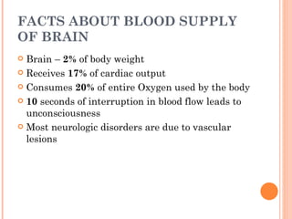
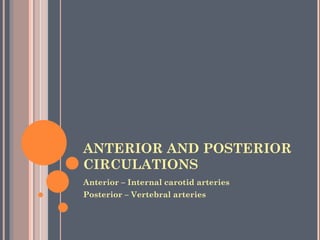
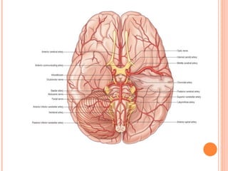
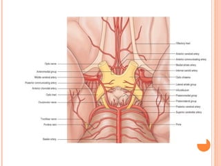
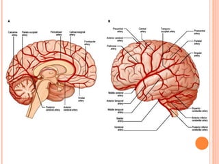
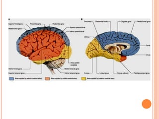
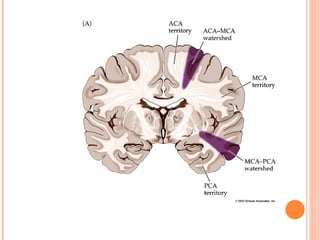
The brain receives a large portion of the body's blood supply and oxygen consumption despite being only 2% of body weight. The internal carotid arteries supply the anterior circulation while the vertebral arteries supply the posterior circulation, with these systems connecting at the circle of Willis. Occlusion of cerebral arteries can cause neurological deficits corresponding to the brain areas supplied, such as hemiplegia from middle cerebral artery occlusion or homonymous hemianopia from posterior cerebral artery occlusion. Proper blood flow is crucial for brain function.
![BRANCHES OF INTERNAL CAROTID
ARTERY[PRIOR TO BIFURCATION]](https://image.slidesharecdn.com/cerebralcirculation-120412075418-phpapp02/85/Cerebral-circulation-by-DR-ARSHAD-8-320.jpg)
![ Alternate route – inadequate, especially in the
elderly [atherosclerosis]
Variations
3. In approximately 33% persons Posterior cerebral
artery arises from Internal carotid artery
4. One Anterior cerebral artery may be small-
anterior communicating artery is wider](https://image.slidesharecdn.com/cerebralcirculation-120412075418-phpapp02/85/Cerebral-circulation-by-DR-ARSHAD-13-320.jpg)
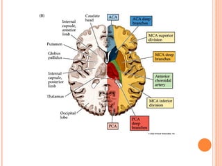
![ANTERIOR CEREBRAL ARTERY
Midline proximity [longitudinal horizontal
fissure]
Joined together by anterior communicating
artery
Branches
4. Medial striate/recurrent artery of
Heubner→ventral part of head of caudate
nucleus, putamen, anterior limb and genu of
internal capsule](https://image.slidesharecdn.com/cerebralcirculation-120412075418-phpapp02/85/Cerebral-circulation-by-DR-ARSHAD-18-320.jpg)
![AREA SUPPLIED
Medial part of orbital surface of frontal lobe
[includes olfactory bulb and tract]
Medial surfaces of frontal and parietal lobes
Corpus callosum
A strip on lateral surface](https://image.slidesharecdn.com/cerebralcirculation-120412075418-phpapp02/85/Cerebral-circulation-by-DR-ARSHAD-20-320.jpg)
![OTHER ASSOCIATED DEFICITS IN
ACA BLOCK
Mental confusion and dysphasia[functional loss
in prefrontal cortex, cingulate gyrus,
supplementary motor area]](https://image.slidesharecdn.com/cerebralcirculation-120412075418-phpapp02/85/Cerebral-circulation-by-DR-ARSHAD-22-320.jpg)
![EFFECTS OF OCCLUSION
Paralysis and sensory deficits in contralateral
leg, perineum
Urinary incontinence [inadequate perineal
sensation, defective cortical control of pelvic floor
muscles]
If obstruction is proximal to anterior
communicating artery [blocked medial striate
artery]
UMN weakness of face, tongue and upper limb
[lesion in or near genu]
Ipsilateral anosmia [maybe]](https://image.slidesharecdn.com/cerebralcirculation-120412075418-phpapp02/85/Cerebral-circulation-by-DR-ARSHAD-23-320.jpg)

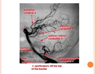
![BRANCHES
Central branches supply
2. Midbrain
3. Pineal gland
4. Lateral geniculate body
5. Lentiform nucleus [partly]
6. Thalamus [partly]](https://image.slidesharecdn.com/cerebralcirculation-120412075418-phpapp02/85/Cerebral-circulation-by-DR-ARSHAD-26-320.jpg)
![SPECIAL CIRCUMSTANCES
Expanding space-occupying lesion in
supratentorial compartment→ herniation of
uncus and midbrain→ compression of one or both
PCA→ necrosis of areas supplied [even after
surgical correction of cause]→ cortical
blindness+inability to form new memories
Traumatic Intracranial hemorrhage can lead to
the same consequences](https://image.slidesharecdn.com/cerebralcirculation-120412075418-phpapp02/85/Cerebral-circulation-by-DR-ARSHAD-30-320.jpg)
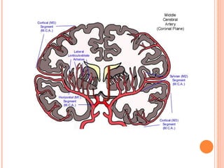
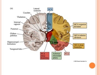
![ Larger, more direct continuation of ICA
Occupies lateral sulcus
Central branches →Head of caudate
nucleus,Putamen, Lateral pallidum, Internal
capsule [anterior limb, genu, posterior limb],
External capsule, Claustrum, Lateral
hypothalamus](https://image.slidesharecdn.com/cerebralcirculation-120412075418-phpapp02/85/Cerebral-circulation-by-DR-ARSHAD-32-320.jpg)
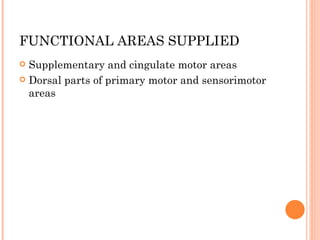
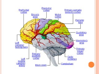
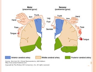
![FUNCTIONAL AREAS SUPPLIED
Most of primary motor and premotor areas
Frontal eye field
Primary somatosensory area
Geniculocalcarine tract
EXCEPT motor and sensory cortex for
lower limb and perineum
In most persons, left MCA supplies all
cortical areas concerned with language
[Wernicke’s,Broca’s]](https://image.slidesharecdn.com/cerebralcirculation-120412075418-phpapp02/85/Cerebral-circulation-by-DR-ARSHAD-33-320.jpg)
![ Cortical neglect, if right hemisphere involved
Conjugate eye movements affected[ at rest, eyes
turn towards the side of lesion]
Hearing unaffected [bilateral representation]
Obstruction to central branches- contralateral
hemiplegia without aphasia](https://image.slidesharecdn.com/cerebralcirculation-120412075418-phpapp02/85/Cerebral-circulation-by-DR-ARSHAD-35-320.jpg)


![Stroke [uncensored] - by MHR Corporation](https://cdn.slidesharecdn.com/ss_thumbnails/mhr4-stroke-101129110104-phpapp01-thumbnail.jpg?width=640&height=640&fit=bounds)


![Circle of willis (finql)[1].pptx anatomy](https://cdn.slidesharecdn.com/ss_thumbnails/circleofwillisfinql1-250913015401-7ca582c5-thumbnail.jpg?width=640&height=640&fit=bounds)


