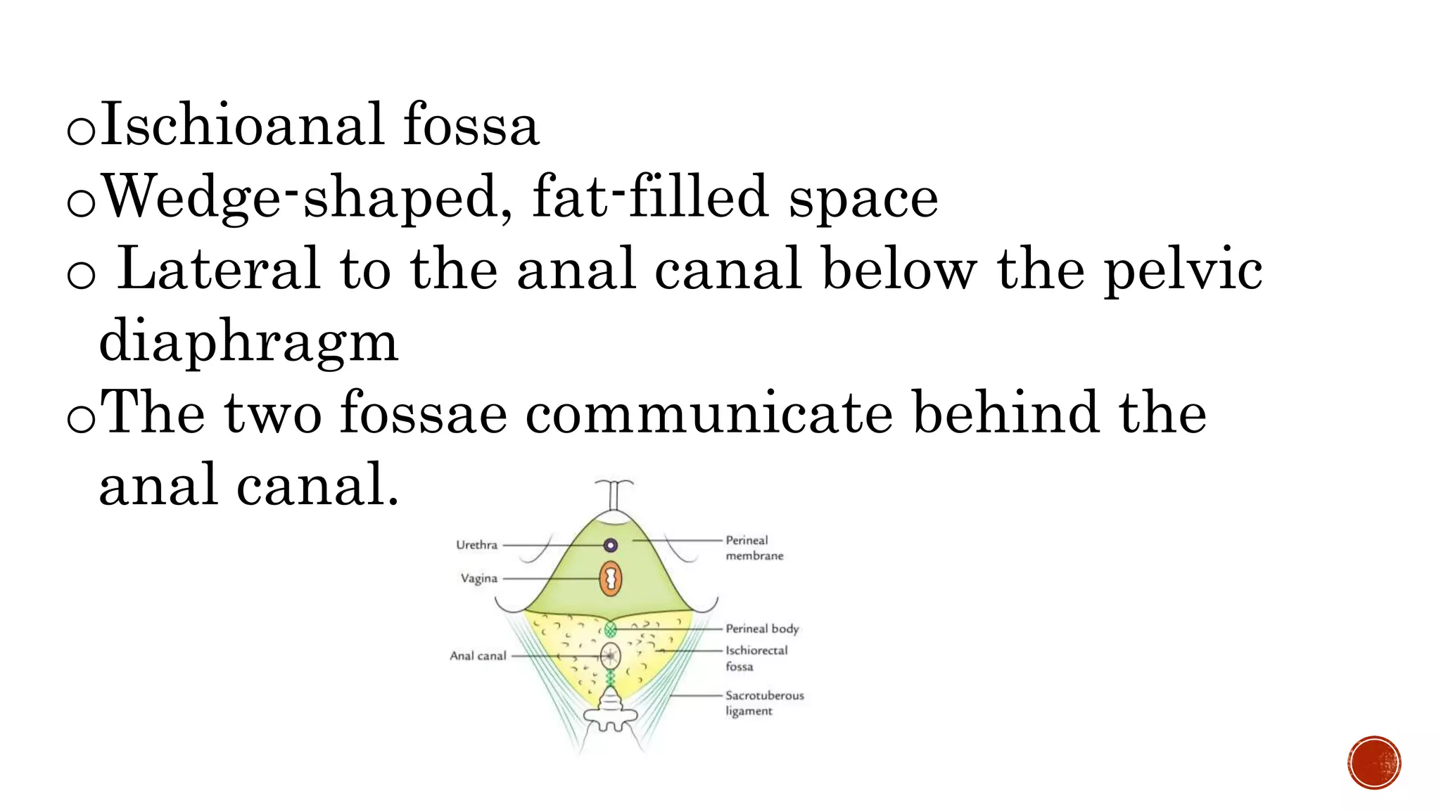
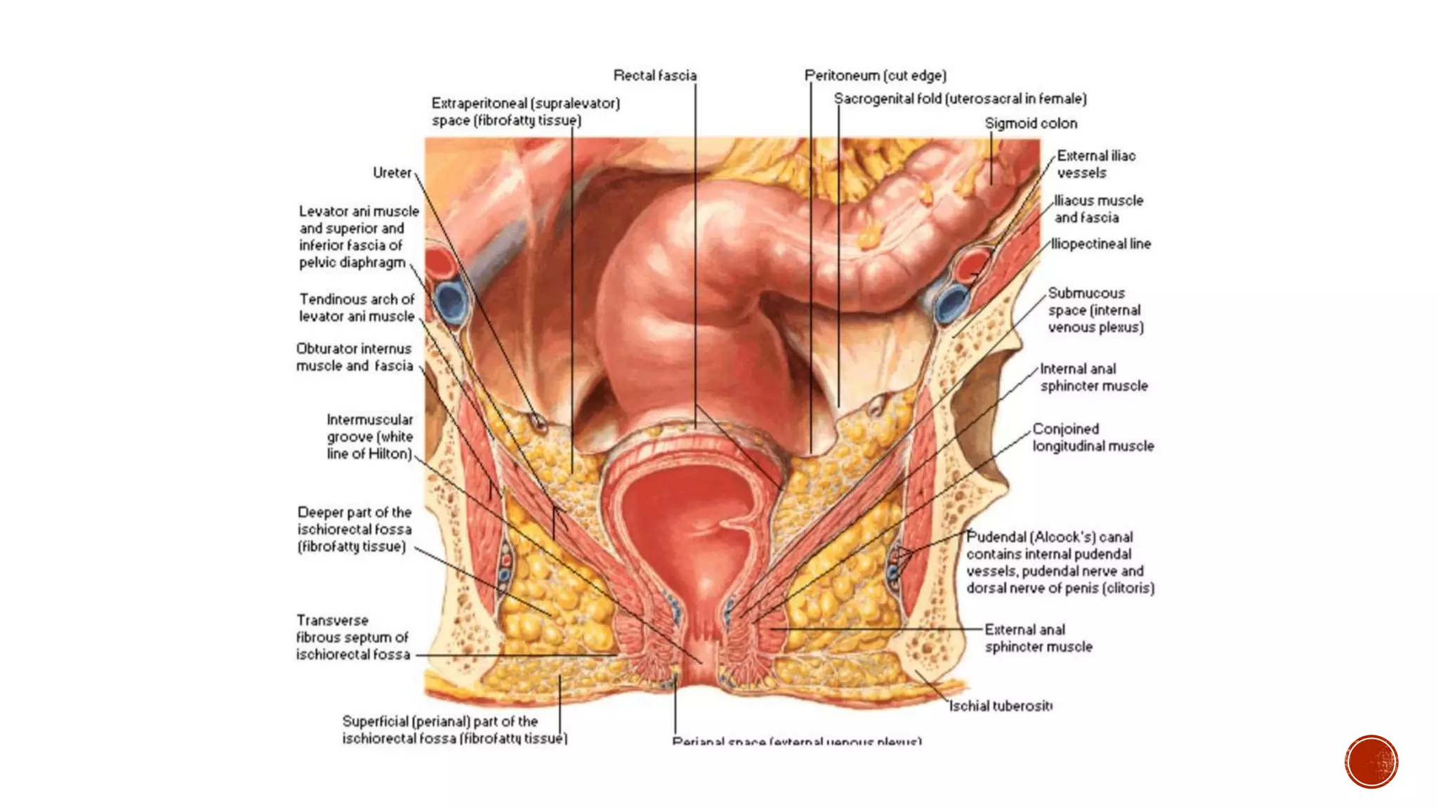
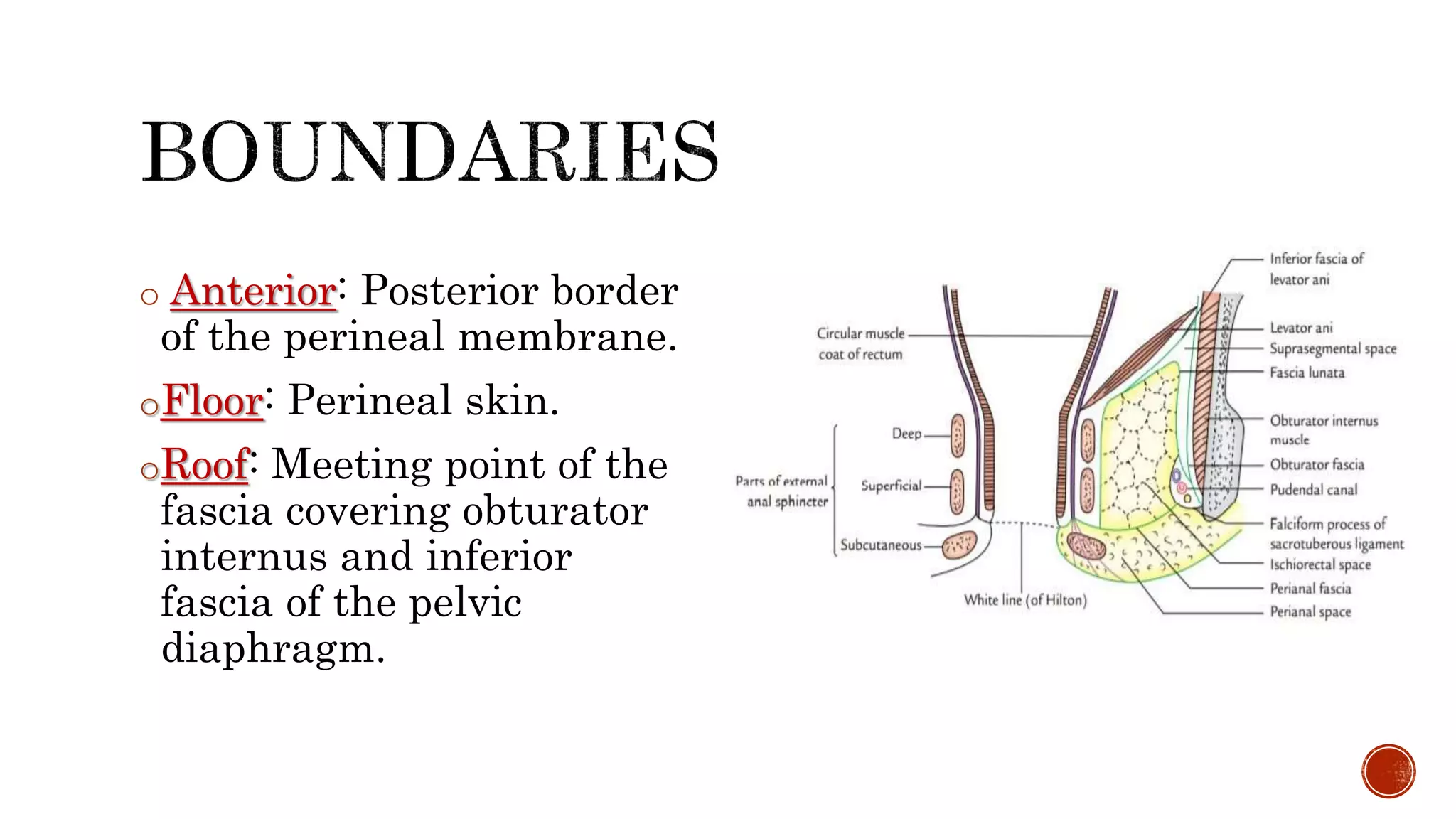
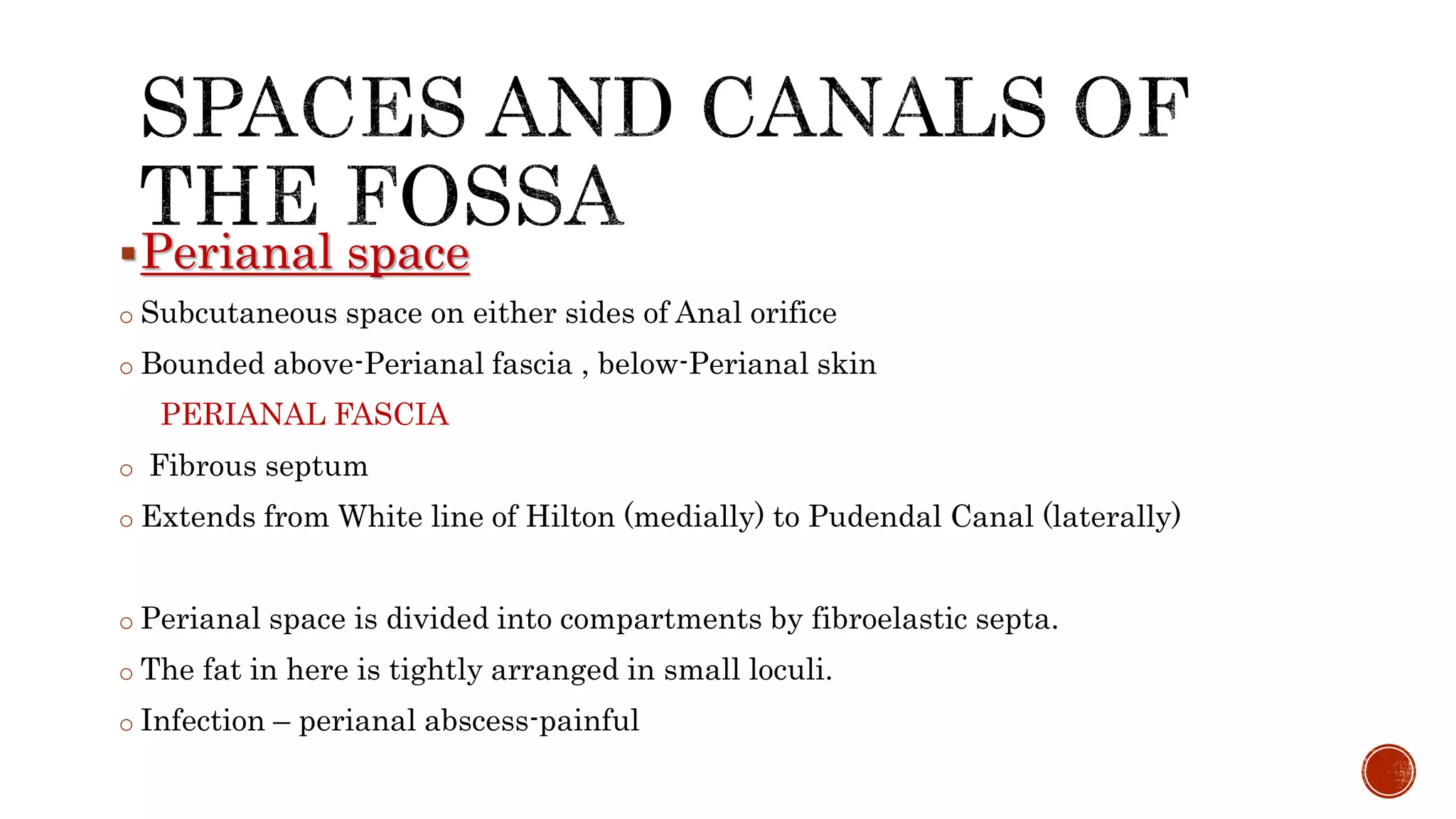
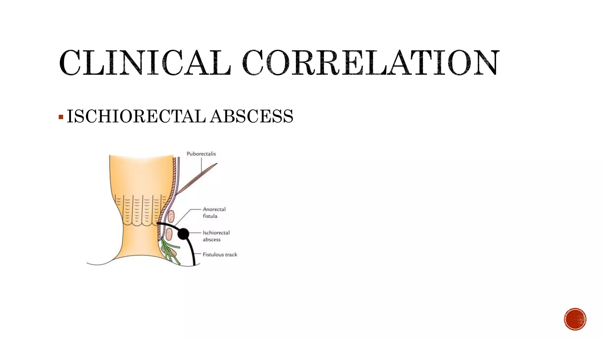
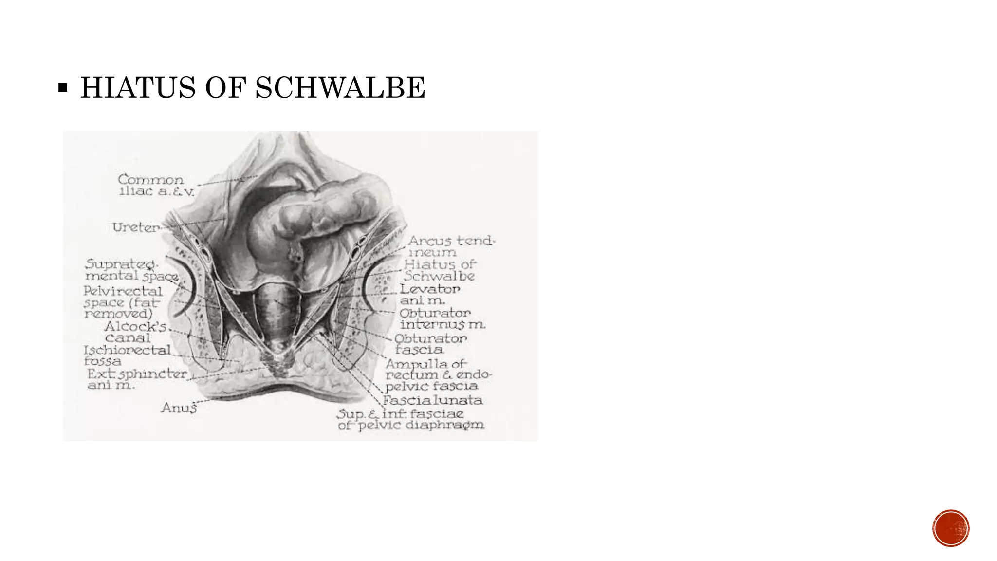
JULIT MATHEW
ROLL NO. 86
oIschioanal fossa
oWedge-shaped, fat-filled space
o Lateral to the anal canal below the pelvic
diaphragm
oThe two fossae communicate behind the
anal canal.
o Base-skin over the anal region of the
perineum
o Apex-directed upward and laterally.
o Measurements-
5cm-5cm-5cm
oLateral: Fascia covering the
obturator internus muscle and
ischial tuberosity.
oMedial: Fascia covering the
levator ani muscle and
external anal sphincter.
oPosterior: Sacrotuberous
ligament, on the posterior
surface of which is gluteus
maximus.
o Anterior: Posterior border
of the perineal membrane.
oFloor: Perineal skin.
oRoof: Meeting point of the
fascia covering obturator
internus and inferior
fascia of the pelvic
diaphragm.
Anterior Recess :Anteriorly
above Urogenital Diaphragm
Posterior Recess :Posteriorly
deep to Sacrotuberous ligament
Horseshoe-shaped Recess :
Connects the two fossa behind
the anal canal
Perianal space
o Subcutaneous space on either sides of Anal orifice
o Bounded above-Perianal fascia , below-Perianal skin
PERIANAL FASCIA
o Fibrous septum
o Extends from White line of Hilton (medially) to Pudendal Canal (laterally)
o Perianal space is divided into compartments by fibroelastic septa.
o The fat in here is tightly arranged in small loculi.
o Infection – perianal abscess-painful
 Ischioanal space
 Large and deep
 Fat here is arranged in large loculi,formed by incomplete
delicate septa
LUNATE FASCIA
 Arches over Ischioanal fat
 Begins laterally at the pudendal canal ,merges medially
with fascia covering deep part of External anal sphincter.
Fascia divides the space into :
Suprategmental space and Tegmental space
o Pudendal canal(Alcock’s canal)
 Fascial canal present in the lateral wall of the ischiorectal fossa about 1
inch (2.5 cm) above the ischial tuberosity
 Extends from the lesser sciatic foramen to the posterior limit of the deep
perineal pouch
 The pudendal canal is formed either by the splitting of the obturator
fascia or by separation between the fascia lunata and the obturator
fascia.
CONTENTS
 1. Pudendal nerve which divides within the canal into the dorsal nerve
of penis and the perineal nerve.
 2. Internal pudendal vessels
 Ischioanal pad of fat
 Inferior rectal nerve and vessels- pass through the fossa from lateral to
medial side. They arch upwards above the fat to supply mucous
membrane, External sphincter, skin around the anus.
 Pudendal canal with its contents. The canal lies along the lateral wall of
the fossa.
 Posterior scrotal(males) or posterior labial(females) nerves and vessels.
They cross the Anterolateral part of the fossa and enter the urogenital
triangle.
 Perineal branch of S4 nerve. It enters the posterior angle of the fossa and
runs over the levator ani to the external anal sphincter.
 Perforating cutaneous branches of nerves S2,S3.They appear at the lower
border of the gluteus maximus, in the posterior part of the fossa
ISCHIORECTAL ABSCESS
 HIATUS OF SCHWALBE
THANK YOU

Ischiorectal fossa

  • 1.
  • 2.
    oIschioanal fossa oWedge-shaped, fat-filledspace o Lateral to the anal canal below the pelvic diaphragm oThe two fossae communicate behind the anal canal.
  • 3.
    o Base-skin overthe anal region of the perineum o Apex-directed upward and laterally. o Measurements- 5cm-5cm-5cm
  • 5.
    oLateral: Fascia coveringthe obturator internus muscle and ischial tuberosity. oMedial: Fascia covering the levator ani muscle and external anal sphincter. oPosterior: Sacrotuberous ligament, on the posterior surface of which is gluteus maximus.
  • 7.
    o Anterior: Posteriorborder of the perineal membrane. oFloor: Perineal skin. oRoof: Meeting point of the fascia covering obturator internus and inferior fascia of the pelvic diaphragm.
  • 8.
    Anterior Recess :Anteriorly aboveUrogenital Diaphragm Posterior Recess :Posteriorly deep to Sacrotuberous ligament Horseshoe-shaped Recess : Connects the two fossa behind the anal canal
  • 10.
    Perianal space o Subcutaneousspace on either sides of Anal orifice o Bounded above-Perianal fascia , below-Perianal skin PERIANAL FASCIA o Fibrous septum o Extends from White line of Hilton (medially) to Pudendal Canal (laterally) o Perianal space is divided into compartments by fibroelastic septa. o The fat in here is tightly arranged in small loculi. o Infection – perianal abscess-painful
  • 12.
     Ischioanal space Large and deep  Fat here is arranged in large loculi,formed by incomplete delicate septa LUNATE FASCIA  Arches over Ischioanal fat  Begins laterally at the pudendal canal ,merges medially with fascia covering deep part of External anal sphincter. Fascia divides the space into : Suprategmental space and Tegmental space
  • 13.
    o Pudendal canal(Alcock’scanal)  Fascial canal present in the lateral wall of the ischiorectal fossa about 1 inch (2.5 cm) above the ischial tuberosity  Extends from the lesser sciatic foramen to the posterior limit of the deep perineal pouch  The pudendal canal is formed either by the splitting of the obturator fascia or by separation between the fascia lunata and the obturator fascia. CONTENTS  1. Pudendal nerve which divides within the canal into the dorsal nerve of penis and the perineal nerve.  2. Internal pudendal vessels
  • 14.
     Ischioanal padof fat  Inferior rectal nerve and vessels- pass through the fossa from lateral to medial side. They arch upwards above the fat to supply mucous membrane, External sphincter, skin around the anus.  Pudendal canal with its contents. The canal lies along the lateral wall of the fossa.  Posterior scrotal(males) or posterior labial(females) nerves and vessels. They cross the Anterolateral part of the fossa and enter the urogenital triangle.  Perineal branch of S4 nerve. It enters the posterior angle of the fossa and runs over the levator ani to the external anal sphincter.  Perforating cutaneous branches of nerves S2,S3.They appear at the lower border of the gluteus maximus, in the posterior part of the fossa
  • 16.
  • 17.
     HIATUS OFSCHWALBE
  • 18.