ANATOMY OF URINARY BLADDER
Prabin Kumar Bam
MBBS student, Chitwan Medical College
Bharatpur, Nepal
Introduction
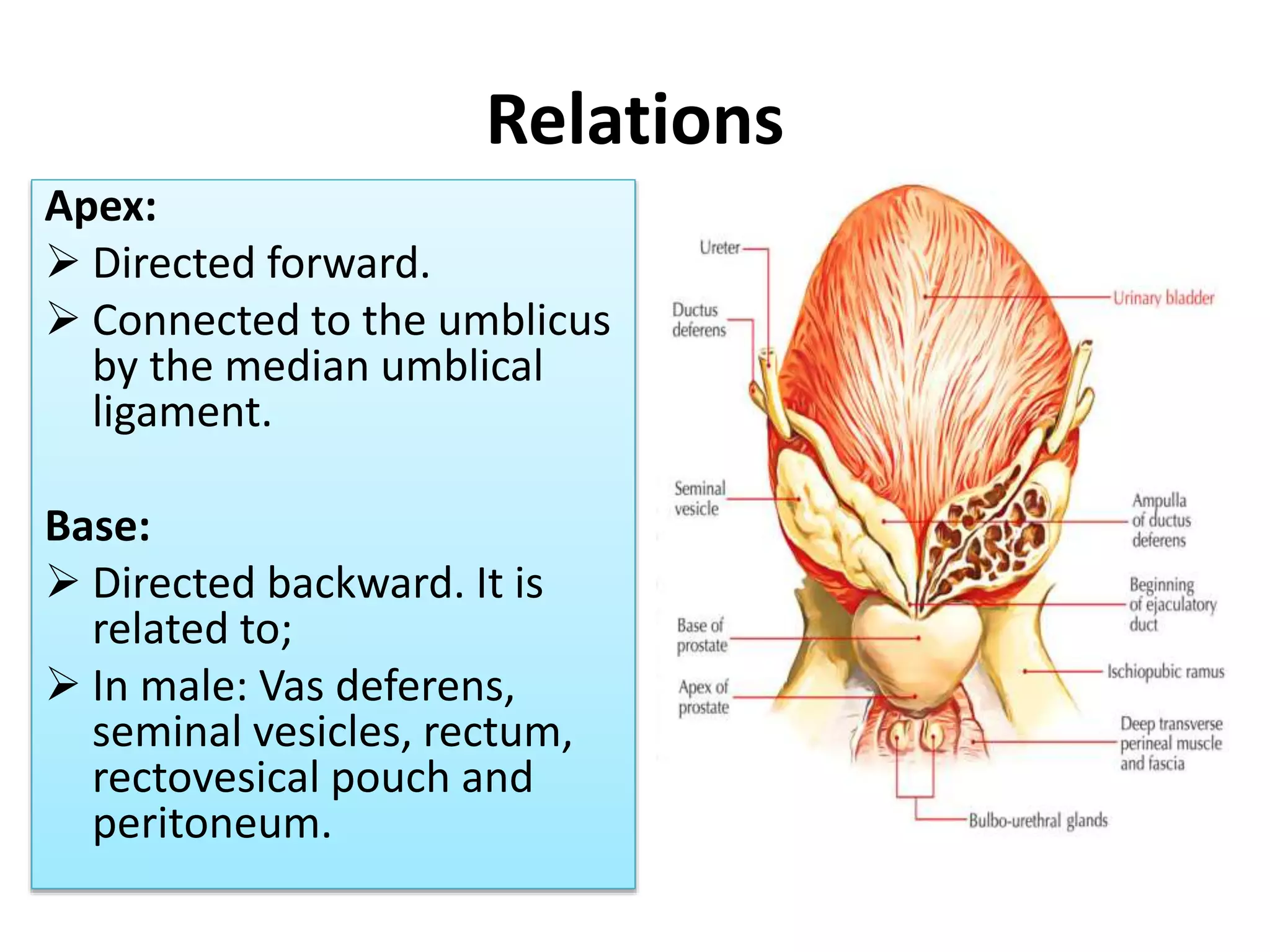
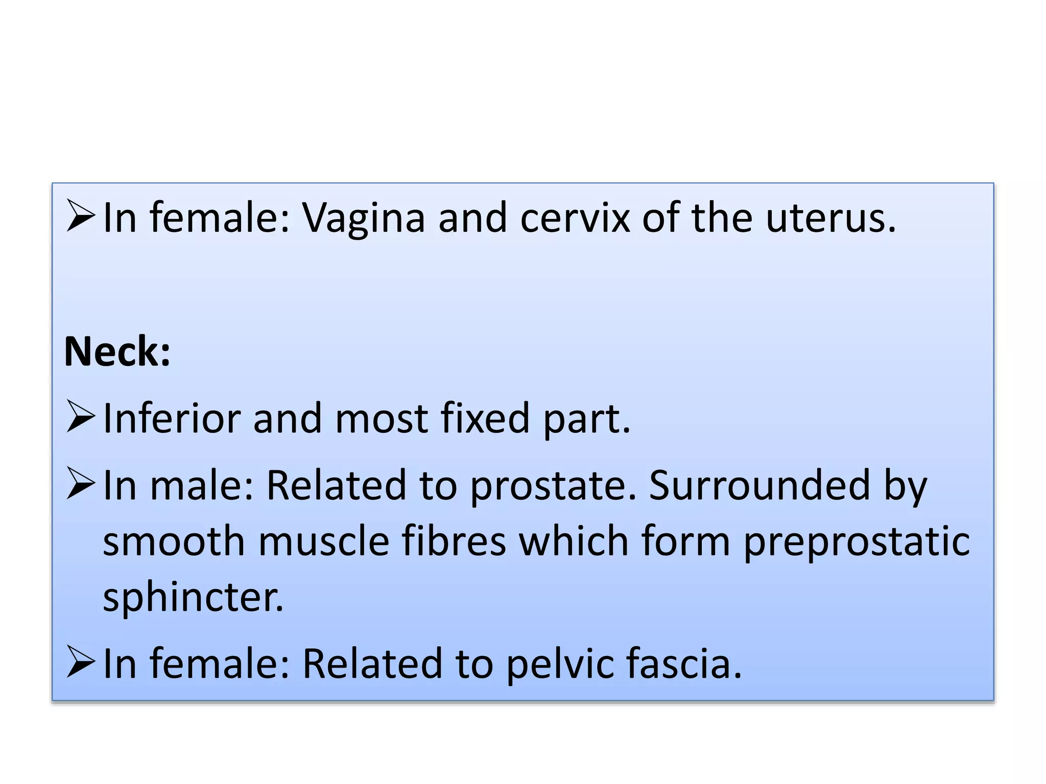
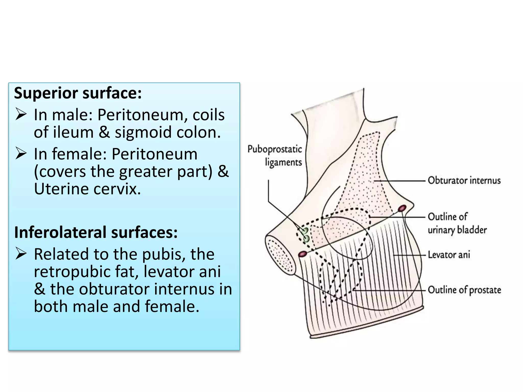
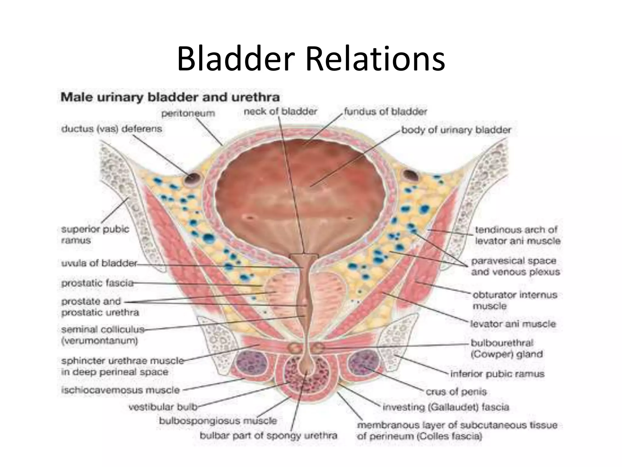
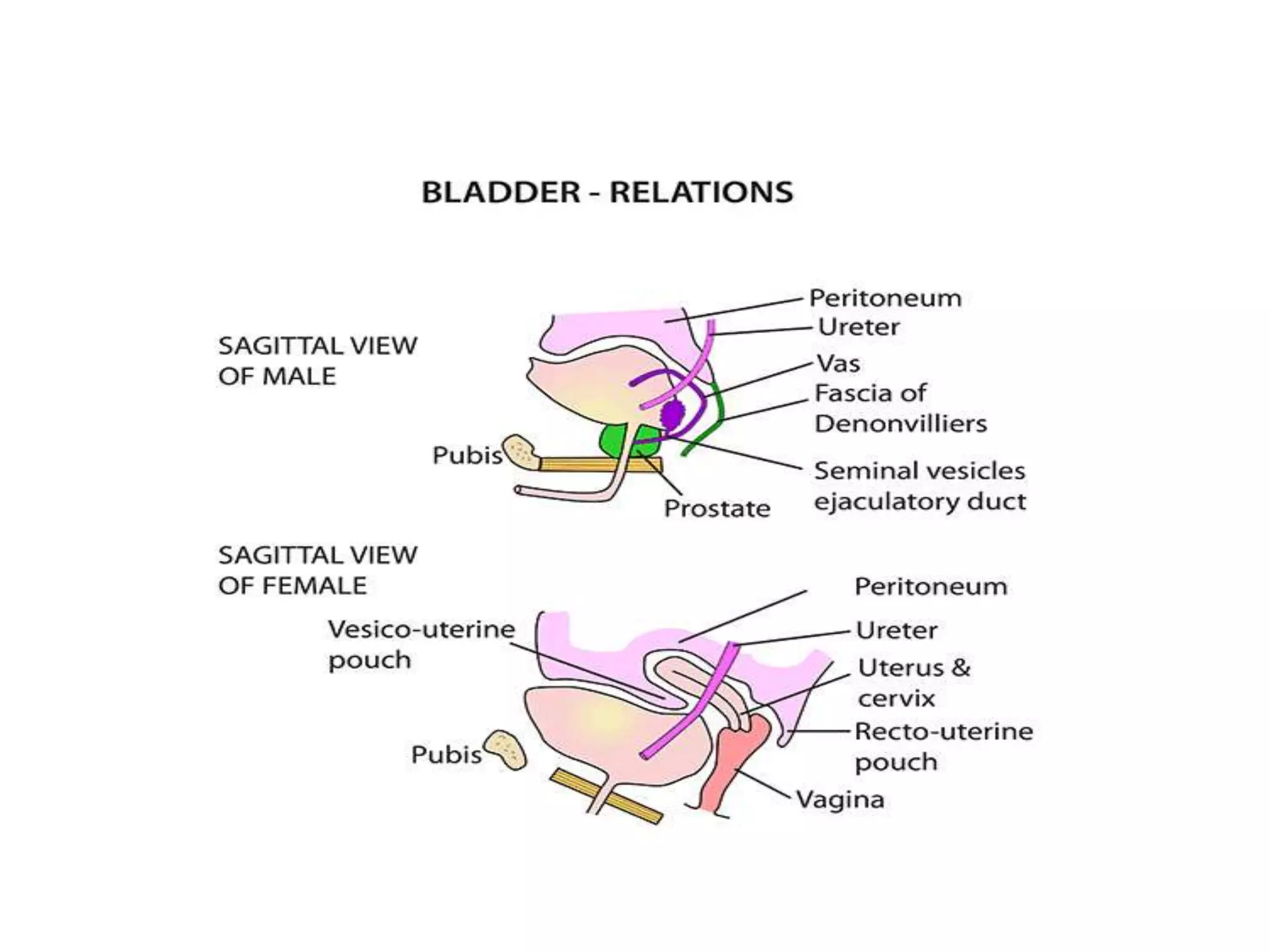
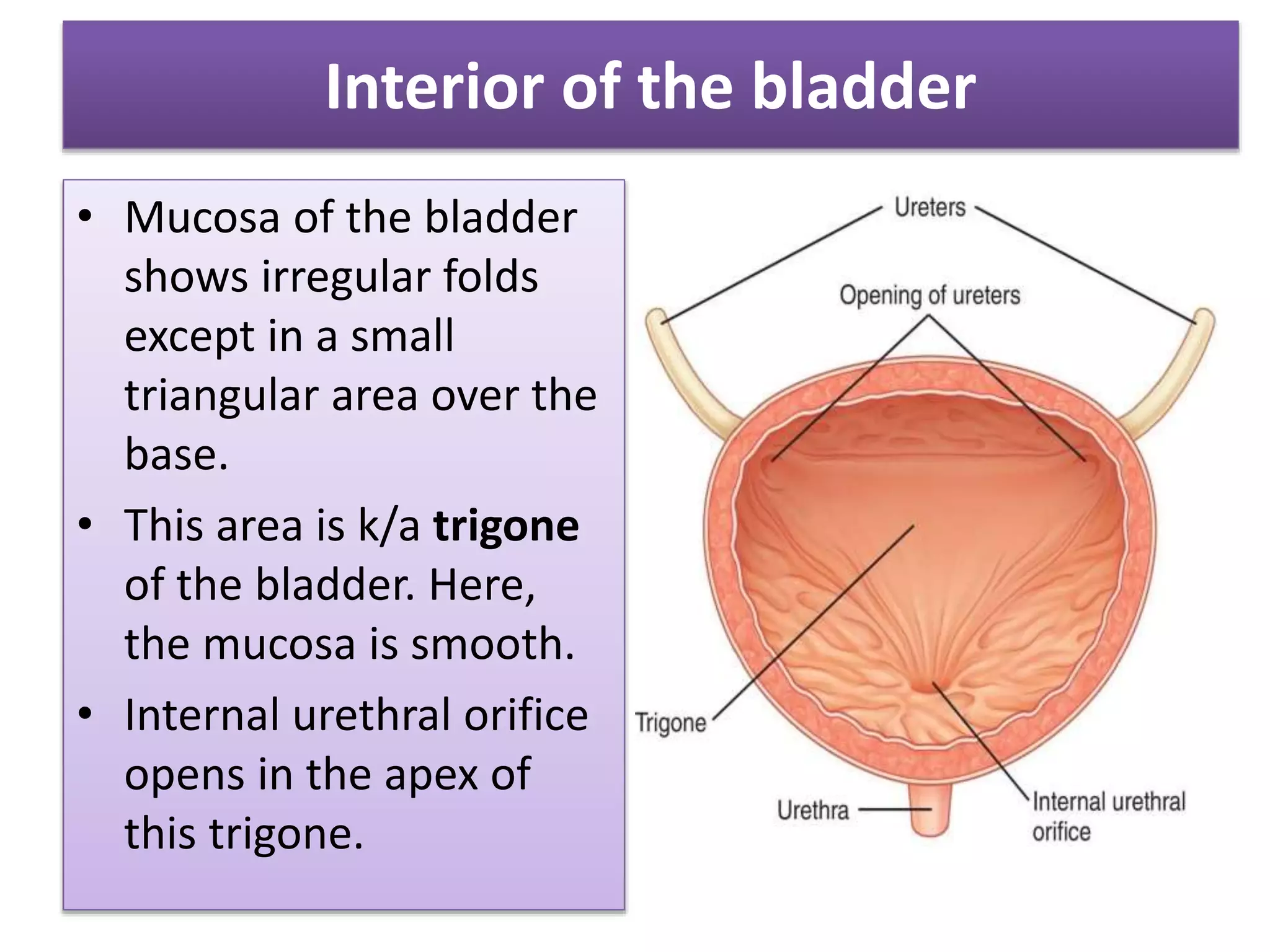
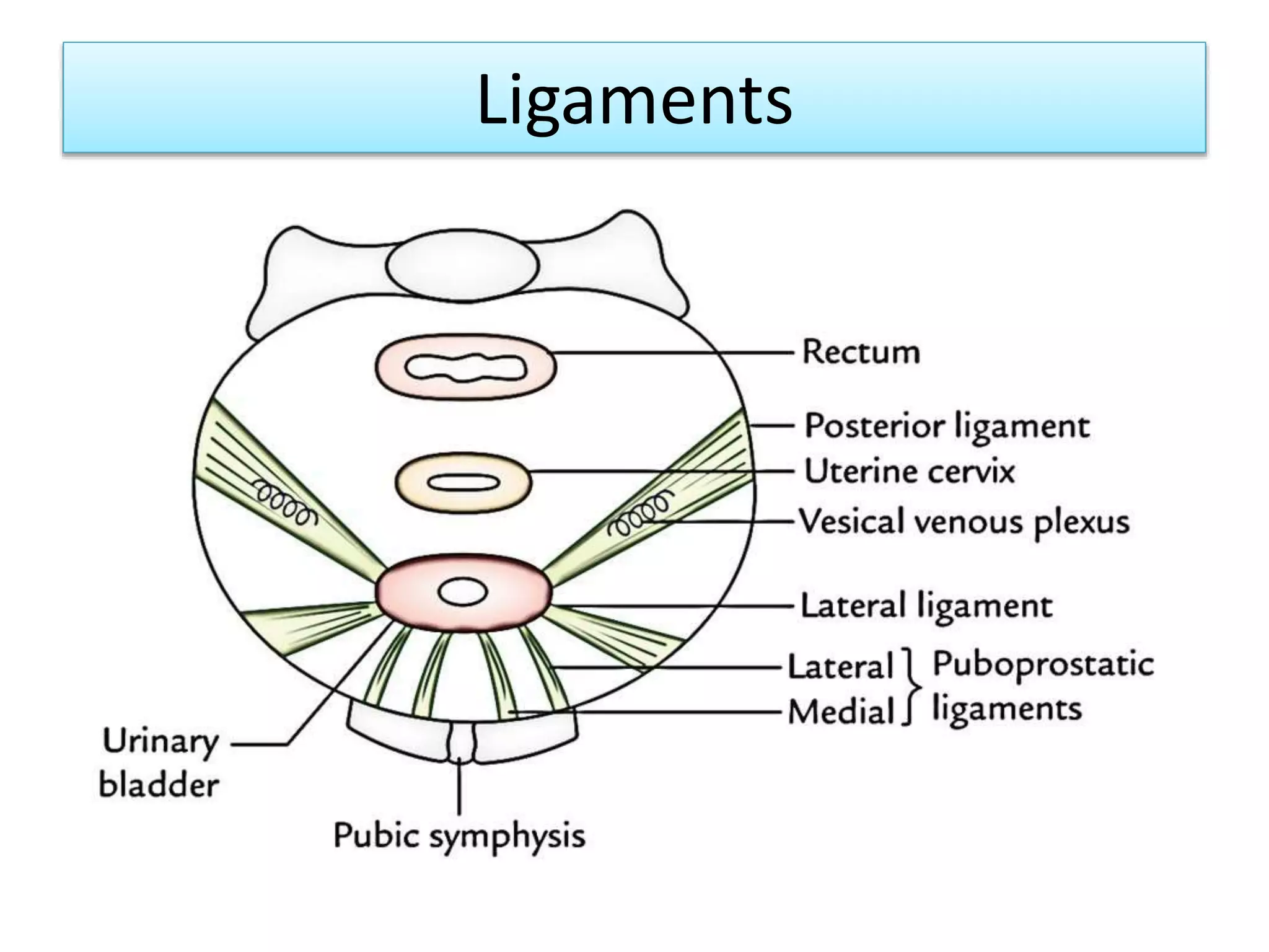
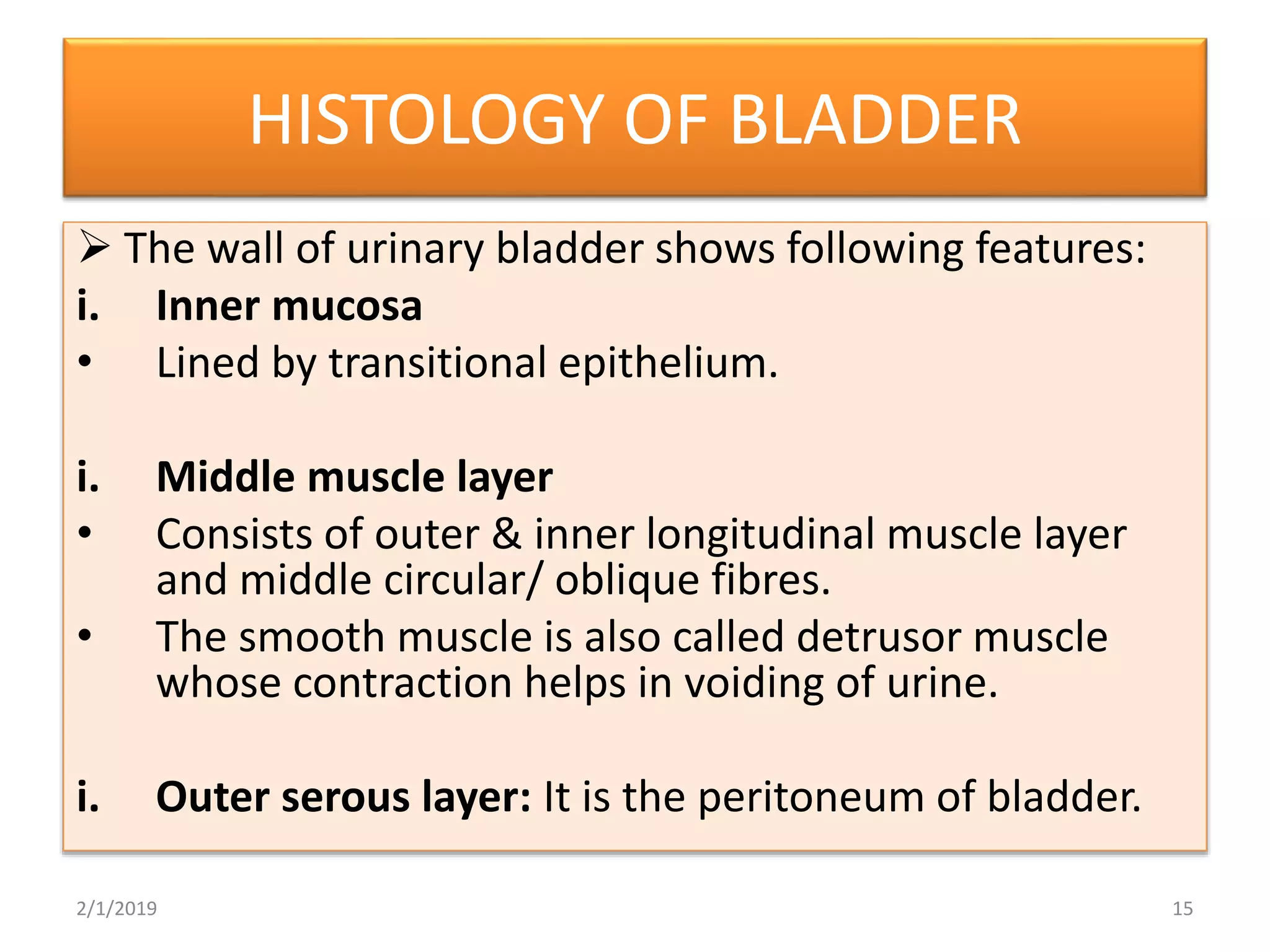
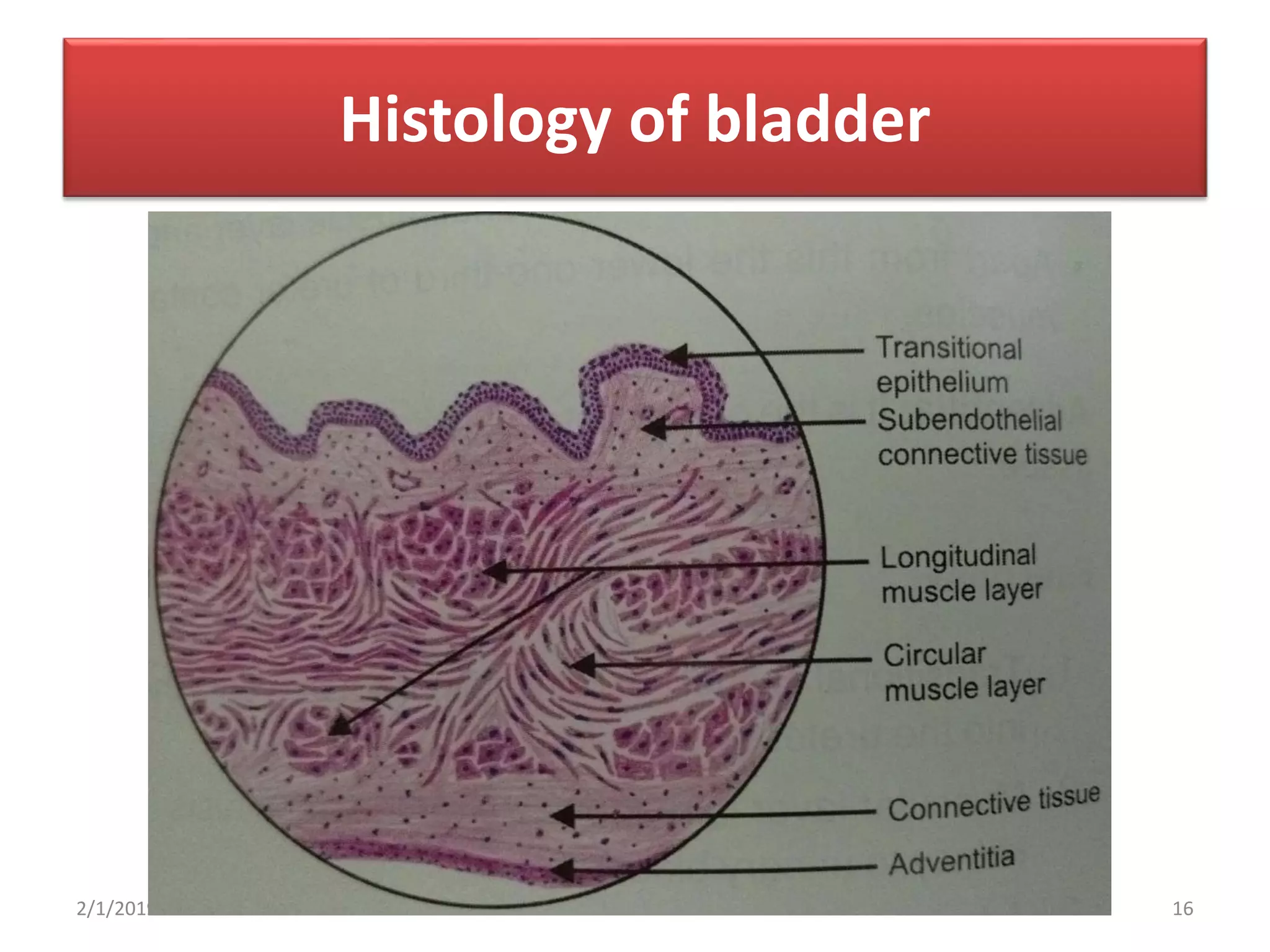
• The urinary bladder is a muscular organ which
lies in the anterior part of pelvic cavity and
helps in the storage of urine.
• Shape, size and position of bladder varies
according to the amount of urine contained in
it.
• An empty bladder lies entirely within the
pelvis but as it fills it extends into abdominal
cavity.
GROSS FEATURES
An empty bladder is tetrahedral in shape & has:
1. Apex
2. Base or fundus
3. Neck
4. 3 surfaces: superior, right & left inferolateral.
5. 4 borders: anterior, posterior & 2 lateral
lateral borders.
Gross Features
2/1/2019 4
Relations
Apex:
 Directed forward.
 Connected to the umblicus
by the median umblical
ligament.
Base:
 Directed backward. It is
related to;
 In male: Vas deferens,
seminal vesicles, rectum,
rectovesical pouch and
peritoneum.
In female: Vagina and cervix of the uterus.
Neck:
Inferior and most fixed part.
In male: Related to prostate. Surrounded by
smooth muscle fibres which form preprostatic
sphincter.
In female: Related to pelvic fascia.
Superior surface:
 In male: Peritoneum, coils
of ileum & sigmoid colon.
 In female: Peritoneum
(covers the greater part) &
Uterine cervix.
Inferolateral surfaces:
 Related to the pubis, the
retropubic fat, levator ani
& the obturator internus in
both male and female.
Puboprostatic ligaments in male &
pubovesical ligaments in female.
Bladder Relations
Interior of the bladder
• Mucosa of the bladder
shows irregular folds
except in a small
triangular area over the
base.
• This area is k/a trigone
of the bladder. Here,
the mucosa is smooth.
• Internal urethral orifice
opens in the apex of
this trigone.
• The ureters open at the posterolateral angle of
the trigone.
• Uvula vesicae: It is a small elevation on the
trigone posterior to the urethral orifice
produced by median lobe of prostate.
Ligaments of the bladder
True ligaments
 They are the condensations of
pelvic fascia.
 They provide support to the
bladder.
1. Lateral true ligament
2. Lateral puboprostatic
ligament
3. Medial puboprostatic
ligament
4. Medial umblical ligament
5. Posterior ligament
2/1/2019 13
False ligaments
 They are the peritoneal
folds.
 They don’t provide
support to the bladder.
1. Median umblical fold
2. Medial umblical fold
3. Lateral false ligament
4. Posterior false
ligament
Ligaments
HISTOLOGY OF BLADDER
 The wall of urinary bladder shows following features:
i. Inner mucosa
• Lined by transitional epithelium.
i. Middle muscle layer
• Consists of outer & inner longitudinal muscle layer
and middle circular/ oblique fibres.
• The smooth muscle is also called detrusor muscle
whose contraction helps in voiding of urine.
i. Outer serous layer: It is the peritoneum of bladder.
2/1/2019 15
Histology of bladder
2/1/2019 16
Urinary bladder (Anatomy)

Urinary bladder (Anatomy)

  • 1.
    ANATOMY OF URINARYBLADDER Prabin Kumar Bam MBBS student, Chitwan Medical College Bharatpur, Nepal
  • 2.
    Introduction • The urinarybladder is a muscular organ which lies in the anterior part of pelvic cavity and helps in the storage of urine. • Shape, size and position of bladder varies according to the amount of urine contained in it. • An empty bladder lies entirely within the pelvis but as it fills it extends into abdominal cavity.
  • 3.
    GROSS FEATURES An emptybladder is tetrahedral in shape & has: 1. Apex 2. Base or fundus 3. Neck 4. 3 surfaces: superior, right & left inferolateral. 5. 4 borders: anterior, posterior & 2 lateral lateral borders.
  • 4.
  • 5.
    Relations Apex:  Directed forward. Connected to the umblicus by the median umblical ligament. Base:  Directed backward. It is related to;  In male: Vas deferens, seminal vesicles, rectum, rectovesical pouch and peritoneum.
  • 6.
    In female: Vaginaand cervix of the uterus. Neck: Inferior and most fixed part. In male: Related to prostate. Surrounded by smooth muscle fibres which form preprostatic sphincter. In female: Related to pelvic fascia.
  • 7.
    Superior surface:  Inmale: Peritoneum, coils of ileum & sigmoid colon.  In female: Peritoneum (covers the greater part) & Uterine cervix. Inferolateral surfaces:  Related to the pubis, the retropubic fat, levator ani & the obturator internus in both male and female.
  • 8.
    Puboprostatic ligaments inmale & pubovesical ligaments in female.
  • 9.
  • 11.
    Interior of thebladder • Mucosa of the bladder shows irregular folds except in a small triangular area over the base. • This area is k/a trigone of the bladder. Here, the mucosa is smooth. • Internal urethral orifice opens in the apex of this trigone.
  • 12.
    • The uretersopen at the posterolateral angle of the trigone. • Uvula vesicae: It is a small elevation on the trigone posterior to the urethral orifice produced by median lobe of prostate.
  • 13.
    Ligaments of thebladder True ligaments  They are the condensations of pelvic fascia.  They provide support to the bladder. 1. Lateral true ligament 2. Lateral puboprostatic ligament 3. Medial puboprostatic ligament 4. Medial umblical ligament 5. Posterior ligament 2/1/2019 13 False ligaments  They are the peritoneal folds.  They don’t provide support to the bladder. 1. Median umblical fold 2. Medial umblical fold 3. Lateral false ligament 4. Posterior false ligament
  • 14.
  • 15.
    HISTOLOGY OF BLADDER The wall of urinary bladder shows following features: i. Inner mucosa • Lined by transitional epithelium. i. Middle muscle layer • Consists of outer & inner longitudinal muscle layer and middle circular/ oblique fibres. • The smooth muscle is also called detrusor muscle whose contraction helps in voiding of urine. i. Outer serous layer: It is the peritoneum of bladder. 2/1/2019 15
  • 16.