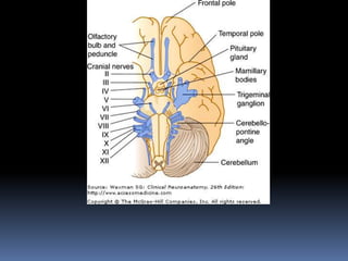
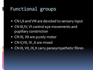
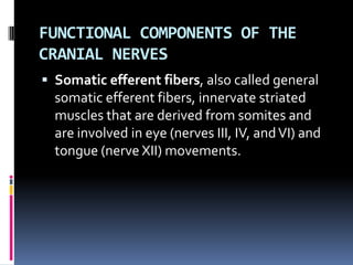
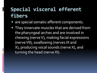
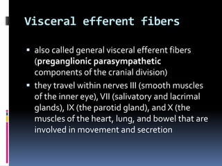
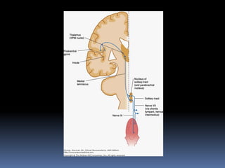
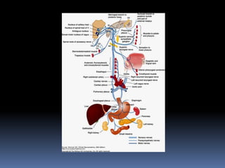
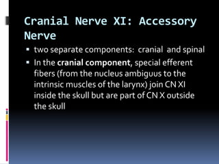
SlideShare a Scribd company logo
CRANIAL NERVES
ORIGIN OF CRANIAL NERVE FIBERS

 Cranial nerve fibers with motor (efferent)
  functions arise from collections of cells
  (motor nuclei) that lie deep within the brain
  stem
 they are homologous to the anterior horn
  cells of the spinal cord.
 Cranial nerve fibers with sensory (afferent)
  functions have their cells of origin (first-order
  nuclei) outside the brain stem, usually in
  ganglia that are homologous to the dorsal
  root ganglia of the spinal nerves
 Second-order sensory nuclei lie within the
  brain stem
Functional groups

 CN I,II and VIII are devoted to sensory input
 CN III,IV, VI control eye movements and
  pupillary constriction
 CN XI, XII are purely motor
 CN V,VII, IX ,X are mixed
 CN III, VII, IX,X carry parasympathetic fibres
FUNCTIONAL COMPONENTS OF THE
CRANIAL NERVES
 Somatic efferent fibers, also called general
  somatic efferent fibers, innervate striated
  muscles that are derived from somites and
  are involved in eye (nerves III, IV, and VI) and
  tongue (nerve XII) movements.
Special visceral efferent
fibers
 are special somatic efferent components.
 They innervate muscles that are derived from
  the pharyngeal arches and are involved in
  chewing (nerve V), making facial expressions
  (nerve VII), swallowing (nerves IX and
  X), producing vocal sounds (nerve X), and
  turning the head (nerve XI).
Visceral efferent fibers

 also called general visceral efferent fibers
  (preganglionic parasympathetic
  components of the cranial division)
 they travel within nerves III (smooth muscles
  of the inner eye), VII (salivatory and lacrimal
  glands), IX (the parotid gland), and X (the
  muscles of the heart, lung, and bowel that are
  involved in movement and secretion
Visceral afferent fibers

 also called general visceral afferent fibers,
  convey sensation from the alimentary tract,
  heart, vessels, and lungs by way of nerves IX
  and X.
 A specialized visceral afferent component is
  involved with the sense of taste; fibers
  carrying gustatory impulses are present in
  cranial nerves VII, IX, and X.
Somatic afferent fibers

 often called general somatic afferent fibers,
  convey sensation from the skin and the
  mucous membranes of the head.
 found mainly in the trigeminal nerve (V).
 A small number of afferent fibers travel with
  the facial (VII), glossopharyngeal (IX), and
  vagus (X) nerves; these fibers terminate on
  trigeminal nuclei in the brain stem.
Special sensory fibers

 found in nerves I (involved in smell), II
  (vision), and VIII (hearing and equilibrium).
Ganglia Related to Cranial Nerves


 Two types
 first type -contains cell bodies of afferent
  (somatic or visceral) axons within the cranial
  nerves.
 somewhat analogous to the dorsal root
  ganglia that contain the cell bodies of sensory
  axons within peripheral nerves.
 The second type contains the synaptic
  terminals of visceral efferent axons, together
  with postsynaptic (parasympathetic) neurons
  that project peripherally
Sensory ganglia

 semilunar (gasserian) ganglion (nerve V)
 geniculate ganglion (nerve VII)
 cochlear and vestibular ganglia (nerve VIII),
 inferior and superior glossopharyngeal
  ganglia (nerve IX)
 superior vagal ganglion (nerve X), and
  inferior vagal (nodose) ganglion (nerve X).
Parasympathetic ganglia

 ciliary ganglion (nerve III)
 the pterygopalatine and submandibular
  ganglia (VII)
 otic ganglion (IX)
 intramural ganglion (X).
 The first four of these ganglia have a close
  association with branches of CN V
 the trigeminal branches may course through
  the autonomic ganglia
Cranial Nerve IX: Glossopharyngeal
Nerve
 contains several types of fibers
 Special visceral efferent fibers from the
  nucleus ambiguus pass to the
  stylopharyngeal muscle.
 Visceral efferent (parasympathetic
  preganglionic) fibers from the inferior
  salivatory nucleus pass through the
  tympanic plexus and lesser petrosal nerve to
  the otic ganglion, from which the
  postganglionic fibers pass to the parotid
  gland.
 The inferior salivatory nucleus receives
  cortical impulses via the dorsal longitudinal
  fasciculus and reflexes from the nucleus of
  the solitary tract.
 Visceral afferent fibers arise from unipolar
  cells in the inferior ganglia.
 Centrally, they terminate in the solitary tract
  and its nucleus, which in turn projects to the
  thalamus (VPM nucleus) and then to the
  cortex
 Peripherally, the visceral afferent axons of
  nerve XI supply general sensation to the
  pharynx, soft palate, posterior third of the
  tongue, fauces, tonsils, auditory tube, and
  tympanic cavity.
 Through the sinus nerve, they supply special
  receptors in the carotid body and carotid
  sinus that are concerned with reflex control
  of respiration, blood pressure, and heart rate.
 Special visceral afferents supply the taste
  buds of the posterior third of the tongue and
  carry impulses via the superior ganglia to the
  gustatory nucleus of the brain stem.
 A few somatic afferent fibers enter by way of
  the glossopharyngeal nerve and end in the
  trigeminal nuclei.
CLINICAL CORRELATIONS

 rarely involved alone by disease processes
  (eg, by neuralgia)
 generally involved with the vagus and
  accessory nerves because of its proximity to
  them.
Pharyngeal (gag) reflex

 Depends on nerve IX for its sensory
  component, whereas nerve X innervates the
  motor component.
 Stroking the affected side of the pharynx
  does not produce gagging if the nerve is
  injured
Carotid sinus reflex

 Depends on nerve IX for its sensory
  component.
 Pressure over the sinus normally produces
  slowing of the heart rate and a fall in blood
  pressure.
Cranial Nerve X: Vagus Nerve

 Special visceral efferent fibers from the
  nucleus ambiguus contribute rootlets to the
  vagus nerve and the cranial component of the
  accessory nerve (XI).
 Those of the vagus nerve pass to the muscles
  of the soft palate and pharynx.
 Those of the accessory nerve join the vagus
  outside the skull and pass, via the recurrent
  laryngeal nerve, to the intrinsic muscles of
  the larynx
Visceral efferent fibers

 From the dorsal motor nucleus of the vagus
  course to the thoracic and abdominal viscera.
 Their postganglionic fibers arise in the
  terminal ganglia within or near the viscera.
 They inhibit heart rate and adrenal secretion
  and stimulate gastrointestinal peristalsis and
  gastric, hepatic, and pancreatic glandular
  activity
Somatic afferent fibers

 from unipolar cells in the superior (formerly
  called the jugular) ganglion send peripheral
  branches
1. via the auricular branch of CN X to the
   external auditory meatus and part of the
   earlobe
2. via the recurrent meningeal branch to the
   dura of the posterior fossa.
 Central branches pass with CN X to the brain
  stem and end in the spinal tract of the
  trigeminal nerve and its nucleus.
Visceral afferent fibers

 From unipolar cells in the inferior (formerly
  nodose) ganglion-
 peripheral branches to the
  pharynx, larynx, trachea, esophagus, and
  thoracic and abdominal viscera
 few special afferent fibers to taste buds in the
  epiglottic region
 Central branches run to the solitary tract and
  terminate in its nucleus.
 The visceral afferent fibers of CN X carry the
  sensations of abdominal distention and
  nausea and the impulses concerned with
  regulating the depth of respiration and
  controlling blood pressure.
 A few special visceral afferent fibers for taste
  from the epiglottis pass via the inferior
  ganglion to the gustatory nucleus of the brain
  stem.
CLINICAL CORRELATIONS

 CN X lesions near the skull base often involve
  CN IX and CN XI and sometimes CN XII as
  well.
 Complete bilateral transection of CN X is
  fatal.
 Unilateral lesions within the cranial vault or
  close to the base of the skull, produce
  widespread dysfunction of the
  palate, pharynx, and larynx
 The soft palate is weak and may be flaccid so
  the voice has a nasal twang.
 Weakness or paralysis of the vocal cord may
  result in hoarseness
 There can be difficulty in swallowing, and
  cardiac arrhythmias may be present.
Damage to the recurrent laryngeal
nerve
 can occur as a result of invasion or
  compression by tumor or as a complication of
  thyroid surgery
 may be accompanied by hoarseness or
  hypophonia but can be asymptomatic.
Cranial Nerve XI: Accessory
Nerve
 two separate components: cranial and spinal
 In the cranial component, special efferent
  fibers (from the nucleus ambiguus to the
  intrinsic muscles of the larynx) join CN XI
  inside the skull but are part of CN X outside
  the skull
 In the spinal component, the special efferent
  fibers from the lateral part of the anterior
  horns of the first 5 or 6 cervical cord
  segments ascend as the spinal root of CN XI
  through the foramen magnum and leave the
  cranial cavity through the jugular foramen
 They supply the sternocleidomastoid muscle
  and partly supply the trapezius muscle.
Central connections of the
spinal component
 These are those of the typical lower motor
  neuron:
 voluntary impulses via the corticospinal
  tracts
 postural impulses via the basal ganglia
 reflexes via the vestibulospinal and
  tectospinal tracts.
CLINICAL CORRELATIONS

Interruption of the spinal component leads to
1. paralysis of the sternocleidomastoid
   muscle, causing the inability to rotate the
   head to the contralateral side
2. paralysis of the upper portion of the
   trapezius muscle, which is characterized by a
   wing-like scapula and the inability to shrug
   the ipsilateral shoulder.
Cranial Nerve XII: Hypoglossal
Nerve
 Somatic efferent fibers from the hypoglossal
  nucleus in the ventromedian portion of the
  gray matter of the medulla emerge between
  the pyramid and the olive to form CN XII
 leaves the skull through the hypoglossal canal
  and passes to the muscles of the tongue
 A few proprioceptive fibers from the tongue
  course in the hypoglossal nerve and end in
  the trigeminal nuclei of the brain stem
 CN XII distributes motor branches to the
  geniohyoid and infrahyoid muscles with
  fibers derived from communicating branches
  of C1 nerve.
 A sensory recurrent meningeal branch of
  CN XII innervates the dura of the posterior
  fossa of the skull.
Cranial nerves by DR.ARSHAD

More Related Content

What's hot

Arteries of head and neck; subclavian vessels
Arteries of head and neck; subclavian vesselsArteries of head and neck; subclavian vessels
Arteries of head and neck; subclavian vessels
Dr. Mohammad Mahmoud
 
Cervical Plexus
Cervical PlexusCervical Plexus
Cervical Plexus
Rashmi Priyem Saravanan
 
Midbrain
MidbrainMidbrain
Midbrain
AMEENAWAHEED
 
The brainstem
The brainstemThe brainstem
The brainstem
Vyjayanthi Kadambi
 
Anatomy of Cerebellum - Dr.Sajith MD RD
Anatomy of Cerebellum - Dr.Sajith MD RD Anatomy of Cerebellum - Dr.Sajith MD RD
Anatomy of Cerebellum - Dr.Sajith MD RD Sajith Selvaganesan
 
Thalamus
ThalamusThalamus
Thalamus
Siva Pesala
 
Gross anatomy of human brain
Gross anatomy of human brainGross anatomy of human brain
Gross anatomy of human brain
ssjjhawar
 
Cerebrum
CerebrumCerebrum
MID BRAIN.pptx
MID BRAIN.pptxMID BRAIN.pptx
MID BRAIN.pptx
Dr. sana yaseen
 
Medulla
MedullaMedulla
Medulla
anatomysrs
 
cervical and brachial plexus
cervical and brachial plexuscervical and brachial plexus
cervical and brachial plexus
Sado Anatomist
 
Functional cortical area & white matter of cerebrum
Functional cortical area & white matter of cerebrumFunctional cortical area & white matter of cerebrum
Functional cortical area & white matter of cerebrum
Prabhakar Yadav
 
NEUROANATOMY OF BRAINSTEM
NEUROANATOMY OF BRAINSTEMNEUROANATOMY OF BRAINSTEM
NEUROANATOMY OF BRAINSTEM
Himani Kaushik
 
Pons Anatomy
Pons AnatomyPons Anatomy
Pons Anatomy
Mehul Tandel
 
Diencephalon 1
Diencephalon 1Diencephalon 1
Diencephalon 1
Dr. sana yaseen
 
Ascending tracts
Ascending tractsAscending tracts
Ascending tracts
Akash Nayak
 
Cerebrum
CerebrumCerebrum
Head and neck anatomy
Head and neck anatomyHead and neck anatomy
Head and neck anatomy
anupamasaneeth
 
Spinal cord
Spinal cord Spinal cord
Spinal cord
Prabhakar Yadav
 
Anatomy Lect 4 Neuroanatomy
Anatomy Lect 4 NeuroanatomyAnatomy Lect 4 Neuroanatomy
Anatomy Lect 4 NeuroanatomyMiami Dade
 

What's hot (20)

Arteries of head and neck; subclavian vessels
Arteries of head and neck; subclavian vesselsArteries of head and neck; subclavian vessels
Arteries of head and neck; subclavian vessels
 
Cervical Plexus
Cervical PlexusCervical Plexus
Cervical Plexus
 
Midbrain
MidbrainMidbrain
Midbrain
 
The brainstem
The brainstemThe brainstem
The brainstem
 
Anatomy of Cerebellum - Dr.Sajith MD RD
Anatomy of Cerebellum - Dr.Sajith MD RD Anatomy of Cerebellum - Dr.Sajith MD RD
Anatomy of Cerebellum - Dr.Sajith MD RD
 
Thalamus
ThalamusThalamus
Thalamus
 
Gross anatomy of human brain
Gross anatomy of human brainGross anatomy of human brain
Gross anatomy of human brain
 
Cerebrum
CerebrumCerebrum
Cerebrum
 
MID BRAIN.pptx
MID BRAIN.pptxMID BRAIN.pptx
MID BRAIN.pptx
 
Medulla
MedullaMedulla
Medulla
 
cervical and brachial plexus
cervical and brachial plexuscervical and brachial plexus
cervical and brachial plexus
 
Functional cortical area & white matter of cerebrum
Functional cortical area & white matter of cerebrumFunctional cortical area & white matter of cerebrum
Functional cortical area & white matter of cerebrum
 
NEUROANATOMY OF BRAINSTEM
NEUROANATOMY OF BRAINSTEMNEUROANATOMY OF BRAINSTEM
NEUROANATOMY OF BRAINSTEM
 
Pons Anatomy
Pons AnatomyPons Anatomy
Pons Anatomy
 
Diencephalon 1
Diencephalon 1Diencephalon 1
Diencephalon 1
 
Ascending tracts
Ascending tractsAscending tracts
Ascending tracts
 
Cerebrum
CerebrumCerebrum
Cerebrum
 
Head and neck anatomy
Head and neck anatomyHead and neck anatomy
Head and neck anatomy
 
Spinal cord
Spinal cord Spinal cord
Spinal cord
 
Anatomy Lect 4 Neuroanatomy
Anatomy Lect 4 NeuroanatomyAnatomy Lect 4 Neuroanatomy
Anatomy Lect 4 Neuroanatomy
 

Viewers also liked

Gastrointestinal physiology
Gastrointestinal physiologyGastrointestinal physiology
Gastrointestinal physiology
Chipego benkele
 
Physio Gi Hormones13.
Physio Gi Hormones13.Physio Gi Hormones13.
Physio Gi Hormones13.
Shaikhani.
 
Gastrointestinal hormones
Gastrointestinal hormonesGastrointestinal hormones
Gastrointestinal hormones
Fathima Mahmood
 
Git hormones-control-
Git hormones-control-Git hormones-control-
Git hormones-control-
Mawara Ali
 
The Gut-Brain Connection: An Inside Look at Depression
The Gut-Brain Connection: An Inside Look at DepressionThe Gut-Brain Connection: An Inside Look at Depression
The Gut-Brain Connection: An Inside Look at Depression
Augustin Bralley
 
Brain gut connections(1)
Brain gut connections(1)Brain gut connections(1)
Brain gut connections(1)
Kate Pastrich-Klemer
 
GIT Hormones
GIT Hormones GIT Hormones
Gastrointestinal hormomes & their role in secretomotor fuction of the gut
Gastrointestinal hormomes & their role in secretomotor fuction of the gutGastrointestinal hormomes & their role in secretomotor fuction of the gut
Gastrointestinal hormomes & their role in secretomotor fuction of the gutRajesh Goit
 
GIT HORMONES
GIT HORMONESGIT HORMONES
GIT HORMONES
Dr Nilesh Kate
 
Gi hormone
Gi hormoneGi hormone
Gi hormone
Atai Rabby
 

Viewers also liked (10)

Gastrointestinal physiology
Gastrointestinal physiologyGastrointestinal physiology
Gastrointestinal physiology
 
Physio Gi Hormones13.
Physio Gi Hormones13.Physio Gi Hormones13.
Physio Gi Hormones13.
 
Gastrointestinal hormones
Gastrointestinal hormonesGastrointestinal hormones
Gastrointestinal hormones
 
Git hormones-control-
Git hormones-control-Git hormones-control-
Git hormones-control-
 
The Gut-Brain Connection: An Inside Look at Depression
The Gut-Brain Connection: An Inside Look at DepressionThe Gut-Brain Connection: An Inside Look at Depression
The Gut-Brain Connection: An Inside Look at Depression
 
Brain gut connections(1)
Brain gut connections(1)Brain gut connections(1)
Brain gut connections(1)
 
GIT Hormones
GIT Hormones GIT Hormones
GIT Hormones
 
Gastrointestinal hormomes & their role in secretomotor fuction of the gut
Gastrointestinal hormomes & their role in secretomotor fuction of the gutGastrointestinal hormomes & their role in secretomotor fuction of the gut
Gastrointestinal hormomes & their role in secretomotor fuction of the gut
 
GIT HORMONES
GIT HORMONESGIT HORMONES
GIT HORMONES
 
Gi hormone
Gi hormoneGi hormone
Gi hormone
 

Similar to Cranial nerves by DR.ARSHAD

Glossopharyngeal nerve
Glossopharyngeal nerveGlossopharyngeal nerve
Glossopharyngeal nerve
RAJAT SINGHAL
 
Cranial nerves x,xi & xii
Cranial nerves x,xi & xiiCranial nerves x,xi & xii
Cranial nerves x,xi & xii
Indian dental academy
 
Vagus nerve
Vagus nerveVagus nerve
Vagus nerve
Shazeena Qaiser
 
Radiologic anatomy of the cranial nerves
Radiologic anatomy of the cranial nervesRadiologic anatomy of the cranial nerves
Radiologic anatomy of the cranial nerves
hazem youssef
 
Radiologic anatomy of the cranial nerves
Radiologic anatomy of the cranial nervesRadiologic anatomy of the cranial nerves
Radiologic anatomy of the cranial nerves
hazem youssef
 
Lower cranial nerves ajay
Lower cranial nerves ajayLower cranial nerves ajay
Lower cranial nerves ajay
drajay02
 
nerves.pdf
nerves.pdfnerves.pdf
nerves.pdf
ssuser91651b1
 
Cranial nerves VIII to XII.pptx
Cranial nerves VIII to XII.pptxCranial nerves VIII to XII.pptx
Cranial nerves VIII to XII.pptx
NikitaSaini11
 
Connecting routes in the central nervous system
Connecting routes in the central nervous systemConnecting routes in the central nervous system
Connecting routes in the central nervous systemmeducationdotnet
 
Cranial nerves
Cranial nerves Cranial nerves
Cranial nerves
Tetyana Knyazevych
 
Chap 10 the peripheral nervous system
Chap 10  the peripheral nervous systemChap 10  the peripheral nervous system
Chap 10 the peripheral nervous system
Sr Anne Ponnattil
 
USMLE NEUROANATOMY 011 CR N 10, 11, 12 VAGUS (X) Accessory nerve (XI) HYPOG...
USMLE   NEUROANATOMY 011 CR N 10, 11, 12 VAGUS (X) Accessory nerve (XI) HYPOG...USMLE   NEUROANATOMY 011 CR N 10, 11, 12 VAGUS (X) Accessory nerve (XI) HYPOG...
USMLE NEUROANATOMY 011 CR N 10, 11, 12 VAGUS (X) Accessory nerve (XI) HYPOG...
AHMED ASHOUR
 
Nerves and the Nervous System
Nerves and the Nervous SystemNerves and the Nervous System
Nerves and the Nervous System
kebnance
 
Anatomy nazeen batch cranial nerves
Anatomy nazeen batch cranial nervesAnatomy nazeen batch cranial nerves
Anatomy nazeen batch cranial nervesMBBS IMS MSU
 
Cranial nerves
Cranial nervesCranial nerves
Cranial nerves
Helao Silas
 
cranial nerves by rabia zulfiqar
 cranial nerves by rabia zulfiqar cranial nerves by rabia zulfiqar
cranial nerves by rabia zulfiqar
rabia zulfiqar
 
Glossopharyngeal nerve & its pathology ppt
Glossopharyngeal nerve & its pathology pptGlossopharyngeal nerve & its pathology ppt
Glossopharyngeal nerve & its pathology ppt
D Venkatesh Kumar
 
Trigeminal nerve maxillary nerve and clinical implication
Trigeminal nerve maxillary nerve and clinical implicationTrigeminal nerve maxillary nerve and clinical implication
Trigeminal nerve maxillary nerve and clinical implication
Dr Ravneet Kour
 
Nervouse system chapter one
Nervouse system chapter oneNervouse system chapter one
Nervouse system chapter one
Dr. Haydar Muneer Salih
 

Similar to Cranial nerves by DR.ARSHAD (20)

Glossopharyngeal nerve
Glossopharyngeal nerveGlossopharyngeal nerve
Glossopharyngeal nerve
 
Cranial nerves x,xi & xii
Cranial nerves x,xi & xiiCranial nerves x,xi & xii
Cranial nerves x,xi & xii
 
Vagus nerve
Vagus nerveVagus nerve
Vagus nerve
 
Radiologic anatomy of the cranial nerves
Radiologic anatomy of the cranial nervesRadiologic anatomy of the cranial nerves
Radiologic anatomy of the cranial nerves
 
Radiologic anatomy of the cranial nerves
Radiologic anatomy of the cranial nervesRadiologic anatomy of the cranial nerves
Radiologic anatomy of the cranial nerves
 
Lower cranial nerves ajay
Lower cranial nerves ajayLower cranial nerves ajay
Lower cranial nerves ajay
 
nerves.pdf
nerves.pdfnerves.pdf
nerves.pdf
 
Cranial nerves VIII to XII.pptx
Cranial nerves VIII to XII.pptxCranial nerves VIII to XII.pptx
Cranial nerves VIII to XII.pptx
 
Connecting routes in the central nervous system
Connecting routes in the central nervous systemConnecting routes in the central nervous system
Connecting routes in the central nervous system
 
Cranial nerves
Cranial nerves Cranial nerves
Cranial nerves
 
Clinical anatomy of 9 th cranial nerve
Clinical  anatomy  of 9 th cranial nerveClinical  anatomy  of 9 th cranial nerve
Clinical anatomy of 9 th cranial nerve
 
Chap 10 the peripheral nervous system
Chap 10  the peripheral nervous systemChap 10  the peripheral nervous system
Chap 10 the peripheral nervous system
 
USMLE NEUROANATOMY 011 CR N 10, 11, 12 VAGUS (X) Accessory nerve (XI) HYPOG...
USMLE   NEUROANATOMY 011 CR N 10, 11, 12 VAGUS (X) Accessory nerve (XI) HYPOG...USMLE   NEUROANATOMY 011 CR N 10, 11, 12 VAGUS (X) Accessory nerve (XI) HYPOG...
USMLE NEUROANATOMY 011 CR N 10, 11, 12 VAGUS (X) Accessory nerve (XI) HYPOG...
 
Nerves and the Nervous System
Nerves and the Nervous SystemNerves and the Nervous System
Nerves and the Nervous System
 
Anatomy nazeen batch cranial nerves
Anatomy nazeen batch cranial nervesAnatomy nazeen batch cranial nerves
Anatomy nazeen batch cranial nerves
 
Cranial nerves
Cranial nervesCranial nerves
Cranial nerves
 
cranial nerves by rabia zulfiqar
 cranial nerves by rabia zulfiqar cranial nerves by rabia zulfiqar
cranial nerves by rabia zulfiqar
 
Glossopharyngeal nerve & its pathology ppt
Glossopharyngeal nerve & its pathology pptGlossopharyngeal nerve & its pathology ppt
Glossopharyngeal nerve & its pathology ppt
 
Trigeminal nerve maxillary nerve and clinical implication
Trigeminal nerve maxillary nerve and clinical implicationTrigeminal nerve maxillary nerve and clinical implication
Trigeminal nerve maxillary nerve and clinical implication
 
Nervouse system chapter one
Nervouse system chapter oneNervouse system chapter one
Nervouse system chapter one
 

More from SMS_2015

Hydrocephalus by DR.ARSHAD
Hydrocephalus by DR.ARSHADHydrocephalus by DR.ARSHAD
Hydrocephalus by DR.ARSHADSMS_2015
 
Development of forebrain by DR.ARSHAD
Development of forebrain by DR.ARSHADDevelopment of forebrain by DR.ARSHAD
Development of forebrain by DR.ARSHADSMS_2015
 
Cerebellum by DR.SIDRA
Cerebellum by DR.SIDRACerebellum by DR.SIDRA
Cerebellum by DR.SIDRASMS_2015
 
meninges, dural venous sinuses part 3
 meninges, dural venous sinuses part 3 meninges, dural venous sinuses part 3
meninges, dural venous sinuses part 3SMS_2015
 
Meninges, dural venous sinuses by DR TATHEER (part-2)
 Meninges, dural venous sinuses by DR TATHEER (part-2) Meninges, dural venous sinuses by DR TATHEER (part-2)
Meninges, dural venous sinuses by DR TATHEER (part-2)SMS_2015
 
Meninges,dural venous sinuses by DR.TATHEER (part 1)
 Meninges,dural venous sinuses by DR.TATHEER (part 1) Meninges,dural venous sinuses by DR.TATHEER (part 1)
Meninges,dural venous sinuses by DR.TATHEER (part 1)SMS_2015
 
Development of brainstem by DR.ARSHAD
Development of brainstem by DR.ARSHADDevelopment of brainstem by DR.ARSHAD
Development of brainstem by DR.ARSHADSMS_2015
 
Cerebrovascular disease by DR.ARSHAD
Cerebrovascular disease by DR.ARSHADCerebrovascular disease by DR.ARSHAD
Cerebrovascular disease by DR.ARSHADSMS_2015
 
Cerebral circulation by DR.ARSHAD
Cerebral circulation by DR.ARSHADCerebral circulation by DR.ARSHAD
Cerebral circulation by DR.ARSHADSMS_2015
 
Cerebrum internal features by DR.ARSHAD
Cerebrum internal features by DR.ARSHADCerebrum internal features by DR.ARSHAD
Cerebrum internal features by DR.ARSHADSMS_2015
 
Sensory systems by DR.ARSHAD
Sensory systems by DR.ARSHADSensory systems by DR.ARSHAD
Sensory systems by DR.ARSHADSMS_2015
 
Thalamus by DR.ARSHAD
Thalamus by DR.ARSHADThalamus by DR.ARSHAD
Thalamus by DR.ARSHADSMS_2015
 
Speech disorders by DR,ARSHAD
Speech disorders by DR,ARSHADSpeech disorders by DR,ARSHAD
Speech disorders by DR,ARSHADSMS_2015
 
Speech and language by DR.ARSHAD
Speech and language by DR.ARSHADSpeech and language by DR.ARSHAD
Speech and language by DR.ARSHADSMS_2015
 
autonomic nervous system by DR.IFRA
 autonomic nervous system by DR.IFRA autonomic nervous system by DR.IFRA
autonomic nervous system by DR.IFRASMS_2015
 
Upper and lower motor neuron lesions by DR.IFRA
Upper and lower motor neuron lesions by DR.IFRAUpper and lower motor neuron lesions by DR.IFRA
Upper and lower motor neuron lesions by DR.IFRASMS_2015
 
Cerebellum by DR.ARSHAD
Cerebellum by DR.ARSHADCerebellum by DR.ARSHAD
Cerebellum by DR.ARSHADSMS_2015
 
Autonomic nervous system by DR.ARSHAD
Autonomic nervous system by DR.ARSHADAutonomic nervous system by DR.ARSHAD
Autonomic nervous system by DR.ARSHADSMS_2015
 
Mid brain by DR.TATHEER
Mid brain by DR.TATHEERMid brain by DR.TATHEER
Mid brain by DR.TATHEERSMS_2015
 
Pons by DR.TATHEER
Pons by DR.TATHEERPons by DR.TATHEER
Pons by DR.TATHEERSMS_2015
 

More from SMS_2015 (20)

Hydrocephalus by DR.ARSHAD
Hydrocephalus by DR.ARSHADHydrocephalus by DR.ARSHAD
Hydrocephalus by DR.ARSHAD
 
Development of forebrain by DR.ARSHAD
Development of forebrain by DR.ARSHADDevelopment of forebrain by DR.ARSHAD
Development of forebrain by DR.ARSHAD
 
Cerebellum by DR.SIDRA
Cerebellum by DR.SIDRACerebellum by DR.SIDRA
Cerebellum by DR.SIDRA
 
meninges, dural venous sinuses part 3
 meninges, dural venous sinuses part 3 meninges, dural venous sinuses part 3
meninges, dural venous sinuses part 3
 
Meninges, dural venous sinuses by DR TATHEER (part-2)
 Meninges, dural venous sinuses by DR TATHEER (part-2) Meninges, dural venous sinuses by DR TATHEER (part-2)
Meninges, dural venous sinuses by DR TATHEER (part-2)
 
Meninges,dural venous sinuses by DR.TATHEER (part 1)
 Meninges,dural venous sinuses by DR.TATHEER (part 1) Meninges,dural venous sinuses by DR.TATHEER (part 1)
Meninges,dural venous sinuses by DR.TATHEER (part 1)
 
Development of brainstem by DR.ARSHAD
Development of brainstem by DR.ARSHADDevelopment of brainstem by DR.ARSHAD
Development of brainstem by DR.ARSHAD
 
Cerebrovascular disease by DR.ARSHAD
Cerebrovascular disease by DR.ARSHADCerebrovascular disease by DR.ARSHAD
Cerebrovascular disease by DR.ARSHAD
 
Cerebral circulation by DR.ARSHAD
Cerebral circulation by DR.ARSHADCerebral circulation by DR.ARSHAD
Cerebral circulation by DR.ARSHAD
 
Cerebrum internal features by DR.ARSHAD
Cerebrum internal features by DR.ARSHADCerebrum internal features by DR.ARSHAD
Cerebrum internal features by DR.ARSHAD
 
Sensory systems by DR.ARSHAD
Sensory systems by DR.ARSHADSensory systems by DR.ARSHAD
Sensory systems by DR.ARSHAD
 
Thalamus by DR.ARSHAD
Thalamus by DR.ARSHADThalamus by DR.ARSHAD
Thalamus by DR.ARSHAD
 
Speech disorders by DR,ARSHAD
Speech disorders by DR,ARSHADSpeech disorders by DR,ARSHAD
Speech disorders by DR,ARSHAD
 
Speech and language by DR.ARSHAD
Speech and language by DR.ARSHADSpeech and language by DR.ARSHAD
Speech and language by DR.ARSHAD
 
autonomic nervous system by DR.IFRA
 autonomic nervous system by DR.IFRA autonomic nervous system by DR.IFRA
autonomic nervous system by DR.IFRA
 
Upper and lower motor neuron lesions by DR.IFRA
Upper and lower motor neuron lesions by DR.IFRAUpper and lower motor neuron lesions by DR.IFRA
Upper and lower motor neuron lesions by DR.IFRA
 
Cerebellum by DR.ARSHAD
Cerebellum by DR.ARSHADCerebellum by DR.ARSHAD
Cerebellum by DR.ARSHAD
 
Autonomic nervous system by DR.ARSHAD
Autonomic nervous system by DR.ARSHADAutonomic nervous system by DR.ARSHAD
Autonomic nervous system by DR.ARSHAD
 
Mid brain by DR.TATHEER
Mid brain by DR.TATHEERMid brain by DR.TATHEER
Mid brain by DR.TATHEER
 
Pons by DR.TATHEER
Pons by DR.TATHEERPons by DR.TATHEER
Pons by DR.TATHEER
 

Recently uploaded

De-mystifying Zero to One: Design Informed Techniques for Greenfield Innovati...
De-mystifying Zero to One: Design Informed Techniques for Greenfield Innovati...De-mystifying Zero to One: Design Informed Techniques for Greenfield Innovati...
De-mystifying Zero to One: Design Informed Techniques for Greenfield Innovati...
Product School
 
Leading Change strategies and insights for effective change management pdf 1.pdf
Leading Change strategies and insights for effective change management pdf 1.pdfLeading Change strategies and insights for effective change management pdf 1.pdf
Leading Change strategies and insights for effective change management pdf 1.pdf
OnBoard
 
State of ICS and IoT Cyber Threat Landscape Report 2024 preview
State of ICS and IoT Cyber Threat Landscape Report 2024 previewState of ICS and IoT Cyber Threat Landscape Report 2024 preview
State of ICS and IoT Cyber Threat Landscape Report 2024 preview
Prayukth K V
 
"Impact of front-end architecture on development cost", Viktor Turskyi
"Impact of front-end architecture on development cost", Viktor Turskyi"Impact of front-end architecture on development cost", Viktor Turskyi
"Impact of front-end architecture on development cost", Viktor Turskyi
Fwdays
 
Slack (or Teams) Automation for Bonterra Impact Management (fka Social Soluti...
Slack (or Teams) Automation for Bonterra Impact Management (fka Social Soluti...Slack (or Teams) Automation for Bonterra Impact Management (fka Social Soluti...
Slack (or Teams) Automation for Bonterra Impact Management (fka Social Soluti...
Jeffrey Haguewood
 
Bits & Pixels using AI for Good.........
Bits & Pixels using AI for Good.........Bits & Pixels using AI for Good.........
Bits & Pixels using AI for Good.........
Alison B. Lowndes
 
IOS-PENTESTING-BEGINNERS-PRACTICAL-GUIDE-.pptx
IOS-PENTESTING-BEGINNERS-PRACTICAL-GUIDE-.pptxIOS-PENTESTING-BEGINNERS-PRACTICAL-GUIDE-.pptx
IOS-PENTESTING-BEGINNERS-PRACTICAL-GUIDE-.pptx
Abida Shariff
 
DevOps and Testing slides at DASA Connect
DevOps and Testing slides at DASA ConnectDevOps and Testing slides at DASA Connect
DevOps and Testing slides at DASA Connect
Kari Kakkonen
 
FIDO Alliance Osaka Seminar: Passkeys at Amazon.pdf
FIDO Alliance Osaka Seminar: Passkeys at Amazon.pdfFIDO Alliance Osaka Seminar: Passkeys at Amazon.pdf
FIDO Alliance Osaka Seminar: Passkeys at Amazon.pdf
FIDO Alliance
 
Connector Corner: Automate dynamic content and events by pushing a button
Connector Corner: Automate dynamic content and events by pushing a buttonConnector Corner: Automate dynamic content and events by pushing a button
Connector Corner: Automate dynamic content and events by pushing a button
DianaGray10
 
Epistemic Interaction - tuning interfaces to provide information for AI support
Epistemic Interaction - tuning interfaces to provide information for AI supportEpistemic Interaction - tuning interfaces to provide information for AI support
Epistemic Interaction - tuning interfaces to provide information for AI support
Alan Dix
 
Neuro-symbolic is not enough, we need neuro-*semantic*
Neuro-symbolic is not enough, we need neuro-*semantic*Neuro-symbolic is not enough, we need neuro-*semantic*
Neuro-symbolic is not enough, we need neuro-*semantic*
Frank van Harmelen
 
ODC, Data Fabric and Architecture User Group
ODC, Data Fabric and Architecture User GroupODC, Data Fabric and Architecture User Group
ODC, Data Fabric and Architecture User Group
CatarinaPereira64715
 
Empowering NextGen Mobility via Large Action Model Infrastructure (LAMI): pav...
Empowering NextGen Mobility via Large Action Model Infrastructure (LAMI): pav...Empowering NextGen Mobility via Large Action Model Infrastructure (LAMI): pav...
Empowering NextGen Mobility via Large Action Model Infrastructure (LAMI): pav...
Thierry Lestable
 
The Art of the Pitch: WordPress Relationships and Sales
The Art of the Pitch: WordPress Relationships and SalesThe Art of the Pitch: WordPress Relationships and Sales
The Art of the Pitch: WordPress Relationships and Sales
Laura Byrne
 
To Graph or Not to Graph Knowledge Graph Architectures and LLMs
To Graph or Not to Graph Knowledge Graph Architectures and LLMsTo Graph or Not to Graph Knowledge Graph Architectures and LLMs
To Graph or Not to Graph Knowledge Graph Architectures and LLMs
Paul Groth
 
GraphRAG is All You need? LLM & Knowledge Graph
GraphRAG is All You need? LLM & Knowledge GraphGraphRAG is All You need? LLM & Knowledge Graph
GraphRAG is All You need? LLM & Knowledge Graph
Guy Korland
 
Knowledge engineering: from people to machines and back
Knowledge engineering: from people to machines and backKnowledge engineering: from people to machines and back
Knowledge engineering: from people to machines and back
Elena Simperl
 
UiPath Test Automation using UiPath Test Suite series, part 4
UiPath Test Automation using UiPath Test Suite series, part 4UiPath Test Automation using UiPath Test Suite series, part 4
UiPath Test Automation using UiPath Test Suite series, part 4
DianaGray10
 
UiPath Test Automation using UiPath Test Suite series, part 3
UiPath Test Automation using UiPath Test Suite series, part 3UiPath Test Automation using UiPath Test Suite series, part 3
UiPath Test Automation using UiPath Test Suite series, part 3
DianaGray10
 

Recently uploaded (20)

De-mystifying Zero to One: Design Informed Techniques for Greenfield Innovati...
De-mystifying Zero to One: Design Informed Techniques for Greenfield Innovati...De-mystifying Zero to One: Design Informed Techniques for Greenfield Innovati...
De-mystifying Zero to One: Design Informed Techniques for Greenfield Innovati...
 
Leading Change strategies and insights for effective change management pdf 1.pdf
Leading Change strategies and insights for effective change management pdf 1.pdfLeading Change strategies and insights for effective change management pdf 1.pdf
Leading Change strategies and insights for effective change management pdf 1.pdf
 
State of ICS and IoT Cyber Threat Landscape Report 2024 preview
State of ICS and IoT Cyber Threat Landscape Report 2024 previewState of ICS and IoT Cyber Threat Landscape Report 2024 preview
State of ICS and IoT Cyber Threat Landscape Report 2024 preview
 
"Impact of front-end architecture on development cost", Viktor Turskyi
"Impact of front-end architecture on development cost", Viktor Turskyi"Impact of front-end architecture on development cost", Viktor Turskyi
"Impact of front-end architecture on development cost", Viktor Turskyi
 
Slack (or Teams) Automation for Bonterra Impact Management (fka Social Soluti...
Slack (or Teams) Automation for Bonterra Impact Management (fka Social Soluti...Slack (or Teams) Automation for Bonterra Impact Management (fka Social Soluti...
Slack (or Teams) Automation for Bonterra Impact Management (fka Social Soluti...
 
Bits & Pixels using AI for Good.........
Bits & Pixels using AI for Good.........Bits & Pixels using AI for Good.........
Bits & Pixels using AI for Good.........
 
IOS-PENTESTING-BEGINNERS-PRACTICAL-GUIDE-.pptx
IOS-PENTESTING-BEGINNERS-PRACTICAL-GUIDE-.pptxIOS-PENTESTING-BEGINNERS-PRACTICAL-GUIDE-.pptx
IOS-PENTESTING-BEGINNERS-PRACTICAL-GUIDE-.pptx
 
DevOps and Testing slides at DASA Connect
DevOps and Testing slides at DASA ConnectDevOps and Testing slides at DASA Connect
DevOps and Testing slides at DASA Connect
 
FIDO Alliance Osaka Seminar: Passkeys at Amazon.pdf
FIDO Alliance Osaka Seminar: Passkeys at Amazon.pdfFIDO Alliance Osaka Seminar: Passkeys at Amazon.pdf
FIDO Alliance Osaka Seminar: Passkeys at Amazon.pdf
 
Connector Corner: Automate dynamic content and events by pushing a button
Connector Corner: Automate dynamic content and events by pushing a buttonConnector Corner: Automate dynamic content and events by pushing a button
Connector Corner: Automate dynamic content and events by pushing a button
 
Epistemic Interaction - tuning interfaces to provide information for AI support
Epistemic Interaction - tuning interfaces to provide information for AI supportEpistemic Interaction - tuning interfaces to provide information for AI support
Epistemic Interaction - tuning interfaces to provide information for AI support
 
Neuro-symbolic is not enough, we need neuro-*semantic*
Neuro-symbolic is not enough, we need neuro-*semantic*Neuro-symbolic is not enough, we need neuro-*semantic*
Neuro-symbolic is not enough, we need neuro-*semantic*
 
ODC, Data Fabric and Architecture User Group
ODC, Data Fabric and Architecture User GroupODC, Data Fabric and Architecture User Group
ODC, Data Fabric and Architecture User Group
 
Empowering NextGen Mobility via Large Action Model Infrastructure (LAMI): pav...
Empowering NextGen Mobility via Large Action Model Infrastructure (LAMI): pav...Empowering NextGen Mobility via Large Action Model Infrastructure (LAMI): pav...
Empowering NextGen Mobility via Large Action Model Infrastructure (LAMI): pav...
 
The Art of the Pitch: WordPress Relationships and Sales
The Art of the Pitch: WordPress Relationships and SalesThe Art of the Pitch: WordPress Relationships and Sales
The Art of the Pitch: WordPress Relationships and Sales
 
To Graph or Not to Graph Knowledge Graph Architectures and LLMs
To Graph or Not to Graph Knowledge Graph Architectures and LLMsTo Graph or Not to Graph Knowledge Graph Architectures and LLMs
To Graph or Not to Graph Knowledge Graph Architectures and LLMs
 
GraphRAG is All You need? LLM & Knowledge Graph
GraphRAG is All You need? LLM & Knowledge GraphGraphRAG is All You need? LLM & Knowledge Graph
GraphRAG is All You need? LLM & Knowledge Graph
 
Knowledge engineering: from people to machines and back
Knowledge engineering: from people to machines and backKnowledge engineering: from people to machines and back
Knowledge engineering: from people to machines and back
 
UiPath Test Automation using UiPath Test Suite series, part 4
UiPath Test Automation using UiPath Test Suite series, part 4UiPath Test Automation using UiPath Test Suite series, part 4
UiPath Test Automation using UiPath Test Suite series, part 4
 
UiPath Test Automation using UiPath Test Suite series, part 3
UiPath Test Automation using UiPath Test Suite series, part 3UiPath Test Automation using UiPath Test Suite series, part 3
UiPath Test Automation using UiPath Test Suite series, part 3
 

Cranial nerves by DR.ARSHAD

  • 2.
  • 3. ORIGIN OF CRANIAL NERVE FIBERS  Cranial nerve fibers with motor (efferent) functions arise from collections of cells (motor nuclei) that lie deep within the brain stem  they are homologous to the anterior horn cells of the spinal cord.
  • 4.  Cranial nerve fibers with sensory (afferent) functions have their cells of origin (first-order nuclei) outside the brain stem, usually in ganglia that are homologous to the dorsal root ganglia of the spinal nerves  Second-order sensory nuclei lie within the brain stem
  • 5. Functional groups  CN I,II and VIII are devoted to sensory input  CN III,IV, VI control eye movements and pupillary constriction  CN XI, XII are purely motor  CN V,VII, IX ,X are mixed  CN III, VII, IX,X carry parasympathetic fibres
  • 6. FUNCTIONAL COMPONENTS OF THE CRANIAL NERVES  Somatic efferent fibers, also called general somatic efferent fibers, innervate striated muscles that are derived from somites and are involved in eye (nerves III, IV, and VI) and tongue (nerve XII) movements.
  • 7. Special visceral efferent fibers  are special somatic efferent components.  They innervate muscles that are derived from the pharyngeal arches and are involved in chewing (nerve V), making facial expressions (nerve VII), swallowing (nerves IX and X), producing vocal sounds (nerve X), and turning the head (nerve XI).
  • 8. Visceral efferent fibers  also called general visceral efferent fibers (preganglionic parasympathetic components of the cranial division)  they travel within nerves III (smooth muscles of the inner eye), VII (salivatory and lacrimal glands), IX (the parotid gland), and X (the muscles of the heart, lung, and bowel that are involved in movement and secretion
  • 9. Visceral afferent fibers  also called general visceral afferent fibers, convey sensation from the alimentary tract, heart, vessels, and lungs by way of nerves IX and X.  A specialized visceral afferent component is involved with the sense of taste; fibers carrying gustatory impulses are present in cranial nerves VII, IX, and X.
  • 10. Somatic afferent fibers  often called general somatic afferent fibers, convey sensation from the skin and the mucous membranes of the head.  found mainly in the trigeminal nerve (V).  A small number of afferent fibers travel with the facial (VII), glossopharyngeal (IX), and vagus (X) nerves; these fibers terminate on trigeminal nuclei in the brain stem.
  • 11. Special sensory fibers  found in nerves I (involved in smell), II (vision), and VIII (hearing and equilibrium).
  • 12. Ganglia Related to Cranial Nerves  Two types  first type -contains cell bodies of afferent (somatic or visceral) axons within the cranial nerves.  somewhat analogous to the dorsal root ganglia that contain the cell bodies of sensory axons within peripheral nerves.
  • 13.  The second type contains the synaptic terminals of visceral efferent axons, together with postsynaptic (parasympathetic) neurons that project peripherally
  • 14. Sensory ganglia  semilunar (gasserian) ganglion (nerve V)  geniculate ganglion (nerve VII)  cochlear and vestibular ganglia (nerve VIII),  inferior and superior glossopharyngeal ganglia (nerve IX)  superior vagal ganglion (nerve X), and inferior vagal (nodose) ganglion (nerve X).
  • 15. Parasympathetic ganglia  ciliary ganglion (nerve III)  the pterygopalatine and submandibular ganglia (VII)  otic ganglion (IX)  intramural ganglion (X).
  • 16.  The first four of these ganglia have a close association with branches of CN V  the trigeminal branches may course through the autonomic ganglia
  • 17. Cranial Nerve IX: Glossopharyngeal Nerve  contains several types of fibers  Special visceral efferent fibers from the nucleus ambiguus pass to the stylopharyngeal muscle.
  • 18.
  • 19.  Visceral efferent (parasympathetic preganglionic) fibers from the inferior salivatory nucleus pass through the tympanic plexus and lesser petrosal nerve to the otic ganglion, from which the postganglionic fibers pass to the parotid gland.
  • 20.  The inferior salivatory nucleus receives cortical impulses via the dorsal longitudinal fasciculus and reflexes from the nucleus of the solitary tract.
  • 21.  Visceral afferent fibers arise from unipolar cells in the inferior ganglia.  Centrally, they terminate in the solitary tract and its nucleus, which in turn projects to the thalamus (VPM nucleus) and then to the cortex
  • 22.  Peripherally, the visceral afferent axons of nerve XI supply general sensation to the pharynx, soft palate, posterior third of the tongue, fauces, tonsils, auditory tube, and tympanic cavity.
  • 23.  Through the sinus nerve, they supply special receptors in the carotid body and carotid sinus that are concerned with reflex control of respiration, blood pressure, and heart rate.
  • 24.  Special visceral afferents supply the taste buds of the posterior third of the tongue and carry impulses via the superior ganglia to the gustatory nucleus of the brain stem.  A few somatic afferent fibers enter by way of the glossopharyngeal nerve and end in the trigeminal nuclei.
  • 25.
  • 26.
  • 27.
  • 28. CLINICAL CORRELATIONS  rarely involved alone by disease processes (eg, by neuralgia)  generally involved with the vagus and accessory nerves because of its proximity to them.
  • 29. Pharyngeal (gag) reflex  Depends on nerve IX for its sensory component, whereas nerve X innervates the motor component.  Stroking the affected side of the pharynx does not produce gagging if the nerve is injured
  • 30. Carotid sinus reflex  Depends on nerve IX for its sensory component.  Pressure over the sinus normally produces slowing of the heart rate and a fall in blood pressure.
  • 31. Cranial Nerve X: Vagus Nerve  Special visceral efferent fibers from the nucleus ambiguus contribute rootlets to the vagus nerve and the cranial component of the accessory nerve (XI).  Those of the vagus nerve pass to the muscles of the soft palate and pharynx.
  • 32.
  • 33.
  • 34.  Those of the accessory nerve join the vagus outside the skull and pass, via the recurrent laryngeal nerve, to the intrinsic muscles of the larynx
  • 35. Visceral efferent fibers  From the dorsal motor nucleus of the vagus course to the thoracic and abdominal viscera.  Their postganglionic fibers arise in the terminal ganglia within or near the viscera.  They inhibit heart rate and adrenal secretion and stimulate gastrointestinal peristalsis and gastric, hepatic, and pancreatic glandular activity
  • 36. Somatic afferent fibers  from unipolar cells in the superior (formerly called the jugular) ganglion send peripheral branches 1. via the auricular branch of CN X to the external auditory meatus and part of the earlobe 2. via the recurrent meningeal branch to the dura of the posterior fossa.
  • 37.  Central branches pass with CN X to the brain stem and end in the spinal tract of the trigeminal nerve and its nucleus.
  • 38. Visceral afferent fibers  From unipolar cells in the inferior (formerly nodose) ganglion-  peripheral branches to the pharynx, larynx, trachea, esophagus, and thoracic and abdominal viscera  few special afferent fibers to taste buds in the epiglottic region
  • 39.  Central branches run to the solitary tract and terminate in its nucleus.  The visceral afferent fibers of CN X carry the sensations of abdominal distention and nausea and the impulses concerned with regulating the depth of respiration and controlling blood pressure.
  • 40.  A few special visceral afferent fibers for taste from the epiglottis pass via the inferior ganglion to the gustatory nucleus of the brain stem.
  • 41. CLINICAL CORRELATIONS  CN X lesions near the skull base often involve CN IX and CN XI and sometimes CN XII as well.  Complete bilateral transection of CN X is fatal.  Unilateral lesions within the cranial vault or close to the base of the skull, produce widespread dysfunction of the palate, pharynx, and larynx
  • 42.  The soft palate is weak and may be flaccid so the voice has a nasal twang.  Weakness or paralysis of the vocal cord may result in hoarseness  There can be difficulty in swallowing, and cardiac arrhythmias may be present.
  • 43. Damage to the recurrent laryngeal nerve  can occur as a result of invasion or compression by tumor or as a complication of thyroid surgery  may be accompanied by hoarseness or hypophonia but can be asymptomatic.
  • 44.
  • 45. Cranial Nerve XI: Accessory Nerve  two separate components: cranial and spinal  In the cranial component, special efferent fibers (from the nucleus ambiguus to the intrinsic muscles of the larynx) join CN XI inside the skull but are part of CN X outside the skull
  • 46.  In the spinal component, the special efferent fibers from the lateral part of the anterior horns of the first 5 or 6 cervical cord segments ascend as the spinal root of CN XI through the foramen magnum and leave the cranial cavity through the jugular foramen  They supply the sternocleidomastoid muscle and partly supply the trapezius muscle.
  • 47. Central connections of the spinal component  These are those of the typical lower motor neuron:  voluntary impulses via the corticospinal tracts  postural impulses via the basal ganglia  reflexes via the vestibulospinal and tectospinal tracts.
  • 48. CLINICAL CORRELATIONS Interruption of the spinal component leads to 1. paralysis of the sternocleidomastoid muscle, causing the inability to rotate the head to the contralateral side 2. paralysis of the upper portion of the trapezius muscle, which is characterized by a wing-like scapula and the inability to shrug the ipsilateral shoulder.
  • 49. Cranial Nerve XII: Hypoglossal Nerve  Somatic efferent fibers from the hypoglossal nucleus in the ventromedian portion of the gray matter of the medulla emerge between the pyramid and the olive to form CN XII  leaves the skull through the hypoglossal canal and passes to the muscles of the tongue
  • 50.  A few proprioceptive fibers from the tongue course in the hypoglossal nerve and end in the trigeminal nuclei of the brain stem  CN XII distributes motor branches to the geniohyoid and infrahyoid muscles with fibers derived from communicating branches of C1 nerve.
  • 51.  A sensory recurrent meningeal branch of CN XII innervates the dura of the posterior fossa of the skull.