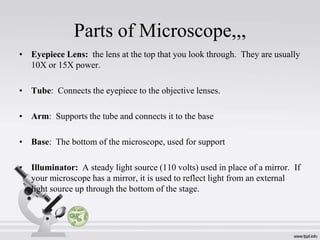
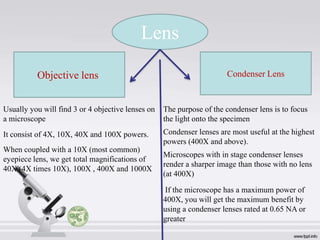
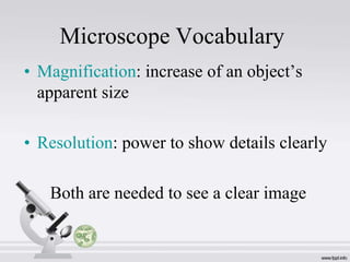
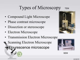
Downloaded 161 times
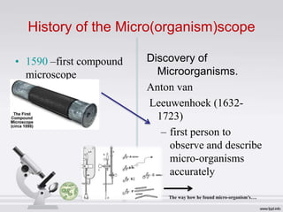
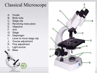

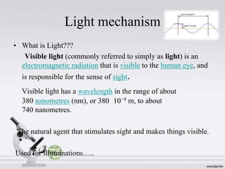

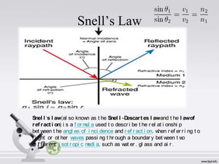
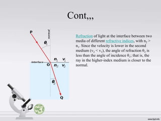
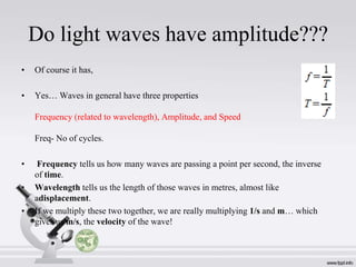
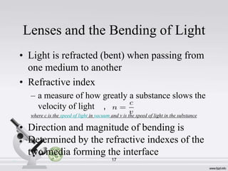
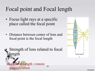
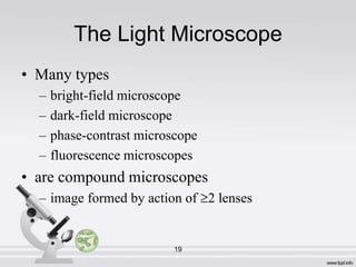
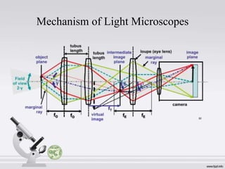
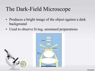
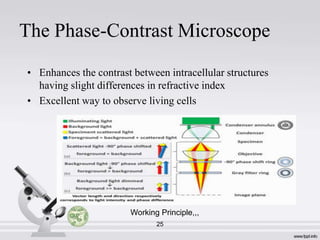
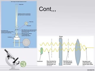
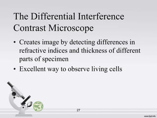

This document provides an introduction to microscopes. It discusses the history of microscopes beginning with Anton van Leeuwenhoek in the 16th century being the first to observe microorganisms. It then describes the basic parts of a classical/light microscope including the ocular lens, stage, objectives, condenser, and illuminator. It also discusses magnification, resolution, working distance, and different types of microscopy including bright field, dark field, phase contrast, and fluorescence microscopes. The document explains how light interacts with lenses and specimens to produce microscope images.