Downloaded 5,656 times

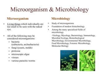
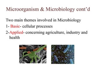
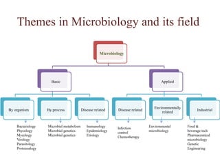
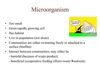
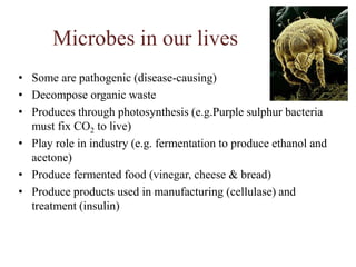
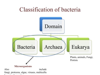
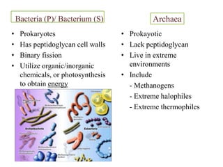
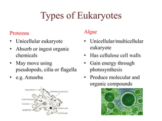
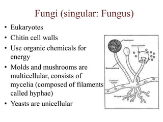
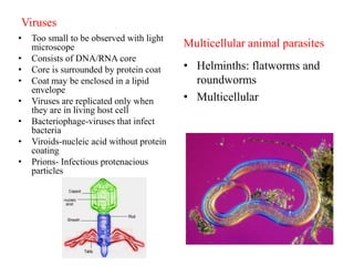
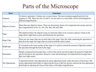
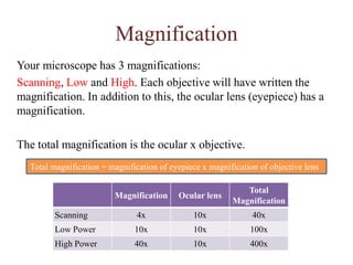
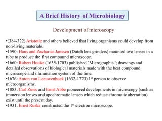
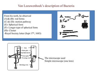


The document provides an overview of microbiology and microorganisms. It discusses that microorganisms are too small to be seen with the naked eye and includes bacteria, fungi, protozoa, algae, and viruses. It also outlines several fields of microbiology like bacteriology, mycology, and virology. The document discusses the roles microorganisms play in various industries like food production and describes how microscopy advanced the study of microbes.