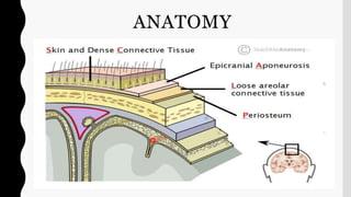
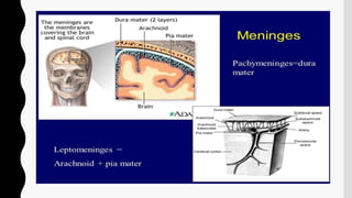
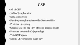
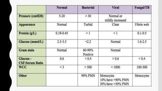
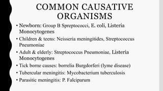
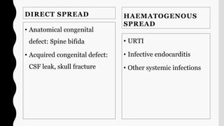
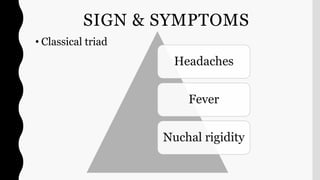
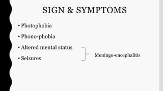
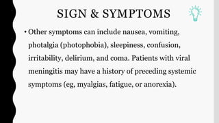
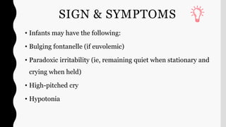
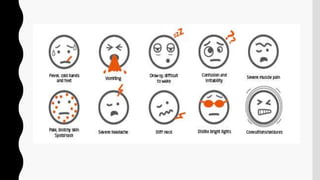
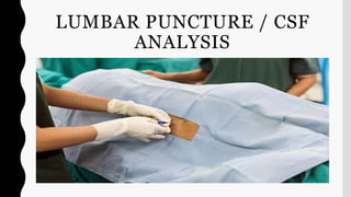
MENINGITIS
Evaluator: Mr L Anand Presenter: Shruti Shirke
[Asso professor, CON AIIMS BBSR] M.Sc Neuroscience Nursing
ANATOMY
CSF
• 1dl of CSF
• 70% of lymphocytes
• 30% Monocytes
• Few Polymorph nuclear cells (Neutrophils)
• Proteins 15 – 50mg
• Glucose 45-100 mg (2/3 of blood glucose level)
• Pressure 200mmh2O (15mmhg)
• Total CSF 150ml
• 500ml CSF produced every day
DEFINITION – MENINGITIS
• Leptomeningitis, which is more commonly referred to as meningitis, represents
inflammation of the subarachnoid space (i.e. arachnoid mater and pia mater) caused
by an infectious or noninfectious process.
DEFINITION – ENCEPHALITIS
MENINGO-ENCEPHALITIS
INCIDENCE
CAUSES/RISK FACTORS
• Autoimmune disease
• Side effects to drugs (intrathecal therapy)
• Infection (most common)
CAUSATIVE ORGANISM
• Bacteria & virus – Acute meningitis
• Fungi – Chronic meningitis
• Streptococcus Pneumoniae (~50%)
• Neisseria meningitidis (~25%)
• Group B Spreptococci (~15%)
• Listeria monocytogenes (~10%)
• Hemophilus influenzae (<10%)
In Aseptic Meningitis the cause
is viral or secondary to
lymphoma, leukemia, HIV,
HSV, MUMPS, Varicella zoster,
Enterovirus
Fungi: Cryptococcus
genuses
Coccidioides genuses
COMMON CAUSATIVE
ORGANISMS
• Newborn: Group B Spreptococci, E. coli, Listeria
Monocytogenes
• Children & teens: Neisseria meningitides, Streptococcus
Pneumoniae
• Adult & elderly: Streptococcus Pneumoniae, Listeria
Monocytogenes
• Tick borne causes: borrelia Burgdorferi (lyme disease)
• Tubercular meningitis: Mycobacterium tuberculosis
• Parasitic meningitis: P. Falciparum
DIRECT SPREAD
• Anatomical congenital
defect: Spine bifida
• Acquired congenital defect:
CSF leak, skull fracture
HAEMATOGENOUS
SPREAD
• URTI
• Infective endocarditis
• Other systemic infections
SIGN & SYMPTOMS
• Classical triad
Headaches
Fever
Nuchal rigidity
SIGN & SYMPTOMS
• Photophobia
• Phono-phobia
• Altered mental status
• Seizures
Meningo-encephalitis
SIGN & SYMPTOMS
• Other symptoms can include nausea, vomiting,
photalgia (photophobia), sleepiness, confusion,
irritability, delirium, and coma. Patients with viral
meningitis may have a history of preceding systemic
symptoms (eg, myalgias, fatigue, or anorexia).
SIGN & SYMPTOMS
• Infants may have the following:
• Bulging fontanelle (if euvolemic)
• Paradoxic irritability (ie, remaining quiet when stationary and
crying when held)
• High-pitched cry
• Hypotonia
PHYSICAL EXAMINATION
• The examination should
evaluate the following:
• Focal neurologic signs
• Signs of meningeal irritation
• Systemic and extracranial
findings
• Level of consciousness
• In chronic meningitis, it is
essential to perform careful
general, systemic, and
neurologic examinations,
looking especially for the
following:
• Lymphadenopathy
• Papilledema
• Meningismus
• Cranial nerve palsies
• Other focal neurological signs
DIAGNOSTIC CRITERIA
• Clinical signs:
• Kernig sign
• Nuckle rigidity
• Jolt maneuver
• Brudzinski sign
DIAGNOSTIC CRITERIA
• Bacterial culture, gram staining of CSF and blood are key diagnostic
tests.
• Gram stain of CSF reveals organism in 70-80% of cases.
• Serum lactic dehydrogenase (LDH) help to differentiate bacterial
form viral infections.
• CBC
• Routine investigations
LUMBAR PUNCTURE / CSF
ANALYSIS
CSF ANALYSIS (BACTERIAL)
CSF ANALYSIS (TB)
CSF ANALYSIS (VIRAL)
DIAGNOSTIC CRITERIA
• PCR: HIV, Enterovirus, HSV, Tuberculosis.
• Western blot test: Borrelia brugdoferi
• Blood smear for malaria
• ANCA/ANA
NEUROIMAGING (CT OF THE
HEAD OR MRI OF THE BRAIN)
• CT
• may be normal
• subtle hydrocephalus
• hyperdensity around basal cisterns
(especially in tuberculosis)
• leptomeningeal enhancement
• complications or sources of the meningitis
• MRI
• T1: may be normal; sulci may appear less
hypointense than normal
• T1 C+ (Gd): leptomeningeal enhancement
• FLAIR: demonstrates hyperintense signal in
CSF space, especially in the sulci
• FLAIR C+ (Gd): has shown to be more
sensitive and specific than T1 C+ (Gd)
sequence in spotting leptomeningeal
enhancement
• MR angiography: arterial narrowing or
occlusion
CT FINDING IN
MENINGITIS
Acute bacterial
meningitis. This axial
nonenhanced computed
tomography scan shows
mild ventriculomegaly
and sulcal effacement.
TREATMENT
• Prompt initiation of empiric antibacterial therapy as
appropriate for patient age and condition
• After identification of the pathogen and determination of
susceptibilities, targeted antibiotic therapy as appropriate for
patient age and condition
• Steroid (typically, dexamethasone) therapy
• In certain patients, consideration of intrathecal antibiotics.
MANAGEMENT
• Fungal meningitis - Cryptococcal (amphotericin B, flucytosine,
fluconazole), Coccidioides immitis (fluconazole, amphotericin B,
itraconazole), Histoplasma capsulatum (liposomal amphotericin B,
itraconazole), or Candida (amphotericin plus 5-flucytosine)
• Tuberculous meningitis (isoniazid, rifampin, pyrazinamide, ethambutol,
streptomycin)
• Lyme meningitis (ceftriaxone; alternatively, penicillin G, doxycycline,
chloramphenicol)
MANAGEMENT
• Dexamethasone has been shown to be beneficial ,if it is
administered 15-20 minutes before the first dose of antibiotic and
every 6 hours for next 4 days.
• Dehydration and shock are treated with fluid volume expanders.
• Seizure which may occur early in the outcome of the disease, are
controlled with phenytoin (Dilantin).
MANAGEMENT
NURSING INTERVENTIONS
• Monitor vital signs and neurological signs.
• Assess for signs of increasing ICP.
• Initiate seizure precautions.
• Monitor for seizure activity.
• Monitor for signs of meningeal irritation.
• Perform cranial nerve assessment.
• Assess peripheral vascular status.
• Maintain isolation precautions as necessary with bacterial meningitis.
• Maintain urine and stool precautions with viral meningitis.
• Maintain respiratory isolation for the client with pneumococcal meningitis.
• Elevate the head of the bed 30 degrees, and avoid neck flexion and extreme hip flexion.
• Prevent stimulation and restrict visitors.
• Administer analgesics as prescribed.
• Administer antibiotics as prescribed.
• Physiological response: Neurological examination; vital signs;
presence of fever; adequacy of airway, breathing, and
circulation
• Fluid and electrolyte balance: Intake and output, body weight,
skin turgor, abnormal serum electrolytes
• Complications: Seizure activity, decreased mental status,
fever, increased ICP
DISCHARGE AND HOME
HEALTHCARE GUIDELINES
• Explain all medications and include the mechanism of action, dosage, route, and side
effects.
• Explain any drug interactions or food interactions.
• Instruct the patient to notify the primary healthcare provider for signs and symptoms
of complications, such as fever, seizures, developmental delays, or behavior changes.
• Provide referrals and teaching specific to the identified neurological deficits.
• Encourage the parents to maintain appropriate activities to facilitate the growth and
development of the child.
BRAIN ABSCESS
S H R U T I S H I R K E
DEFINITION
• A brain abscess is a collection of infectious material within the
tissue of the brain.
• Brain abscess is a focal area of necrosis with a surrounding
membrane within the brain parenchyma, usually resulting from an
infectious process or rarely from a traumatic process.

Meningitis and brain abscess

  • 1.
    MENINGITIS Evaluator: Mr LAnand Presenter: Shruti Shirke [Asso professor, CON AIIMS BBSR] M.Sc Neuroscience Nursing
  • 2.
  • 4.
    CSF • 1dl ofCSF • 70% of lymphocytes • 30% Monocytes • Few Polymorph nuclear cells (Neutrophils) • Proteins 15 – 50mg • Glucose 45-100 mg (2/3 of blood glucose level) • Pressure 200mmh2O (15mmhg) • Total CSF 150ml • 500ml CSF produced every day
  • 7.
    DEFINITION – MENINGITIS •Leptomeningitis, which is more commonly referred to as meningitis, represents inflammation of the subarachnoid space (i.e. arachnoid mater and pia mater) caused by an infectious or noninfectious process.
  • 8.
  • 9.
  • 10.
  • 11.
    CAUSES/RISK FACTORS • Autoimmunedisease • Side effects to drugs (intrathecal therapy) • Infection (most common)
  • 13.
    CAUSATIVE ORGANISM • Bacteria& virus – Acute meningitis • Fungi – Chronic meningitis • Streptococcus Pneumoniae (~50%) • Neisseria meningitidis (~25%) • Group B Spreptococci (~15%) • Listeria monocytogenes (~10%) • Hemophilus influenzae (<10%) In Aseptic Meningitis the cause is viral or secondary to lymphoma, leukemia, HIV, HSV, MUMPS, Varicella zoster, Enterovirus Fungi: Cryptococcus genuses Coccidioides genuses
  • 14.
    COMMON CAUSATIVE ORGANISMS • Newborn:Group B Spreptococci, E. coli, Listeria Monocytogenes • Children & teens: Neisseria meningitides, Streptococcus Pneumoniae • Adult & elderly: Streptococcus Pneumoniae, Listeria Monocytogenes • Tick borne causes: borrelia Burgdorferi (lyme disease) • Tubercular meningitis: Mycobacterium tuberculosis • Parasitic meningitis: P. Falciparum
  • 15.
    DIRECT SPREAD • Anatomicalcongenital defect: Spine bifida • Acquired congenital defect: CSF leak, skull fracture HAEMATOGENOUS SPREAD • URTI • Infective endocarditis • Other systemic infections
  • 16.
    SIGN & SYMPTOMS •Classical triad Headaches Fever Nuchal rigidity
  • 18.
    SIGN & SYMPTOMS •Photophobia • Phono-phobia • Altered mental status • Seizures Meningo-encephalitis
  • 19.
    SIGN & SYMPTOMS •Other symptoms can include nausea, vomiting, photalgia (photophobia), sleepiness, confusion, irritability, delirium, and coma. Patients with viral meningitis may have a history of preceding systemic symptoms (eg, myalgias, fatigue, or anorexia).
  • 20.
    SIGN & SYMPTOMS •Infants may have the following: • Bulging fontanelle (if euvolemic) • Paradoxic irritability (ie, remaining quiet when stationary and crying when held) • High-pitched cry • Hypotonia
  • 22.
    PHYSICAL EXAMINATION • Theexamination should evaluate the following: • Focal neurologic signs • Signs of meningeal irritation • Systemic and extracranial findings • Level of consciousness • In chronic meningitis, it is essential to perform careful general, systemic, and neurologic examinations, looking especially for the following: • Lymphadenopathy • Papilledema • Meningismus • Cranial nerve palsies • Other focal neurological signs
  • 23.
    DIAGNOSTIC CRITERIA • Clinicalsigns: • Kernig sign • Nuckle rigidity • Jolt maneuver • Brudzinski sign
  • 25.
    DIAGNOSTIC CRITERIA • Bacterialculture, gram staining of CSF and blood are key diagnostic tests. • Gram stain of CSF reveals organism in 70-80% of cases. • Serum lactic dehydrogenase (LDH) help to differentiate bacterial form viral infections. • CBC • Routine investigations
  • 26.
    LUMBAR PUNCTURE /CSF ANALYSIS
  • 27.
  • 28.
  • 29.
  • 30.
    DIAGNOSTIC CRITERIA • PCR:HIV, Enterovirus, HSV, Tuberculosis. • Western blot test: Borrelia brugdoferi • Blood smear for malaria • ANCA/ANA
  • 31.
    NEUROIMAGING (CT OFTHE HEAD OR MRI OF THE BRAIN) • CT • may be normal • subtle hydrocephalus • hyperdensity around basal cisterns (especially in tuberculosis) • leptomeningeal enhancement • complications or sources of the meningitis • MRI • T1: may be normal; sulci may appear less hypointense than normal • T1 C+ (Gd): leptomeningeal enhancement • FLAIR: demonstrates hyperintense signal in CSF space, especially in the sulci • FLAIR C+ (Gd): has shown to be more sensitive and specific than T1 C+ (Gd) sequence in spotting leptomeningeal enhancement • MR angiography: arterial narrowing or occlusion
  • 32.
    CT FINDING IN MENINGITIS Acutebacterial meningitis. This axial nonenhanced computed tomography scan shows mild ventriculomegaly and sulcal effacement.
  • 34.
    TREATMENT • Prompt initiationof empiric antibacterial therapy as appropriate for patient age and condition • After identification of the pathogen and determination of susceptibilities, targeted antibiotic therapy as appropriate for patient age and condition • Steroid (typically, dexamethasone) therapy • In certain patients, consideration of intrathecal antibiotics.
  • 35.
    MANAGEMENT • Fungal meningitis- Cryptococcal (amphotericin B, flucytosine, fluconazole), Coccidioides immitis (fluconazole, amphotericin B, itraconazole), Histoplasma capsulatum (liposomal amphotericin B, itraconazole), or Candida (amphotericin plus 5-flucytosine) • Tuberculous meningitis (isoniazid, rifampin, pyrazinamide, ethambutol, streptomycin) • Lyme meningitis (ceftriaxone; alternatively, penicillin G, doxycycline, chloramphenicol)
  • 36.
    MANAGEMENT • Dexamethasone hasbeen shown to be beneficial ,if it is administered 15-20 minutes before the first dose of antibiotic and every 6 hours for next 4 days. • Dehydration and shock are treated with fluid volume expanders. • Seizure which may occur early in the outcome of the disease, are controlled with phenytoin (Dilantin).
  • 37.
  • 38.
    NURSING INTERVENTIONS • Monitorvital signs and neurological signs. • Assess for signs of increasing ICP. • Initiate seizure precautions. • Monitor for seizure activity. • Monitor for signs of meningeal irritation. • Perform cranial nerve assessment. • Assess peripheral vascular status. • Maintain isolation precautions as necessary with bacterial meningitis. • Maintain urine and stool precautions with viral meningitis. • Maintain respiratory isolation for the client with pneumococcal meningitis. • Elevate the head of the bed 30 degrees, and avoid neck flexion and extreme hip flexion. • Prevent stimulation and restrict visitors. • Administer analgesics as prescribed. • Administer antibiotics as prescribed.
  • 39.
    • Physiological response:Neurological examination; vital signs; presence of fever; adequacy of airway, breathing, and circulation • Fluid and electrolyte balance: Intake and output, body weight, skin turgor, abnormal serum electrolytes • Complications: Seizure activity, decreased mental status, fever, increased ICP
  • 40.
    DISCHARGE AND HOME HEALTHCAREGUIDELINES • Explain all medications and include the mechanism of action, dosage, route, and side effects. • Explain any drug interactions or food interactions. • Instruct the patient to notify the primary healthcare provider for signs and symptoms of complications, such as fever, seizures, developmental delays, or behavior changes. • Provide referrals and teaching specific to the identified neurological deficits. • Encourage the parents to maintain appropriate activities to facilitate the growth and development of the child.
  • 41.
    BRAIN ABSCESS S HR U T I S H I R K E
  • 42.
    DEFINITION • A brainabscess is a collection of infectious material within the tissue of the brain. • Brain abscess is a focal area of necrosis with a surrounding membrane within the brain parenchyma, usually resulting from an infectious process or rarely from a traumatic process.