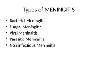
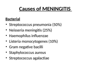
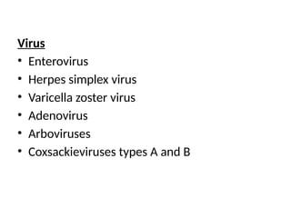
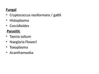
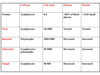
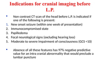
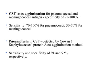
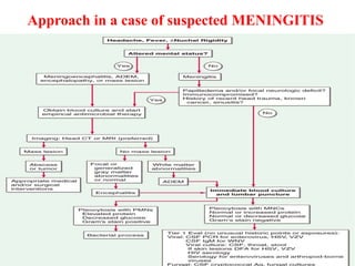
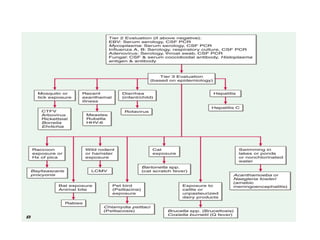
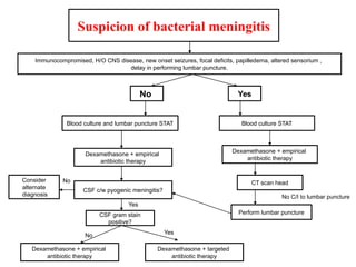
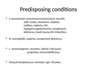
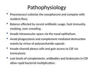
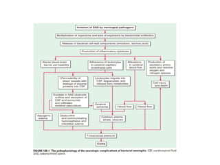
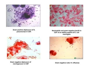
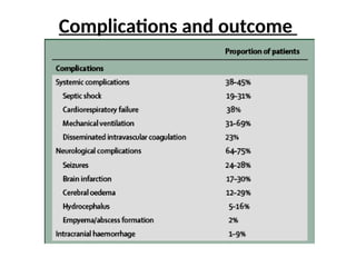
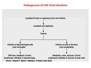
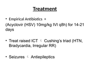
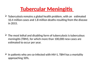
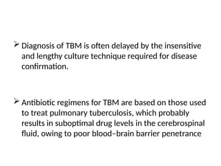
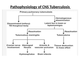
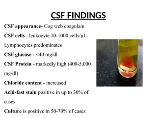
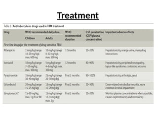
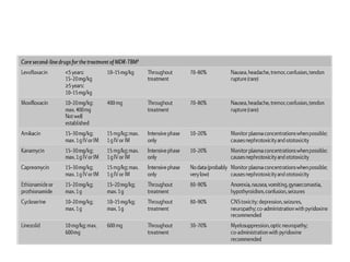
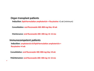
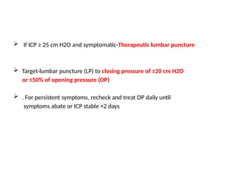
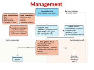
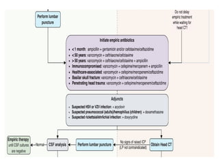
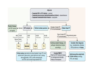
PRESENTED BY
Dr.Neelima second year resident
Dr.Y. M.M. Ganesh first year resident
Dr.S. Karthik first year resident
MENINGITIS
• Introduction
• Types of meningitis
• Causes
• Clinical presentation
• Investigations
• Lumbar puncture procedure
• Approach in case of suspected meningitis
• Management Algorithm
• Brief overveiw of different types of meningitis
DEFINITION
Meningitis is an
inflammation of the
leptomeninges
(arachnoid and pia
mater) surrounding
the brain and spinal
cord, with involvement
of the subarachnoid
space
Introduction
Based on duration
⮚ Acute meningitis
⮚ Subacute meningitis
⮚ Chronic meningitis/Recurrent meningitis
Based on blood culture
⮚ Septic meningitis
⮚ Aseptic meningitis
Types of MENINGITIS
• Bacterial Meningitis
• Fungal Meningitis
• Viral Meningitis
• Parasitic Meningitis
• Non infectious Meningitis
Bacterial
• Streptococcus pneumonia (50%)
• Neisseria meningitis (25%)
• Haemophilus influenzae
• Listeria monocytogenes (10%)
• Gram negative bacilli
• Staphylococcus aureus
• Streptococcus agalactiae
Causes of MENINGITIS
Virus
• Enterovirus
• Herpes simplex virus
• Varicella zoster virus
• Adenovirus
• Arboviruses
• Coxsackieviruses types A and B
Fungal
• Cryptococcus neoformans / gattii
• Histoplasma
• Coccidioides
Parasitic
• Taenia solium
• Naeglaria Floweri
• Toxoplasma
• Acanthamoeba
Drug
• Anti-CD3 monoclonal antibody
• Azathioprine
• Ibuprofen
• Other NSAIDs
• Trimethoprim-sulfamethoxazole
Malignancy
• Leukemia
• Lymphoma
• Metastatic carcinomas and adenocarcinomas
Autoimmune
• Sarcoidosis
• Systemic lyupus erythematosus
• Vogt-Koyanagi-Harada syndrome
Other
• Epidermoid cyst
• Postvaccination
Lancet Neurol 2008; 7: 637–48
Common signs and symptoms of acute
bacterial meningitis
Clinical presentation
• Classic triad of fever, neck stiffness, and headache seen in
only 2/3rd
of adults.
• More common in pneumococcal than other bacterial
meningitis.
• 1 of the 3 elements present in almost all patients.
• Absence of all 3 rules out acute meningitis with sensitivity of
99-100%.
• Usually history is of 24 to 48 hours.
 Rash most commonly seen with meningococcal
disease (92% of meningitis cases with rash).
 Presence of shock, rash or clustering of cases should
raise suspicion of meningococcemia.
Petechial skin rash that accompanies
meningitis due to Neisseria meningitidis.
Fine petechial rash in disseminated infection and
meningitis due to Staphylococcus aureus
 Kernig’s and Brudzinski’s sign have poor
sensitivity (5%) with high specificity (95%)
 Nuchal rigidity has low sensitivity (30%) and
specificity (68%).
 ↑ TLC (mean 10,600/cc vs 8900/cc).
 ↓Platelet counts- systemic infection, sepsis.
 **Hyponatremia (serum Na < 135mEq/l) seen in 30% cases.
- Severe (Na <130) in 6%. (resolves spontaneously)
- More common with L. monocytogenes and in
patients with symptoms > 24 hours.
 Blood culture- should be taken immediately, before starting
antibiotics. Positive in 74% cases.
**QJ Med 2007;100:37-
Investigations
▪ Acute inflammatory markers - ESR, CRP and Procalcitonin
elevated : distinguish acute bacterial from non bacterial
meningitis.
▪ Diagnostic lumbar puncture - performed in all cases unless
specific contraindications present-
i) local infection at puncture site
ii) subdural abscess
iii) bleeding diathesis
iv) septic shock- diastolic BP < 60 mm of Hg
v) mass lesion/ ventricular obstruction/
brain shift on cranial imaging
Cell type Cell count Glucose Protein
Normal Lymphocytes 0-4 >60% of blood
glucose
< 0.45 mg/dl
Viral Lymphocytes 10-2000 Normal Normal
Bacterial Polymorphs 1000-5000 Decreased Increased
Tubercular Lymphocytes/
polymorphs
50-5000 Decreased Increased
Fungal Lymphocytes 50-500 Decreased Increased
Indications for cranial imaging before
L.P.
 Non contrast CT scan of the head before L.P. is indicated if
one of the following is present:
1. New onset seizure (within one week of presentation)
2. Immunocompromised state
3. Papilledema
4. Focal neurological signs (excluding hearing loss)
5. Moderate to severe impairment of consciousness (GCS <10)
 Absence of all these features has 97% negative predictive
value for an intra cranial abnormality that would preclude a
lumbar puncture
• CSF latex agglutination for pneumococcal and
meningococcal antigen - specificity of 95-100%.
• Sensitivity 70-100% for pneumococci, 30-70% for
meningococcci.
• Pneumolysin in CSF - detected by Cowan 1
Staphylococccal protein A co-agglutination method.
• Sensitivity and specificity of 91 and 92%
respectively.
Approach in a case of suspected MENINGITIS
Suspicion of bacterial meningitis
Immunocompromised, H/O CNS disease, new onset seizures, focal deficits, papilledema, altered sensorium ,
delay in performing lumbar puncture.
Blood culture STAT
Dexamethasone + empirical
antibiotic therapy
CT scan head
Perform lumbar puncture
No C/I to lumbar puncture
Yes
No
Blood culture and lumbar puncture STAT
Dexamethasone + empirical
antibiotic therapy
CSF c/w pyogenic meningitis?
Consider
alternate
diagnosis
No
Yes
CSF gram stain
positive?
Dexamethasone + empirical
antibiotic therapy
Dexamethasone + targeted
antibiotic therapy
No Yes
 Definition- Acute purulent infection within the subarachnoid space.
 Meninges, subarachnoid space and brain parenchyma frequently
involved together-meningoencephalitis.
 Incidence- 0.6-4/1,00,000 adults in developed nations.
 Upto 10 times higher in developing nations.
 Mortality 16% to 37% despite modern antibiotics.
Lancet Infect Dis 2007; 7:191–200
Lancet Neurol 2006; 5: 332–42
BACTERIAL MENINGITIS
 In developed world MC organism in both children and adults is S.
pneumoniae ≈ 50%
 N. meningitidis ≈ 25%
 Group B Streptococcus ≈ 15%
 Listeria monocytogenes ≈10%
 H. influenzae < 10%
 Staphylococcal species and gram negative bacilli in special circumstances.
Tubercular and cryptococcal meningitis common in HIV
 Maybe difficult to distinguish from acute bacterial meningitis.
Etiology
Predisposing conditions
 S. pneumoniae: pneumococcal pneumonia, sinusitis
otitis media, alcoholism, diabetes
mellitus, asplenia, HIV,
hypogammaglobulinemia, complement
deficiency, head trauma,CSF rhinorrhea.
 N. meningitidis: asplenia, complement deficiency.
 L. monocytogenes: neonates, elderly (>60 years),
pregnancy, immunodeficiency.
 Group B streptococcus: neonates, age> 50 years.
 Pneumococci colonize the nasopharynx and compete with
resident flora.
 Balance effected by recent antibiotic usage, host immunity,
smoking, over crowding.
 Invade intravascular space via the nasal epithelium.
 Avoid phagocytosis and complement mediated destruction
mainly by virtue of polysaccharide capsule.
 Invade choroid plexus cells and gain access to CSF via
transcytosis.
 Low levels of complements, antibodies and leukocytes in CSF
allow rapid bacterial multiplication.
Pathophysiology
Gram positive diplococci of S.
pneumoniae in CSF.
Neutrophils and gram negative bacilli in
CSF of an elderly patient of E. coli
meningitis
Gram negative rods- H. influenza
Gram negative diplococci of
meningococci in CSF
Turbid CSF with fibrous spider clot after 1 hour in pyogenic meningitis
Lancet Neurol 2008; 7: 637–48
Clinical Infectious Diseases 2004; 39:1267–84
 WHO recommendation – 5 day antibiotic therapy
 To be extended if-
– Immunocompromise
– Persistent fever
– Persistent seizures
– Coma
Duration of antibiotic therapy
 Mortality and rate of neurological sequelae remain high
despite appropriate antimicrobial therapy.
 Due to adverse effects of inflammatory cytokines.
 Corticosteroids act by
– inhibiting synthesis of IL-1 and TNF at m-RNA level
– decreasing CSF outflow resistance and
– stabilizing the BBB.
 Once macrophages and microglia activated and TNF
production induced, steroids have less effect.
Role of corticosteroids
 Hence steroids to be given early, with or before 1st
dose of
antibiotics to have maximum effect.
 Duration of this window of opportunity not described.
 Dose- Dexamethasone 0.15 mg/kg I.V. 6th
hourly for 4 days is
the most widely recommended dose.
 Treatment of raised ICP –
- Head end elevation (30°-45°)
- IV mannitol (25-100 g 4th
hourly)
- Intubation and hyperventilation (PaCO2 25-30 mm of
Hg). Maintain ICP below 20 mm of Hg.
 Maintain blood pressure and urine output.
 Aggressive fluid resuscitation to be avoided for fear
of hyponatremia.
Supportive treatment
Lancet Neurol 2006; 5: 332–42
Complications and outcome
 Viral infection of the nervous system can
result in a myriad of clinical
presentations occurring separately or in
combinations including acute or
chronic meningitis, encephalitis,
myelitis, ganglionitis, and
polyradiculitis.
 Viruses may also incite para- or post-
infectious CNS inflammatory or
autoimmune syndromes such as acute
disseminated encephalomyelitis
(ADEM) or encephalitis associated with
autoantibodies
VIRAL MENINGITIS
Etiology
• Leading cause of viral meningitis
– Neonates- fever, vomiting, anorexia, rash, upper respiratory tract
symptoms, meningeal signs (nuchal rigidity, bulging antetrior
fontanelle) +/-
Severe form with hepatic necrosis, myocarditis, necrotizing
enterocolitis, encephalitis
– Children, adults- fever, headache, neck stiffness, photophobia
Anorexia, vomiting, rash, diarrhoea, cough, pharyngitis, myalgia
• h/o community enteroviral outbreaks, rash, conjunctivitis,
pleurodynia, pericarditis, herpangina
HSV 2 meningitis
• Neurological complications- urinary
retention, dysaesthesis, paraesthesia,
neuralgia, motor weakness,
paraparesis, difficulty in concentration,
impaired hearing, usually resolve in 3-
6 months
EBV meningitis
• Associated with pharyngitis, lymph
adenopathy, splenomegaly
VZV meningitis
• Associated with diffuse vesicular rash
Pathogenesis of CNS Viral infections
• Empirical Antibiotics +
(Acyclovir (HSV) 10mg/kg IVI q8h) for 14-21
days
• Treat raised ICT  Cushing’s triad (HTN,
Bradycardia, Irregular RR)
• Seizures  Antiepileptics
Treatment
 Tuberculosis remains a global health problem, with an estimated
10.4 million cases and 1.8 million deaths resulting from the disease
in 2015.
 The most lethal and disabling form of tuberculosis is tuberculous
meningitis (TBM), for which more than 100,000 new cases are
estimated to occur per year.
 In patients who are co-infected with HIV-1, TBM has a mortality
approaching 50%.
Tubercular Meningitis
 Diagnosis of TBM is often delayed by the insensitive
and lengthy culture technique required for disease
confirmation.
 Antibiotic regimens for TBM are based on those used
to treat pulmonary tuberculosis, which probably
results in suboptimal drug levels in the cerebrospinal
fluid, owing to poor blood–brain barrier penetrance
Pathophysiology of CNS Tuberculosis
CSF appearance- Cog web coagulam
CSF cells - leukocyte 10-1000 cells/µl -
Lymphocytes predominates
CSF glucose - <40 mg/dl
CSF Protein - markedly high (400-5,000
mg/dl)
Chloride content - increased
Acid-fast stain positive in up to 30% of
cases
Culture is positive in 50-70% of cases
CSF FINDINGS
Treatment
A 6-month course of therapy is acceptable, but therapy should
be prolonged for 9–12 months in patients who have-
• an inadequate resolution of symptoms of meningitis
• positive mycobacterial cultures of CSF during the course of
therapy.
• Dexamethasone therapy is recommended for patients with
tuberculous meningitis. The dose is 12–16 mg/d for 3
weeks,and then tapered over 3 weeks
• Typically acquired by the inhalation of airborne
fungal spores.
• The initial pulmonary infection may be
asymptomatic or present with fever, cough,
sputum production, and chest pain and may
later disseminate to
CNS(immunocompromised hosts)
Fungal Meningitis
Mode of Infection
MOMOMODEmm
The most common pathogen causing fungal meningitis is C.
neoformans
HIV patients
 Induction: liposomal amphotericin B + flucytosine x 2 weeks
followed by 1 wk of fluconazole (1200 mg/day, adult)
 Alternate induction:
fluconazole (400-1200 mg/day, adult) + flucytosine ×2 wk
 Consolidation: oral fluconazole 800 mg/day ×8 wk (minimum)
 Maintenance/secondary prophylaxis: oral fluconazole 200 mg/day
Corticosteroids: not recommended during induction
Antiretroviral therapy (ART) initiation: defer for 4–6 wk from start of antifungal
treatment
Treatment
Organ transplant patients
Induction: lipid-formulation amphotericin + flucytosine ×2 wk (minimum)
Consolidation: oral fluconazole 400–800 mg/day ×8 wk
Maintenance: oral fluconazole 200–400 mg ×6–12 mo
Immunocompetent patients
Induction: amphotericin B/lipid-formulation amphotericin +
flucytosine ×4 wk
Consolidation: oral fluconazole 400–800 mg/day ×8 wk
Maintenance: oral fluconazole 200–400 mg ×6–12 mo
•
 If ICP ≥ 25 cm H2O and symptomatic-Therapeutic lumbar puncture
 Target-lumbar puncture (LP) to closing pressure of ≤20 cm H2O
or ≤50% of opening pressure (OP)
 . For persistent symptoms, recheck and treat OP daily until
symptoms abate or ICP stable ×2 days
Management
THANK YOU

acute meningitis .pptxbxkxmcmcmcmccmcmmcm

  • 1.
    PRESENTED BY Dr.Neelima secondyear resident Dr.Y. M.M. Ganesh first year resident Dr.S. Karthik first year resident MENINGITIS
  • 2.
    • Introduction • Typesof meningitis • Causes • Clinical presentation • Investigations • Lumbar puncture procedure • Approach in case of suspected meningitis • Management Algorithm • Brief overveiw of different types of meningitis
  • 3.
    DEFINITION Meningitis is an inflammationof the leptomeninges (arachnoid and pia mater) surrounding the brain and spinal cord, with involvement of the subarachnoid space Introduction
  • 4.
    Based on duration ⮚Acute meningitis ⮚ Subacute meningitis ⮚ Chronic meningitis/Recurrent meningitis Based on blood culture ⮚ Septic meningitis ⮚ Aseptic meningitis
  • 5.
    Types of MENINGITIS •Bacterial Meningitis • Fungal Meningitis • Viral Meningitis • Parasitic Meningitis • Non infectious Meningitis
  • 6.
    Bacterial • Streptococcus pneumonia(50%) • Neisseria meningitis (25%) • Haemophilus influenzae • Listeria monocytogenes (10%) • Gram negative bacilli • Staphylococcus aureus • Streptococcus agalactiae Causes of MENINGITIS
  • 7.
    Virus • Enterovirus • Herpessimplex virus • Varicella zoster virus • Adenovirus • Arboviruses • Coxsackieviruses types A and B
  • 8.
    Fungal • Cryptococcus neoformans/ gattii • Histoplasma • Coccidioides Parasitic • Taenia solium • Naeglaria Floweri • Toxoplasma • Acanthamoeba
  • 9.
    Drug • Anti-CD3 monoclonalantibody • Azathioprine • Ibuprofen • Other NSAIDs • Trimethoprim-sulfamethoxazole Malignancy • Leukemia • Lymphoma • Metastatic carcinomas and adenocarcinomas
  • 10.
    Autoimmune • Sarcoidosis • Systemiclyupus erythematosus • Vogt-Koyanagi-Harada syndrome Other • Epidermoid cyst • Postvaccination
  • 11.
    Lancet Neurol 2008;7: 637–48 Common signs and symptoms of acute bacterial meningitis Clinical presentation
  • 12.
    • Classic triadof fever, neck stiffness, and headache seen in only 2/3rd of adults. • More common in pneumococcal than other bacterial meningitis. • 1 of the 3 elements present in almost all patients. • Absence of all 3 rules out acute meningitis with sensitivity of 99-100%. • Usually history is of 24 to 48 hours.
  • 13.
     Rash mostcommonly seen with meningococcal disease (92% of meningitis cases with rash).  Presence of shock, rash or clustering of cases should raise suspicion of meningococcemia. Petechial skin rash that accompanies meningitis due to Neisseria meningitidis. Fine petechial rash in disseminated infection and meningitis due to Staphylococcus aureus
  • 14.
     Kernig’s andBrudzinski’s sign have poor sensitivity (5%) with high specificity (95%)  Nuchal rigidity has low sensitivity (30%) and specificity (68%).
  • 15.
     ↑ TLC(mean 10,600/cc vs 8900/cc).  ↓Platelet counts- systemic infection, sepsis.  **Hyponatremia (serum Na < 135mEq/l) seen in 30% cases. - Severe (Na <130) in 6%. (resolves spontaneously) - More common with L. monocytogenes and in patients with symptoms > 24 hours.  Blood culture- should be taken immediately, before starting antibiotics. Positive in 74% cases. **QJ Med 2007;100:37- Investigations
  • 16.
    ▪ Acute inflammatorymarkers - ESR, CRP and Procalcitonin elevated : distinguish acute bacterial from non bacterial meningitis. ▪ Diagnostic lumbar puncture - performed in all cases unless specific contraindications present- i) local infection at puncture site ii) subdural abscess iii) bleeding diathesis iv) septic shock- diastolic BP < 60 mm of Hg v) mass lesion/ ventricular obstruction/ brain shift on cranial imaging
  • 17.
    Cell type Cellcount Glucose Protein Normal Lymphocytes 0-4 >60% of blood glucose < 0.45 mg/dl Viral Lymphocytes 10-2000 Normal Normal Bacterial Polymorphs 1000-5000 Decreased Increased Tubercular Lymphocytes/ polymorphs 50-5000 Decreased Increased Fungal Lymphocytes 50-500 Decreased Increased
  • 18.
    Indications for cranialimaging before L.P.  Non contrast CT scan of the head before L.P. is indicated if one of the following is present: 1. New onset seizure (within one week of presentation) 2. Immunocompromised state 3. Papilledema 4. Focal neurological signs (excluding hearing loss) 5. Moderate to severe impairment of consciousness (GCS <10)  Absence of all these features has 97% negative predictive value for an intra cranial abnormality that would preclude a lumbar puncture
  • 19.
    • CSF latexagglutination for pneumococcal and meningococcal antigen - specificity of 95-100%. • Sensitivity 70-100% for pneumococci, 30-70% for meningococcci. • Pneumolysin in CSF - detected by Cowan 1 Staphylococccal protein A co-agglutination method. • Sensitivity and specificity of 91 and 92% respectively.
  • 20.
    Approach in acase of suspected MENINGITIS
  • 22.
    Suspicion of bacterialmeningitis Immunocompromised, H/O CNS disease, new onset seizures, focal deficits, papilledema, altered sensorium , delay in performing lumbar puncture. Blood culture STAT Dexamethasone + empirical antibiotic therapy CT scan head Perform lumbar puncture No C/I to lumbar puncture Yes No Blood culture and lumbar puncture STAT Dexamethasone + empirical antibiotic therapy CSF c/w pyogenic meningitis? Consider alternate diagnosis No Yes CSF gram stain positive? Dexamethasone + empirical antibiotic therapy Dexamethasone + targeted antibiotic therapy No Yes
  • 23.
     Definition- Acutepurulent infection within the subarachnoid space.  Meninges, subarachnoid space and brain parenchyma frequently involved together-meningoencephalitis.  Incidence- 0.6-4/1,00,000 adults in developed nations.  Upto 10 times higher in developing nations.  Mortality 16% to 37% despite modern antibiotics. Lancet Infect Dis 2007; 7:191–200 Lancet Neurol 2006; 5: 332–42 BACTERIAL MENINGITIS
  • 24.
     In developedworld MC organism in both children and adults is S. pneumoniae ≈ 50%  N. meningitidis ≈ 25%  Group B Streptococcus ≈ 15%  Listeria monocytogenes ≈10%  H. influenzae < 10%  Staphylococcal species and gram negative bacilli in special circumstances. Tubercular and cryptococcal meningitis common in HIV  Maybe difficult to distinguish from acute bacterial meningitis. Etiology
  • 25.
    Predisposing conditions  S.pneumoniae: pneumococcal pneumonia, sinusitis otitis media, alcoholism, diabetes mellitus, asplenia, HIV, hypogammaglobulinemia, complement deficiency, head trauma,CSF rhinorrhea.  N. meningitidis: asplenia, complement deficiency.  L. monocytogenes: neonates, elderly (>60 years), pregnancy, immunodeficiency.  Group B streptococcus: neonates, age> 50 years.
  • 26.
     Pneumococci colonizethe nasopharynx and compete with resident flora.  Balance effected by recent antibiotic usage, host immunity, smoking, over crowding.  Invade intravascular space via the nasal epithelium.  Avoid phagocytosis and complement mediated destruction mainly by virtue of polysaccharide capsule.  Invade choroid plexus cells and gain access to CSF via transcytosis.  Low levels of complements, antibodies and leukocytes in CSF allow rapid bacterial multiplication. Pathophysiology
  • 28.
    Gram positive diplococciof S. pneumoniae in CSF. Neutrophils and gram negative bacilli in CSF of an elderly patient of E. coli meningitis Gram negative rods- H. influenza Gram negative diplococci of meningococci in CSF
  • 29.
    Turbid CSF withfibrous spider clot after 1 hour in pyogenic meningitis
  • 32.
    Lancet Neurol 2008;7: 637–48 Clinical Infectious Diseases 2004; 39:1267–84  WHO recommendation – 5 day antibiotic therapy  To be extended if- – Immunocompromise – Persistent fever – Persistent seizures – Coma Duration of antibiotic therapy
  • 33.
     Mortality andrate of neurological sequelae remain high despite appropriate antimicrobial therapy.  Due to adverse effects of inflammatory cytokines.  Corticosteroids act by – inhibiting synthesis of IL-1 and TNF at m-RNA level – decreasing CSF outflow resistance and – stabilizing the BBB.  Once macrophages and microglia activated and TNF production induced, steroids have less effect. Role of corticosteroids
  • 34.
     Hence steroidsto be given early, with or before 1st dose of antibiotics to have maximum effect.  Duration of this window of opportunity not described.  Dose- Dexamethasone 0.15 mg/kg I.V. 6th hourly for 4 days is the most widely recommended dose.
  • 35.
     Treatment ofraised ICP – - Head end elevation (30°-45°) - IV mannitol (25-100 g 4th hourly) - Intubation and hyperventilation (PaCO2 25-30 mm of Hg). Maintain ICP below 20 mm of Hg.  Maintain blood pressure and urine output.  Aggressive fluid resuscitation to be avoided for fear of hyponatremia. Supportive treatment
  • 36.
    Lancet Neurol 2006;5: 332–42 Complications and outcome
  • 37.
     Viral infectionof the nervous system can result in a myriad of clinical presentations occurring separately or in combinations including acute or chronic meningitis, encephalitis, myelitis, ganglionitis, and polyradiculitis.  Viruses may also incite para- or post- infectious CNS inflammatory or autoimmune syndromes such as acute disseminated encephalomyelitis (ADEM) or encephalitis associated with autoantibodies VIRAL MENINGITIS
  • 38.
  • 39.
    • Leading causeof viral meningitis – Neonates- fever, vomiting, anorexia, rash, upper respiratory tract symptoms, meningeal signs (nuchal rigidity, bulging antetrior fontanelle) +/- Severe form with hepatic necrosis, myocarditis, necrotizing enterocolitis, encephalitis – Children, adults- fever, headache, neck stiffness, photophobia Anorexia, vomiting, rash, diarrhoea, cough, pharyngitis, myalgia • h/o community enteroviral outbreaks, rash, conjunctivitis, pleurodynia, pericarditis, herpangina
  • 40.
    HSV 2 meningitis •Neurological complications- urinary retention, dysaesthesis, paraesthesia, neuralgia, motor weakness, paraparesis, difficulty in concentration, impaired hearing, usually resolve in 3- 6 months EBV meningitis • Associated with pharyngitis, lymph adenopathy, splenomegaly VZV meningitis • Associated with diffuse vesicular rash
  • 41.
    Pathogenesis of CNSViral infections
  • 42.
    • Empirical Antibiotics+ (Acyclovir (HSV) 10mg/kg IVI q8h) for 14-21 days • Treat raised ICT  Cushing’s triad (HTN, Bradycardia, Irregular RR) • Seizures  Antiepileptics Treatment
  • 43.
     Tuberculosis remainsa global health problem, with an estimated 10.4 million cases and 1.8 million deaths resulting from the disease in 2015.  The most lethal and disabling form of tuberculosis is tuberculous meningitis (TBM), for which more than 100,000 new cases are estimated to occur per year.  In patients who are co-infected with HIV-1, TBM has a mortality approaching 50%. Tubercular Meningitis
  • 44.
     Diagnosis ofTBM is often delayed by the insensitive and lengthy culture technique required for disease confirmation.  Antibiotic regimens for TBM are based on those used to treat pulmonary tuberculosis, which probably results in suboptimal drug levels in the cerebrospinal fluid, owing to poor blood–brain barrier penetrance
  • 45.
  • 46.
    CSF appearance- Cogweb coagulam CSF cells - leukocyte 10-1000 cells/µl - Lymphocytes predominates CSF glucose - <40 mg/dl CSF Protein - markedly high (400-5,000 mg/dl) Chloride content - increased Acid-fast stain positive in up to 30% of cases Culture is positive in 50-70% of cases CSF FINDINGS
  • 47.
  • 49.
    A 6-month courseof therapy is acceptable, but therapy should be prolonged for 9–12 months in patients who have- • an inadequate resolution of symptoms of meningitis • positive mycobacterial cultures of CSF during the course of therapy. • Dexamethasone therapy is recommended for patients with tuberculous meningitis. The dose is 12–16 mg/d for 3 weeks,and then tapered over 3 weeks
  • 50.
    • Typically acquiredby the inhalation of airborne fungal spores. • The initial pulmonary infection may be asymptomatic or present with fever, cough, sputum production, and chest pain and may later disseminate to CNS(immunocompromised hosts) Fungal Meningitis
  • 51.
    Mode of Infection MOMOMODEmm Themost common pathogen causing fungal meningitis is C. neoformans
  • 52.
    HIV patients  Induction:liposomal amphotericin B + flucytosine x 2 weeks followed by 1 wk of fluconazole (1200 mg/day, adult)  Alternate induction: fluconazole (400-1200 mg/day, adult) + flucytosine ×2 wk  Consolidation: oral fluconazole 800 mg/day ×8 wk (minimum)  Maintenance/secondary prophylaxis: oral fluconazole 200 mg/day Corticosteroids: not recommended during induction Antiretroviral therapy (ART) initiation: defer for 4–6 wk from start of antifungal treatment Treatment
  • 53.
    Organ transplant patients Induction:lipid-formulation amphotericin + flucytosine ×2 wk (minimum) Consolidation: oral fluconazole 400–800 mg/day ×8 wk Maintenance: oral fluconazole 200–400 mg ×6–12 mo Immunocompetent patients Induction: amphotericin B/lipid-formulation amphotericin + flucytosine ×4 wk Consolidation: oral fluconazole 400–800 mg/day ×8 wk Maintenance: oral fluconazole 200–400 mg ×6–12 mo •
  • 54.
     If ICP≥ 25 cm H2O and symptomatic-Therapeutic lumbar puncture  Target-lumbar puncture (LP) to closing pressure of ≤20 cm H2O or ≤50% of opening pressure (OP)  . For persistent symptoms, recheck and treat OP daily until symptoms abate or ICP stable ×2 days
  • 55.
  • 59.