Definition
• Meningitis
is
an
inflammation ofthe brain
and spinal cord that may
be caused by either
bacterial or viral infection.
Any microorganism that
enters the body can result
in meningitis.
• Bacterial meningitis is a
serious infection that
is spread by direct
contact with
discharge from the
respiratory tract of an
infected person.
3.
Etiology
• Bacterial
– Neisseriameningitidis
– Streptococcus pneumoniae
– Haemophilus influenzae type b (Hib)
– Listeria monocytogenes
• Viral
– Herpes simplex virus
– HIV, mumps
– West nile virus
• Fungal meningitis
4.
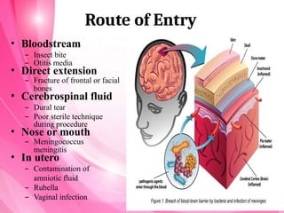
Route of Entry
•Bloodstream
– Insect bite
– Otitis media
• Direct extension
– Fracture of frontal or facial
bones
• Cerebrospinal fluid
– Dural tear
– Poor sterile technique
during procedure
• Nose or mouth
– Meningococcus
meningitis
• In utero
– Contamination of
amniotic fluid
– Rubella
– Vaginal infection
5.
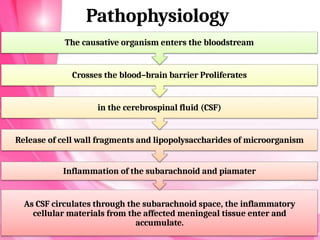
Pathophysiology
The causative organismenters the bloodstream
Crosses the blood–brain barrier Proliferates
in the cerebrospinal fluid (CSF)
Release of cell wall fragments and lipopolysaccharides of microorganism
Inflammation of the subarachnoid and piamater
As CSF circulates through the subarachnoid space, the inflammatory
cellular materials from the affected meningeal tissue enter and
accumulate.
6.
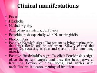
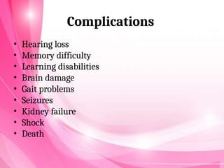
Clinical manifestations
• Fever
•Headache
• Nuchal rigidity
• Altered mental status, confusion
• Petechial rash especially with N. meningitidis.
• Photophobia
• Positive Kernig’s sign: The patient is lying supine with
the thigh flexed on the abdomen. Slowly extend the
upper leg, resulting in pain and spasm of the hamstring
muscle.
• Positive Brudzinski’s sign: To elicit Brudzinski's sign,
place the patient supine and flex the head upward.
Resulting flexion of hips, knees, and ankles with
neck flexion indicates meningeal irritation.
Diagnostic Evaluation
• Historycollection
• Physical examination
• Complete blood count (CBC)
• Blood cultures are obtained to indicate the organism.
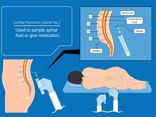
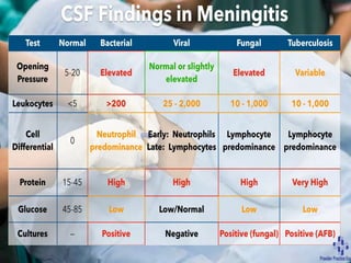
• Lumbar puncture : CSF evaluation for pressure, leukocytes,
protein, glucose CSF normally has five or fewer lymphocytes or
mononuclear cells/mm3.
– In acute bacterial meningitis, the CSF may indicate elevated
pressure, elevated leukocytes (several thousand), elevated
protein, elevated glucose. A culture and smear will identify
the organism. WBC differential should be done by a stained
smear of sediment.
• MRI/CT scan with and without contrast rules out other
disorders.
• Serological test such as Latex agglutination may be positive for
antigens in meningitis.
13.
Management
• Early administrationof an antibiotic that crosses the
blood– brain barrier into the subarachnoid space
in sufficient concentration to reduce the multiplication
of bacteria.
• Vancomycin hydrochloride in combination with one
of the cephalosporins (eg, ceftriaxone sodium,
cefotaxime sodium) is administered intravenously (IV)
• Antiviral drugs
• Dexamethasone (Decadron) is administered 15 to 20
minutes before the first dose of antibiotic and every 6
hours for the next 4 days.
• Antipyretics
• Dehydration and shock are treated with fluid volume
expanders.
• Seizures, are controlled with Phenytoin.
14.
Nursing management
• NursingAssessment
• Obtain a history of recent infections such as
upper respiratory infection, and exposure to
causative agents.
• Assess neurologic status and vital signs.
• Evaluate for signs of meningeal irritation.
• Assess sensorineural hearing loss (vision and
hearing), cranial nerve damage (eg. facial nerve
palsy), and diminished cognitive function.
15.
Nursing Diagnoses
• Hyperthermiarelated to the infectious process and
cerebral edema
• Ineffective Tissue Perfusion (cerebral) related to
infectious process and cerebral edema
• Acute Pain related to meningeal irritation or nuchal
rigidity
• Impaired Physical Mobility related to prolonged bed
rest.
• Risk for Imbalanced Fluid Volume related to fever
and decreased intake
• Risk for injury related to positive culture in CSF
16.
Nursing Interventions
• ReducingFever
– Administer antimicrobial agents on time to maintain optimal blood levels.
– Monitor temperature frequently or continuously, and administer antipyretics as
ordered.
– Institute other cooling measures, such as a hypothermia blanket, as indicated.
• Maintaining Fluid Balance
– Prevent I.V. fluid overload, which may worsen cerebral edema.
– Monitor intake and output closely.
– Monitor CVP frequently.
• Enhancing Cerebral Perfusion
– Assess LOC, vital signs, and neurologic parameters frequently. Observe for signs and
symptoms of ICP (eg, decreased LOC, dilated pupils, widening pulse pressure).
– Maintain a quiet, calm environment to prevent agitation, which may cause an
increased ICP.
– Prepare patient for a lumbar puncture for CSF evaluation, and repeat spinal tap, if
indicated.
– Notify the health care provider of signs of deterioration: increasing temperature,
decreasing LOC, seizure activity, or altered respirations.
17.
• Reducing Pain
–Administer analgesics as ordered; monitor for
response and adverse reactions. Avoid
opioids, which may mask a decreasing LOC.
– Darken the room if photophobia is present.
– Assist with position of comfort for neck
stiffness, and turn patient slowly and
carefully with head and neck in alignment.
– Elevate the head of the bed to decrease ICP and
reduce pain.


![growthanddevelopment2-190402170040[1].pptx](https://cdn.slidesharecdn.com/ss_thumbnails/growthanddevelopment2-1904021700401-241125091944-49fc96da-thumbnail.jpg?width=640&height=640&fit=bounds)


