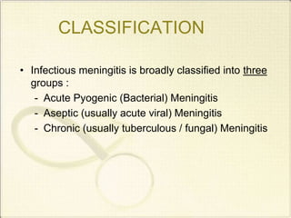
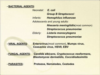
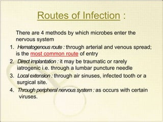
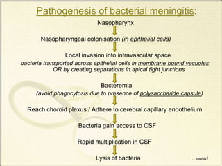
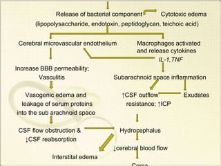
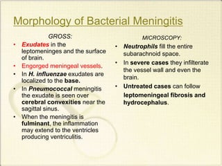
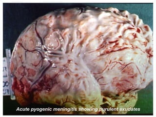
Meningitis
Group- 5 Tutor:Dr.Rajiv Shrestha
Group Members
• Sagun Baral
• Ashkal Basi
• Ashutosh Ghimire
• Kamal Ghimire
• Anup Subedi
• Samir Raut
• Prasesh Dhakal
• Prastuti Shrestha
• Saugat Lamichhane
• Kriti Pandey
• Shravya Rayilla
What is meningitis??
• Meningitis refers to an inflammatory process of
leptomeninges and CSF within the sub-arachnoid
space.
• Meningitis is generally caused by an infection, but
chemical meningitis may also occur in response to a
bacterial irritant injected into the sub-arachnoid
space.
CLASSIFICATION
• Infectious meningitis is broadly classified into three
groups :
- Acute Pyogenic (Bacterial) Meningitis
- Aseptic (usually acute viral) Meningitis
- Chronic (usually tuberculous / fungal) Meningitis
CAUSATIVE AGENTS
OF
MENINGITIS
- BACTERIAL AGENTS:
Neonatal: E. coli
Group B Streptococci
Infants: Hemophilus influenzae
Adolescents and young adults:
Niesseria meningitidis(most common)
Streptococcus pneumoniae
Elderly: Listeria monocytogens
Streptococcus pneumoniae
- VIRAL AGENTS: Enterovirus(most common), Mumps virus,
Coxsackie virus, HSVII, EBV
- FUNGAL AGENTS: Candida albicans, Cryptococcus neoformans,
Blastomyces dermatidis, Coccidiodesimitis
- PARASITES: Protozoa, Nematodes, Cestodes
Routes of Infection :
There are 4 methods by which microbes enter the
nervous system
1. Hematogenous route : through arterial and venous spread;
is the most common route of entry
2. Direct implantation : it may be traumatic or rarely
iatrogenic i.e. through a lumbar puncture needle
3. Local extension : through air sinuses, infected tooth or a
surgical site.
4. Through peripheral nervous system : as occurs with certain
viruses.
Pathogenesis of bacterial meningitis:
Nasopharynx
Nasopharyngeal colonisation (in epithelial cells)
Local invasion into intravascular space
bacteria transported across epithelial cells in membrane bound vacuoles
OR by creating separations in apical tight junctions
Bacteremia
(avoid phagocytosis due to presence of polysaccharide capsule)
Reach choroid plexus / Adhere to cerebral capillary endothelium
Bacteria gain access to CSF
Rapid multiplication in CSF
Lysis of bacteria …contd
Release of bacterial component Cytotoxic edema
(lipopolysaccharide, endotoxin, peptidoglycan, teichoic acid)
Cerebral microvascular endothelium Macrophages activated
and release cytokines
IL-1,TNF
Increase BBB permeability;
Vasculitis Subarachnoid space inflammation
Vasogenic edema and ↑CSF outflow Exudates
leakage of serum proteins resistance; ↑ICP
into the sub arachnoid space
CSF flow obstruction & Hydrocephalus
↓CSF reabsorption
↓cerebral blood flow
Interstital edema
Morphology of Bacterial Meningitis
GROSS:
• Exudates in the
leptomeninges and the surface
of brain.
• Engorged meningeal vessels.
• In H. influenzae exudates are
localized to the base.
• In Pneumococcal meningitis
the exudate is seen over
cerebral convexities near the
sagittal sinus.
• When the meningitis is
fulminant, the inflammation
may extend to the ventricles
producing ventriculitis.
MICROSCOPY:
• Neutrophils fill the entire
subarachnoid space.
• In severe cases they infilterate
the vessel wall and even the
brain.
• Untreated cases can follow
leptomeningeal fibrosis and
hydrocephalus.
Acute pyogenic meningitis showing purulent exudates
Clinical Features
• Fever, chills and rigor
• Headache, nausea, vomiting
• Seizures, cranial nerve palsies
• Signs of meningeal irritation
– Neck rigidity
– Photophobia
Signs
Complications:
• Bacterial : - Waterhouse-Friderichsen syndrome
- obstructive hydrocephalus
- chronic adhesive arachnoiditis
- focal cerebritis
- phlebitis leading to venous occlusion and
hemorrage of underlying brain.
Cerobrospinal Fluid (CSF) in Normal individuals and in different types of meningitis
Characteristics Normal CSF Acute pyogenic
meningitis
Tuberculous
meningitis
Viral meningitis
Pressure Normal
(<20cm H20)
Highly
increased
Moderately
increased
Slightly
increased
Direct examination
A. Cell
count/cumm and
predominant cell
1-3
Lymphocytes
1,000-20,000
Neutrophils
(90-95%)
50-500
Lymphocytes
(90%)
10-500
Lymphocytes
B. Biochemical
analysis
1. Protein
(mg%)
2. Sugars
(mg%)
30-45
40-80
Highly
increased
(100-600)
Diminished
(10-20)
Moderately
increased
(80-120)
Diminished
(10-20)
Slightly
increased
(60-80)
Normal
Bacteriological examination
A. Microscopy
Gram stain
ZN-staining
Nil
Nil
GPC,GNC,GN
B,GPB etc
Nil
-
Acid fast bacilli
-
B. Culture Nil Specific
medium
In LJ medium Cell cultures
References
Harrison’s Principles Of Internal Medicine
Pathologic Basis Of Disease
Robbins and Cotran
K YOU

THANK
YOU

meningitis

  • 1.
  • 2.
    Group Members • SagunBaral • Ashkal Basi • Ashutosh Ghimire • Kamal Ghimire • Anup Subedi • Samir Raut • Prasesh Dhakal • Prastuti Shrestha • Saugat Lamichhane • Kriti Pandey • Shravya Rayilla
  • 3.
    What is meningitis?? •Meningitis refers to an inflammatory process of leptomeninges and CSF within the sub-arachnoid space. • Meningitis is generally caused by an infection, but chemical meningitis may also occur in response to a bacterial irritant injected into the sub-arachnoid space.
  • 4.
    CLASSIFICATION • Infectious meningitisis broadly classified into three groups : - Acute Pyogenic (Bacterial) Meningitis - Aseptic (usually acute viral) Meningitis - Chronic (usually tuberculous / fungal) Meningitis
  • 5.
  • 6.
    - BACTERIAL AGENTS: Neonatal:E. coli Group B Streptococci Infants: Hemophilus influenzae Adolescents and young adults: Niesseria meningitidis(most common) Streptococcus pneumoniae Elderly: Listeria monocytogens Streptococcus pneumoniae - VIRAL AGENTS: Enterovirus(most common), Mumps virus, Coxsackie virus, HSVII, EBV - FUNGAL AGENTS: Candida albicans, Cryptococcus neoformans, Blastomyces dermatidis, Coccidiodesimitis - PARASITES: Protozoa, Nematodes, Cestodes
  • 7.
    Routes of Infection: There are 4 methods by which microbes enter the nervous system 1. Hematogenous route : through arterial and venous spread; is the most common route of entry 2. Direct implantation : it may be traumatic or rarely iatrogenic i.e. through a lumbar puncture needle 3. Local extension : through air sinuses, infected tooth or a surgical site. 4. Through peripheral nervous system : as occurs with certain viruses.
  • 8.
    Pathogenesis of bacterialmeningitis: Nasopharynx Nasopharyngeal colonisation (in epithelial cells) Local invasion into intravascular space bacteria transported across epithelial cells in membrane bound vacuoles OR by creating separations in apical tight junctions Bacteremia (avoid phagocytosis due to presence of polysaccharide capsule) Reach choroid plexus / Adhere to cerebral capillary endothelium Bacteria gain access to CSF Rapid multiplication in CSF Lysis of bacteria …contd
  • 9.
    Release of bacterialcomponent Cytotoxic edema (lipopolysaccharide, endotoxin, peptidoglycan, teichoic acid) Cerebral microvascular endothelium Macrophages activated and release cytokines IL-1,TNF Increase BBB permeability; Vasculitis Subarachnoid space inflammation Vasogenic edema and ↑CSF outflow Exudates leakage of serum proteins resistance; ↑ICP into the sub arachnoid space CSF flow obstruction & Hydrocephalus ↓CSF reabsorption ↓cerebral blood flow Interstital edema
  • 10.
    Morphology of BacterialMeningitis GROSS: • Exudates in the leptomeninges and the surface of brain. • Engorged meningeal vessels. • In H. influenzae exudates are localized to the base. • In Pneumococcal meningitis the exudate is seen over cerebral convexities near the sagittal sinus. • When the meningitis is fulminant, the inflammation may extend to the ventricles producing ventriculitis. MICROSCOPY: • Neutrophils fill the entire subarachnoid space. • In severe cases they infilterate the vessel wall and even the brain. • Untreated cases can follow leptomeningeal fibrosis and hydrocephalus.
  • 11.
    Acute pyogenic meningitisshowing purulent exudates
  • 12.
    Clinical Features • Fever,chills and rigor • Headache, nausea, vomiting • Seizures, cranial nerve palsies • Signs of meningeal irritation – Neck rigidity – Photophobia
  • 13.
  • 14.
    Complications: • Bacterial :- Waterhouse-Friderichsen syndrome - obstructive hydrocephalus - chronic adhesive arachnoiditis - focal cerebritis - phlebitis leading to venous occlusion and hemorrage of underlying brain.
  • 15.
    Cerobrospinal Fluid (CSF)in Normal individuals and in different types of meningitis Characteristics Normal CSF Acute pyogenic meningitis Tuberculous meningitis Viral meningitis Pressure Normal (<20cm H20) Highly increased Moderately increased Slightly increased Direct examination A. Cell count/cumm and predominant cell 1-3 Lymphocytes 1,000-20,000 Neutrophils (90-95%) 50-500 Lymphocytes (90%) 10-500 Lymphocytes B. Biochemical analysis 1. Protein (mg%) 2. Sugars (mg%) 30-45 40-80 Highly increased (100-600) Diminished (10-20) Moderately increased (80-120) Diminished (10-20) Slightly increased (60-80) Normal Bacteriological examination A. Microscopy Gram stain ZN-staining Nil Nil GPC,GNC,GN B,GPB etc Nil - Acid fast bacilli - B. Culture Nil Specific medium In LJ medium Cell cultures
  • 16.
    References Harrison’s Principles OfInternal Medicine Pathologic Basis Of Disease Robbins and Cotran
  • 17.