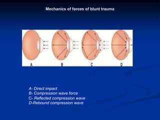
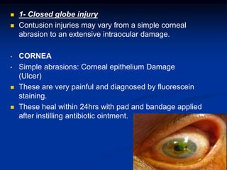
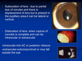
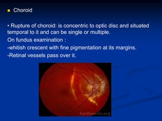
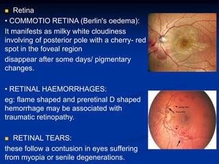
SlideShare a Scribd company logo
Mechanical Ocular
Trauma
General Considerations
 Ocular trauma constitude about 6% of all injuries,
but eyes set up only 0,1% from the surface of
human body.
 Ocular trauma can result in a wide spectrum of
tissue lesions of the globe, optic nerve and adnexa,
ranging from relatively superficial to vision
threatening.
 Consequently, the socioeconomic impact of ocular
trauma can hardly be overstimated. Those affected
often have to face:
 loss of career opportunities
 major lifestyle changes
 permanent physical disfigurement
Ocular Trauma Epidemiology
Who is at Risk
 approximately 80% of
injured are males
The Site
 the workplace has been
most common site
 domestic injuries has been
increasing
The Source
 blunt objects (rocks, fists,
wood branches, baseballs.
champagne corks)
 work-related injuries
 sports and recreational
activities
 hammering on metal and
nails
 firearms and fireworks
Terminology of Ocular Trauma
Without a standardized terminology of eye injury
types, it is impossible to communicate between
ophthalmologists.
BETT – Birmingham Eye Trauma Terminology
BETT satisfies all criteria for standard terminology
by providing a clear definition for all injury types
and placing each injury type within the
framework of all comprehensive system.
BETT
Injury
Closed Globe Open Globe
Contusion Lamellar Laceration Laceration Rupture
Penetrating
IOFB
Perforating
Terms and Definitions in BETT
 Eyewall
Sclera and cornea.
 Closed Globe Injury
No full-thickness wound of the
eyewall.
 Open Globe Injury
Full-thickness wound of the eyewall.
 Contusion
No wound.
The injury is due to either direct
energy delivery by the object or
changes in the shape of the
globe.
 Lamellar laceration
Partial-thickness wound of the
eyewall.
 Rupture
Full-thickness wound of the
eyewall caused by a blunt
object.
Because the eye is filled with
incompressible liquid, the
impact results in momentary
increase in intraocular pressure
(IOP). The eyewall yields at its
weakest point. The actual
wound is produced by an
„inside-out“ mechanism.
Terms and Definitions in BETT
 Laceration
Full-thickness wound of the
eyewall caused by a sharp
object.
The wound occurs at the
impact site by an „outside-
in“ mechanism.
 Penetrating injury
Entrance wound of the
eyewall.
If more than one wound is
present, each must have
been caused by a different
agent.
 IOFB
Retained foreign object.
 Perforating injury
Entrance and „exit“ wound.
Both wounds caused by the
same agent.
BLUNT TRAUMA
 Modes of trauma
• Direct blow to the eyeball by fist, ball or blunt instruments
like sticks, and big stones.
• Accidental blunt trauma to eyeball may also occur in
roadside accidents, injuries by instruments/ machines and
fall upon the projecting blunt objects.
Mechanics of forces of blunt trauma
A- Direct impact
B- Compression wave force
C- Reflected compression wave
D-Rebound compression wave
 Mechanisms of damage
l. Mechanical tearing of the tissues of eyeball.
2. Damage to the tissue cells sufficient to cause
disruption of their physiological activity.
3. Vascular damage leading to ischaemia, oedema and
haemorrhages.
4. Trophic changes due to disturbances of the nerve
supply.
5. Delayed complications of blunt trauma such as
secondary glaucoma, haemophthalmitis, late rosette
cataract and retinal detachment.
 Traumatic lesions of blunt trauma
1. CLOSED GLOBE INJURY
2. GLOBE RUPTURE
3. EXTRAOCULAR LESIONS
 1- Closed globe injury
 Contusion injuries may vary from a simple corneal
abrasion to an extensive intraocular damage.
• CORNEA
• Simple abrasions: Corneal epithelium Damage
(Ulcer)
 These are very painful and diagnosed by fluorescein
staining.
 These heal within 24hrs with pad and bandage applied
after instilling antibiotic ointment.
 Recurrent corneal erosions
(recurrent keractalgia)
Caused by fingernail trauma. Patient
presents with recurrent attacks of
acute pain and lacrimation on
opening eye in the morning and is
due to abnormally loose attachment
of epithelium to Bowman's
membrane.
 Partial corneal tears ( lamellar
corneal lacerations )
 BLOOD STAINING OF CORNEA:
hyphaema + Raised IOP
cornea is reddish brown or greenish in
color and in later stages lead to dislocation
of lens into anterior chamber.
It clears very slowly from periphery
towards the center and may take up to 2
yrs.
 • DEEP CORNEAL OPACITY:
* Oedema of corneal stroma
folds in the Descemet's membrane.
 Sclera
Partial thickness scleral wounds occur
alone or with other lesions of closed globe
type.
 Anterior chamber
-TRAUMATIC HYPHAEMA or blood in anterior
chamber which occurs due to injury to iris or
anterior ciliary vessels.
-EXUDATES: these collect here following
traumatic uveitis
 Iris, pupil and ciliary body
• TRAUMATIC MIOSIS:
occurs initially due to irritation of ciliary nerves or with
spasm of accommodation.
• TRAUMATIC MYDRIASIS (iridoplegia):
It is permanent and is associated with traumatic
cycloplegia.
• RUPTURE OF PUPILARY MARGIN
• RADIATING TEARS IN THE IRIS STROMA
• IRIDODIALYSIS:
detachment of iris from its root at ciliary body which results
in 'D' shaped pupil
 TRAUMATIC ANIRIDIA:
the completely torn iris from ciliary body sinks
to the bottom of anterior chamber in the form of
a small ball.
 ANGLE RECESSION:
refers to tear between the longitudinal and
circular muscle fires of ciliary body,
characterized by deepening of ant chamber
and widening of ciliary body on gonioscopy and
leads to glaucoma.
 INFLAMMATORY CHANGES:
these include traumatic iridocyclitis, post
traumatic iris atrophy etc.
TTT: consist of atropine, antibiotics and
steroids.
 Lens
• VOSSIUS RING :
It is a circular ring of brown pigment seen
on the anterior capsule
It occurs due to striking of contracted
pupillary margin against the lens.
•CONCUSSION CATARACT:
due to imbibition of aqueous and due to
direct mechanical effects of the injury on
lens fibers
• Subluxation of lens : due to partial
tear of zonules and there is
displacement of lens but is present in
the pupillary area-it can be lateral or
vertical.
• Dislocation of lens: when rupture of
zonules is complete and can be
intraocular or extraocular.
-intraocular-into AC or posterior vitreous
-extraocular-subconjunctival or may fall
outside the eye
 VITREOUS
• Liquefaction and appearance
of clouds of fine pigmentary
opacities
• Detachment of the vitreous
either anterior or posterior
• Vitreous hemorrhage
• Vitreous herniation in anterior
chamber may occur with
subluxation or dislocation of
lens
 Choroid
• Rupture of choroid: is concentric to optic disc and situated
temporal to it and can be single or multiple.
On fundus examination :
-whitish crescent with fine pigmentation at its margins.
-Retinal vessels pass over it.
• Choroidal hemorrhage may occur under retina
or may even enter the vitreous if retina is torn.
• Choroidal detachment
• Traumatic choroiditis :
-seen on fundus examination as patches of
pigmentation and discoloration after eye becomes
silent
 Retina
• COMMOTIO RETINA (Berlin's oedema):
It manifests as milky white cloudiness
involving of posterior pole with a cherry- red
spot in the foveal region
disappear after some days/ pigmentary
changes.
• RETINAL HAEMORRHAGES:
eg: flame shaped and preretinal D shaped
hemorrhage may be associated with
traumatic retinopathy.
 RETINAL TEARS:
these follow a contusion in eyes suffering
from myopia or senile degenerations.
• TRAUMATIC PROLIFERATIVE
RETINOPATHY :
occur secondary to vitreous
haemorrhage.
• RETINAL DETACHMENT:
follows retinal tears or vitreo-retinal
tractional bands.
 CONCUSSION CHANGES AT
MACULA: traumatic macular oedema
followed by pigmentary degeneration.
Sometimes, a macular cyst is formed ,
which on rupture is converted to a
lamellar or full thickness macular hole.
 lOP changes in closed globe injury
• TRAUMATIC GLAUCOMA
• TRAUMATIC HYPOTONY
it may follow damage to the ciliary body.
 Traumatic changes in refraction
• Myopia may follow ciliary spasm or rupture
of zonules or anterior shift of lens
• Hypermetropia and loss of accommodation
may result from damage to the ciliary body
(cycloplegia).
 2- GLOBE RUPTURE
It is a full-thickness wound of eye-wall caused by blunt object
and can occur in 2 ways:
• DIRECT RUPTURE: at the site of injury.
• INDIRECT RUPTURE: occurs because of compression
force. The impact results in momentary increase in IOP
and inside out injury at the weakest part of the eye wall
TREATMENT:
 A badly damaged globe should be enucleated .
 In less severe cases it can be repaired under general
anesthesia.
 3- Extraocular lesions
Open Globe Injuries
Globe Rupture
Etiology:
Full-thickness wound of the
eyewall caused by a blunt
object.
Clinical findings:
 Cover rupture - prolapsus
of intraocular tissue is
beneath conjunctiva
 Uncover rupture –
prolapsus is over
conjunctiva
Globe Rupture
Clinical findings:
Low visual acuity,
hypotony of the eye,
bleeding in anterior
chamber and vitreous
body, perilimbal
wound, prolapsus of
intracoular tissue (iris,
lens, vitreous body…).
Examination:
Visual acuity, slit lamp,
ultrasound
Treatment:
Only surgical with
systemic and topical
antibiotics.
Globe laceration– penetrating injury
Etiology:
One entrance wound of the
eyewall (section and slash
wound of conjunctiva,
cornea, sclera).
Clinical findings:
 with prolapsus of intraocular
tissue
 without prolapsus of
intraocular tissue
Laceration – penetrating injury
Examination:
Visual acuity, slit lamp, opththalmoscopy, ultrasound,
CT or x-ray (for elimination of the foreign body).
Treatment:
 surgical – suture immediately
 medical - contact lens (only if the wound is small
and edges of the wound are adapted) + systemic
and topical antibiotics
Open Globe
Laceration + IOFB
IntraOcular Foreign Body is defined as
intraocularly retained material.
Cause:
Hammering in 80%, power or machine tools in
25%, weapon-related in 20%.
Terminology of IOFB:
 metallic or non metallic
 x-ray contrast or x-ray noncontrast
 magnetic or non magnetic
IOFB
Clinical findings:
Depends on localization
of foreign body inside
the eye.
Clinical features range
between no visual
impairment to
blindness.
IOFB
Examination:
Slit lamp, ophthalmoscopy,
ultrasound, x-ray, CT scan.
Detection of IOFB by x-ray
method:
 anterior and lateral
projection with prosthesis
on the cornea
 Comberg-Baltin method =
exact calculation of
localization IOFB inside the
eye.
IOFB
Ocular damage
 Entrance Wound
The IOFB must possess certain
energy to perforate the eye ´s
protective wall. The length of
the entry wound is predictive of
the risk of retinal damage: the
shorter the wound, the less
energy to be lost during
penetration.
 Mechanical Intraocular Damage
Little or no damage is expected if
the IOFB has completely lost its
kinectic energy upon entry. The
primary impact may be followed
by additional impaction via
ricocheting.
 Inflammation
Breach of the eyewall, intraocular
haemorrhage and vitreous/lens
admixture incite an inflammatory
response.
 Chemical Implications
Metallic IOFB ´s are rarely pure.
 Siderosis
IOFB - related corrosion is caused by
interaction between trivalent iron
ions and proteins in the eye ´s
epithelial cells. The cytotoxicity
involves enzyme liberation leading
to cell degeneration. The ferric
iron is thought to be toxic by
generating free radicals.
IOFB
Ocular damage
 Siderosis
Siderotic changes include the
following clinical findings:
 Chronic open-angle glaucoma
 Brownish discoloration of the
iris
 Dilated, nonreactive pupil
 Yellow cataract with brown
deposits on the anterior capsule
 Pigmentary retinal degeneration
withg visual field loss
 Visual impairment
The clinical diagnosis is confirmed by
characteristic ERG changes such
as:
 Increased A wave initially
 Progressive reduction of the B
wave subsequently
IOFB
Ocular damage
 Chalcosis
Copper IOFBs cause rapid,
sterile endophthalmitis-
like reaction including
corneal/scleral melting,
hypopyon (inflammatory
exudation in anterior
chamber) and retinal
detachment.
Copper tends to deposit in
membranes and causes
destruction by increasing
lipid peroxidation.
The typical clinical findings
include:
 Green discoloration of the
iris
 Kayser-fleischer ring
 Greenish/brown – colored
cataract with spokes of
copper deposits(sunflower)
 Copper particles in the
vitreous and copper particles
on the retinal surface
IOFB
Treatment:
All the IOFB ´s must be
removed from the eye!
Retained IOFB = high
risk of
endophthalmitis and
siderosis
Timing of removal:
 Immediately
 Delayed – between 5
and 10 days after injury
 Surgical treatment –
extraction by pars plana
vitrectomy with forceps
or intraocular magnet
 Medical treatment –
systemic antibiotics,
topic antibiotics and
corticosteroids,
mydriatics.
Open Globe
Laceration – perforating injury
Entrance and „exit“
wound.
Both wounds caused by
the same agent.
Etiology:
Section, puncture, splash
wound (f.e.wire, knife)
or entrance and exit
wound caused by
IOFB.
Specifity of perforating
injury
 Entrance wound is
smaller than exit
wound (cases caused
by IOFB)
 In most cases exit
wound is technically
impossible to suture
(exit wound located in
posterior pole)
Perforating injury
Clinical findings:
Same as the penetrating
injury
Examination:
Slit lamp, IOP
measurement,
ophthalmoscopy,
ultrasound, x-ray, CT
Treatment:
Suture of the entrance
wound (exit wound if
it is technically
possible), cataract
extraction, pars plana
vitrectomy…
Thank You

More Related Content

What's hot

Vitrectomy: Development And Steps
Vitrectomy: Development And StepsVitrectomy: Development And Steps
Vitrectomy: Development And Steps
Dr Samarth Mishra
 
Corneal degenerations
Corneal degenerationsCorneal degenerations
Corneal degenerations
drkvasantha
 
Ocular viscoelastic devices(OVD)
Ocular viscoelastic devices(OVD)Ocular viscoelastic devices(OVD)
Ocular viscoelastic devices(OVD)
Sivateja Challa
 
Herpetic eye disease
Herpetic eye diseaseHerpetic eye disease
Herpetic eye disease
Othman Al-Abbadi
 
Phakic iol ppt
Phakic iol pptPhakic iol ppt
Phakic iol ppt
DrArino John
 
Epiretinal membrane
Epiretinal membraneEpiretinal membrane
Epiretinal membrane
Pushkal Katara
 
Keratoconus
KeratoconusKeratoconus
Peripheral ulcerative keratitis (puk)
Peripheral ulcerative keratitis (puk)Peripheral ulcerative keratitis (puk)
Peripheral ulcerative keratitis (puk)
Desta Genete
 
Pigmentary glaucoma - Dr Shylesh B Dabke
Pigmentary glaucoma - Dr Shylesh B DabkePigmentary glaucoma - Dr Shylesh B Dabke
Pigmentary glaucoma - Dr Shylesh B Dabke
Shylesh Dabke
 
Cystoid macular oedema
Cystoid macular oedemaCystoid macular oedema
Cystoid macular oedema
Laxmi Eye Institute
 
Malignant glaucoma
Malignant glaucomaMalignant glaucoma
Malignant glaucomaJi Young Lee
 
Lamellar Keratoplasty in ophthalmology
Lamellar Keratoplasty in ophthalmologyLamellar Keratoplasty in ophthalmology
Lamellar Keratoplasty in ophthalmology
pratik mohod
 
Infantile esotropia
Infantile esotropia Infantile esotropia
Infantile esotropia
Anisha Rathod
 
Normal tension glaucoma
Normal tension glaucomaNormal tension glaucoma
Normal tension glaucoma
Laxmi Eye Institute
 
Vitreous substitutes
Vitreous substitutesVitreous substitutes
Vitreous substitutes
sri kiran eye institue
 
Diabetic macular edema
Diabetic macular edemaDiabetic macular edema
Diabetic macular edema
drkvasantha
 
Ocular surface squamous neoplasia(ossn)
Ocular surface squamous neoplasia(ossn)Ocular surface squamous neoplasia(ossn)
Ocular surface squamous neoplasia(ossn)
SSSIHMS-PG
 
Keratoplasty associated complications
Keratoplasty associated complicationsKeratoplasty associated complications
Keratoplasty associated complications
Karan Bhatia
 
Giant Retinal Tears
Giant Retinal TearsGiant Retinal Tears
Giant Retinal Tears
Duleepa Baranage
 
Disc anomalies, pits and treatment of associated
Disc anomalies, pits and treatment of associatedDisc anomalies, pits and treatment of associated
Disc anomalies, pits and treatment of associated
abhishek ghelani
 

What's hot (20)

Vitrectomy: Development And Steps
Vitrectomy: Development And StepsVitrectomy: Development And Steps
Vitrectomy: Development And Steps
 
Corneal degenerations
Corneal degenerationsCorneal degenerations
Corneal degenerations
 
Ocular viscoelastic devices(OVD)
Ocular viscoelastic devices(OVD)Ocular viscoelastic devices(OVD)
Ocular viscoelastic devices(OVD)
 
Herpetic eye disease
Herpetic eye diseaseHerpetic eye disease
Herpetic eye disease
 
Phakic iol ppt
Phakic iol pptPhakic iol ppt
Phakic iol ppt
 
Epiretinal membrane
Epiretinal membraneEpiretinal membrane
Epiretinal membrane
 
Keratoconus
KeratoconusKeratoconus
Keratoconus
 
Peripheral ulcerative keratitis (puk)
Peripheral ulcerative keratitis (puk)Peripheral ulcerative keratitis (puk)
Peripheral ulcerative keratitis (puk)
 
Pigmentary glaucoma - Dr Shylesh B Dabke
Pigmentary glaucoma - Dr Shylesh B DabkePigmentary glaucoma - Dr Shylesh B Dabke
Pigmentary glaucoma - Dr Shylesh B Dabke
 
Cystoid macular oedema
Cystoid macular oedemaCystoid macular oedema
Cystoid macular oedema
 
Malignant glaucoma
Malignant glaucomaMalignant glaucoma
Malignant glaucoma
 
Lamellar Keratoplasty in ophthalmology
Lamellar Keratoplasty in ophthalmologyLamellar Keratoplasty in ophthalmology
Lamellar Keratoplasty in ophthalmology
 
Infantile esotropia
Infantile esotropia Infantile esotropia
Infantile esotropia
 
Normal tension glaucoma
Normal tension glaucomaNormal tension glaucoma
Normal tension glaucoma
 
Vitreous substitutes
Vitreous substitutesVitreous substitutes
Vitreous substitutes
 
Diabetic macular edema
Diabetic macular edemaDiabetic macular edema
Diabetic macular edema
 
Ocular surface squamous neoplasia(ossn)
Ocular surface squamous neoplasia(ossn)Ocular surface squamous neoplasia(ossn)
Ocular surface squamous neoplasia(ossn)
 
Keratoplasty associated complications
Keratoplasty associated complicationsKeratoplasty associated complications
Keratoplasty associated complications
 
Giant Retinal Tears
Giant Retinal TearsGiant Retinal Tears
Giant Retinal Tears
 
Disc anomalies, pits and treatment of associated
Disc anomalies, pits and treatment of associatedDisc anomalies, pits and treatment of associated
Disc anomalies, pits and treatment of associated
 

Similar to Mechanical_ocular_trauma.pptx

Blunt trauma of eye
Blunt trauma of eyeBlunt trauma of eye
Blunt trauma of eye
challenger klvk
 
Ocular trauma
Ocular traumaOcular trauma
Ocular trauma
Supun Dhanasekara
 
Ocular injuries new
Ocular injuries newOcular injuries new
Ocular injuries new
Deogratias George
 
Ocular trauma
Ocular traumaOcular trauma
Ocular trauma
SIDESH HENDAVITHARANA
 
OCULAR TRAUMA - Classification of mechanical injuries, clinical features and ...
OCULAR TRAUMA - Classification of mechanical injuries, clinical features and ...OCULAR TRAUMA - Classification of mechanical injuries, clinical features and ...
OCULAR TRAUMA - Classification of mechanical injuries, clinical features and ...
rahul ramesh
 
Posterior segment manifestations of blunt trauma-1.pptx
Posterior segment manifestations of blunt trauma-1.pptxPosterior segment manifestations of blunt trauma-1.pptx
Posterior segment manifestations of blunt trauma-1.pptx
Mahfuz Shohag
 
Penetrating Ocular Injuries
Penetrating Ocular InjuriesPenetrating Ocular Injuries
Penetrating Ocular Injuriesjohn xxx
 
Eye Injuries
Eye InjuriesEye Injuries
Eye Injuries
laraib jameel
 
Ocular trauma
Ocular traumaOcular trauma
Ocular trauma
Arshad Ali Awan
 
Blunt trauma to eye
Blunt trauma to eyeBlunt trauma to eye
Blunt trauma to eye
emirates741
 
OCULAR INJURIES
OCULAR INJURIESOCULAR INJURIES
OCULAR INJURIES
Dr. Lucky Narain
 
eye trauma approach and management
eye trauma approach and management eye trauma approach and management
eye trauma approach and management
Abbas W Abbas
 
penetrating injury to eye
penetrating injury to eyepenetrating injury to eye
penetrating injury to eye
DARSHAN S M
 
Penetrating Oc Injury
Penetrating Oc InjuryPenetrating Oc Injury
Penetrating Oc Injuryjohn xxx
 
Penetrating Oc Injury
Penetrating Oc InjuryPenetrating Oc Injury
Penetrating Oc Injuryjohn xxx
 
Common eye injury
Common eye injuryCommon eye injury
Common eye injury
Azizul Islam
 
Trauma to eye REVISION NOTES
Trauma to eye REVISION NOTES Trauma to eye REVISION NOTES
Trauma to eye REVISION NOTES
TONY SCARIA
 
Ocular trauma
Ocular traumaOcular trauma
Ocular trauma
Niwar Ameen
 
Eye injuries
Eye injuriesEye injuries
Eye injuries
Amr Mounir
 
eyeinjuries-170615112226.pdf
eyeinjuries-170615112226.pdfeyeinjuries-170615112226.pdf
eyeinjuries-170615112226.pdf
MsellemKhamis
 

Similar to Mechanical_ocular_trauma.pptx (20)

Blunt trauma of eye
Blunt trauma of eyeBlunt trauma of eye
Blunt trauma of eye
 
Ocular trauma
Ocular traumaOcular trauma
Ocular trauma
 
Ocular injuries new
Ocular injuries newOcular injuries new
Ocular injuries new
 
Ocular trauma
Ocular traumaOcular trauma
Ocular trauma
 
OCULAR TRAUMA - Classification of mechanical injuries, clinical features and ...
OCULAR TRAUMA - Classification of mechanical injuries, clinical features and ...OCULAR TRAUMA - Classification of mechanical injuries, clinical features and ...
OCULAR TRAUMA - Classification of mechanical injuries, clinical features and ...
 
Posterior segment manifestations of blunt trauma-1.pptx
Posterior segment manifestations of blunt trauma-1.pptxPosterior segment manifestations of blunt trauma-1.pptx
Posterior segment manifestations of blunt trauma-1.pptx
 
Penetrating Ocular Injuries
Penetrating Ocular InjuriesPenetrating Ocular Injuries
Penetrating Ocular Injuries
 
Eye Injuries
Eye InjuriesEye Injuries
Eye Injuries
 
Ocular trauma
Ocular traumaOcular trauma
Ocular trauma
 
Blunt trauma to eye
Blunt trauma to eyeBlunt trauma to eye
Blunt trauma to eye
 
OCULAR INJURIES
OCULAR INJURIESOCULAR INJURIES
OCULAR INJURIES
 
eye trauma approach and management
eye trauma approach and management eye trauma approach and management
eye trauma approach and management
 
penetrating injury to eye
penetrating injury to eyepenetrating injury to eye
penetrating injury to eye
 
Penetrating Oc Injury
Penetrating Oc InjuryPenetrating Oc Injury
Penetrating Oc Injury
 
Penetrating Oc Injury
Penetrating Oc InjuryPenetrating Oc Injury
Penetrating Oc Injury
 
Common eye injury
Common eye injuryCommon eye injury
Common eye injury
 
Trauma to eye REVISION NOTES
Trauma to eye REVISION NOTES Trauma to eye REVISION NOTES
Trauma to eye REVISION NOTES
 
Ocular trauma
Ocular traumaOcular trauma
Ocular trauma
 
Eye injuries
Eye injuriesEye injuries
Eye injuries
 
eyeinjuries-170615112226.pdf
eyeinjuries-170615112226.pdfeyeinjuries-170615112226.pdf
eyeinjuries-170615112226.pdf
 

More from Harshika Malik

artifical intelligence in ophthalmology.pptx
artifical intelligence in ophthalmology.pptxartifical intelligence in ophthalmology.pptx
artifical intelligence in ophthalmology.pptx
Harshika Malik
 
Retinal Dystrophy.pptx vvvvvvvvvvvvvvvvv
Retinal Dystrophy.pptx vvvvvvvvvvvvvvvvvRetinal Dystrophy.pptx vvvvvvvvvvvvvvvvv
Retinal Dystrophy.pptx vvvvvvvvvvvvvvvvv
Harshika Malik
 
ECTROPION^JENTROPION AND THEIR MANAGEMENT 2.pptx
ECTROPION^JENTROPION AND THEIR MANAGEMENT 2.pptxECTROPION^JENTROPION AND THEIR MANAGEMENT 2.pptx
ECTROPION^JENTROPION AND THEIR MANAGEMENT 2.pptx
Harshika Malik
 
EPISCLERITIS AND SCLERITIS.pptx bbbbbbbb
EPISCLERITIS AND SCLERITIS.pptx bbbbbbbbEPISCLERITIS AND SCLERITIS.pptx bbbbbbbb
EPISCLERITIS AND SCLERITIS.pptx bbbbbbbb
Harshika Malik
 
PENETRATING KERATOPLASTY - Cmmmmopy.pptx
PENETRATING KERATOPLASTY - Cmmmmopy.pptxPENETRATING KERATOPLASTY - Cmmmmopy.pptx
PENETRATING KERATOPLASTY - Cmmmmopy.pptx
Harshika Malik
 
LAMELLAR KERATOPLASTY.pptx vvvvvvvvvvvvvv
LAMELLAR KERATOPLASTY.pptx vvvvvvvvvvvvvvLAMELLAR KERATOPLASTY.pptx vvvvvvvvvvvvvv
LAMELLAR KERATOPLASTY.pptx vvvvvvvvvvvvvv
Harshika Malik
 
rop.pptx
rop.pptxrop.pptx
rop.pptx
Harshika Malik
 
TED.pptx
TED.pptxTED.pptx
TED.pptx
Harshika Malik
 
proptosis.pptx
proptosis.pptxproptosis.pptx
proptosis.pptx
Harshika Malik
 
retinal_detachment 4.pptx
retinal_detachment 4.pptxretinal_detachment 4.pptx
retinal_detachment 4.pptx
Harshika Malik
 
squint copy.pptx
squint copy.pptxsquint copy.pptx
squint copy.pptx
Harshika Malik
 
diabetic_retinopathy copy.pptx
diabetic_retinopathy copy.pptxdiabetic_retinopathy copy.pptx
diabetic_retinopathy copy.pptx
Harshika Malik
 
830_Optic_nerve_final copy 2.pptx
830_Optic_nerve_final copy 2.pptx830_Optic_nerve_final copy 2.pptx
830_Optic_nerve_final copy 2.pptx
Harshika Malik
 
retinoblastoma 2.pptx
retinoblastoma 2.pptxretinoblastoma 2.pptx
retinoblastoma 2.pptx
Harshika Malik
 
Secondary-glaucoma-Final.pptx
Secondary-glaucoma-Final.pptxSecondary-glaucoma-Final.pptx
Secondary-glaucoma-Final.pptx
Harshika Malik
 
approach to padiatric cataract 2.pptx
approach to padiatric cataract 2.pptxapproach to padiatric cataract 2.pptx
approach to padiatric cataract 2.pptx
Harshika Malik
 
lacrimal apparatus.pptx
lacrimal apparatus.pptxlacrimal apparatus.pptx
lacrimal apparatus.pptx
Harshika Malik
 
catarcact-ug1 2.pptx
catarcact-ug1 2.pptxcatarcact-ug1 2.pptx
catarcact-ug1 2.pptx
Harshika Malik
 
1167_Anterior-Uveitis 2.pptx
1167_Anterior-Uveitis 2.pptx1167_Anterior-Uveitis 2.pptx
1167_Anterior-Uveitis 2.pptx
Harshika Malik
 

More from Harshika Malik (20)

artifical intelligence in ophthalmology.pptx
artifical intelligence in ophthalmology.pptxartifical intelligence in ophthalmology.pptx
artifical intelligence in ophthalmology.pptx
 
Retinal Dystrophy.pptx vvvvvvvvvvvvvvvvv
Retinal Dystrophy.pptx vvvvvvvvvvvvvvvvvRetinal Dystrophy.pptx vvvvvvvvvvvvvvvvv
Retinal Dystrophy.pptx vvvvvvvvvvvvvvvvv
 
ECTROPION^JENTROPION AND THEIR MANAGEMENT 2.pptx
ECTROPION^JENTROPION AND THEIR MANAGEMENT 2.pptxECTROPION^JENTROPION AND THEIR MANAGEMENT 2.pptx
ECTROPION^JENTROPION AND THEIR MANAGEMENT 2.pptx
 
EPISCLERITIS AND SCLERITIS.pptx bbbbbbbb
EPISCLERITIS AND SCLERITIS.pptx bbbbbbbbEPISCLERITIS AND SCLERITIS.pptx bbbbbbbb
EPISCLERITIS AND SCLERITIS.pptx bbbbbbbb
 
PENETRATING KERATOPLASTY - Cmmmmopy.pptx
PENETRATING KERATOPLASTY - Cmmmmopy.pptxPENETRATING KERATOPLASTY - Cmmmmopy.pptx
PENETRATING KERATOPLASTY - Cmmmmopy.pptx
 
LAMELLAR KERATOPLASTY.pptx vvvvvvvvvvvvvv
LAMELLAR KERATOPLASTY.pptx vvvvvvvvvvvvvvLAMELLAR KERATOPLASTY.pptx vvvvvvvvvvvvvv
LAMELLAR KERATOPLASTY.pptx vvvvvvvvvvvvvv
 
rop.pptx
rop.pptxrop.pptx
rop.pptx
 
TED.pptx
TED.pptxTED.pptx
TED.pptx
 
proptosis.pptx
proptosis.pptxproptosis.pptx
proptosis.pptx
 
retinal_detachment 4.pptx
retinal_detachment 4.pptxretinal_detachment 4.pptx
retinal_detachment 4.pptx
 
squint copy.pptx
squint copy.pptxsquint copy.pptx
squint copy.pptx
 
diabetic_retinopathy copy.pptx
diabetic_retinopathy copy.pptxdiabetic_retinopathy copy.pptx
diabetic_retinopathy copy.pptx
 
830_Optic_nerve_final copy 2.pptx
830_Optic_nerve_final copy 2.pptx830_Optic_nerve_final copy 2.pptx
830_Optic_nerve_final copy 2.pptx
 
retinoblastoma 2.pptx
retinoblastoma 2.pptxretinoblastoma 2.pptx
retinoblastoma 2.pptx
 
Secondary-glaucoma-Final.pptx
Secondary-glaucoma-Final.pptxSecondary-glaucoma-Final.pptx
Secondary-glaucoma-Final.pptx
 
approach to padiatric cataract 2.pptx
approach to padiatric cataract 2.pptxapproach to padiatric cataract 2.pptx
approach to padiatric cataract 2.pptx
 
aion.pptx
aion.pptxaion.pptx
aion.pptx
 
lacrimal apparatus.pptx
lacrimal apparatus.pptxlacrimal apparatus.pptx
lacrimal apparatus.pptx
 
catarcact-ug1 2.pptx
catarcact-ug1 2.pptxcatarcact-ug1 2.pptx
catarcact-ug1 2.pptx
 
1167_Anterior-Uveitis 2.pptx
1167_Anterior-Uveitis 2.pptx1167_Anterior-Uveitis 2.pptx
1167_Anterior-Uveitis 2.pptx
 

Recently uploaded

Couples presenting to the infertility clinic- Do they really have infertility...
Couples presenting to the infertility clinic- Do they really have infertility...Couples presenting to the infertility clinic- Do they really have infertility...
Couples presenting to the infertility clinic- Do they really have infertility...
Sujoy Dasgupta
 
TEST BANK for Operations Management, 14th Edition by William J. Stevenson, Ve...
TEST BANK for Operations Management, 14th Edition by William J. Stevenson, Ve...TEST BANK for Operations Management, 14th Edition by William J. Stevenson, Ve...
TEST BANK for Operations Management, 14th Edition by William J. Stevenson, Ve...
kevinkariuki227
 
NVBDCP.pptx Nation vector borne disease control program
NVBDCP.pptx Nation vector borne disease control programNVBDCP.pptx Nation vector borne disease control program
NVBDCP.pptx Nation vector borne disease control program
Sapna Thakur
 
Knee anatomy and clinical tests 2024.pdf
Knee anatomy and clinical tests 2024.pdfKnee anatomy and clinical tests 2024.pdf
Knee anatomy and clinical tests 2024.pdf
vimalpl1234
 
ARTIFICIAL INTELLIGENCE IN HEALTHCARE.pdf
ARTIFICIAL INTELLIGENCE IN  HEALTHCARE.pdfARTIFICIAL INTELLIGENCE IN  HEALTHCARE.pdf
ARTIFICIAL INTELLIGENCE IN HEALTHCARE.pdf
Anujkumaranit
 
ACUTE SCROTUM.....pdf. ACUTE SCROTAL CONDITIOND
ACUTE SCROTUM.....pdf. ACUTE SCROTAL CONDITIONDACUTE SCROTUM.....pdf. ACUTE SCROTAL CONDITIOND
ACUTE SCROTUM.....pdf. ACUTE SCROTAL CONDITIOND
DR SETH JOTHAM
 
Ozempic: Preoperative Management of Patients on GLP-1 Receptor Agonists
Ozempic: Preoperative Management of Patients on GLP-1 Receptor Agonists  Ozempic: Preoperative Management of Patients on GLP-1 Receptor Agonists
Ozempic: Preoperative Management of Patients on GLP-1 Receptor Agonists
Saeid Safari
 
micro teaching on communication m.sc nursing.pdf
micro teaching on communication m.sc nursing.pdfmicro teaching on communication m.sc nursing.pdf
micro teaching on communication m.sc nursing.pdf
Anurag Sharma
 
263778731218 Abortion Clinic /Pills In Harare ,
263778731218 Abortion Clinic /Pills In Harare ,263778731218 Abortion Clinic /Pills In Harare ,
263778731218 Abortion Clinic /Pills In Harare ,
sisternakatoto
 
heat stroke and heat exhaustion in children
heat stroke and heat exhaustion in childrenheat stroke and heat exhaustion in children
heat stroke and heat exhaustion in children
SumeraAhmad5
 
Ocular injury ppt Upendra pal optometrist upums saifai etawah
Ocular injury  ppt  Upendra pal  optometrist upums saifai etawahOcular injury  ppt  Upendra pal  optometrist upums saifai etawah
Ocular injury ppt Upendra pal optometrist upums saifai etawah
pal078100
 
Physiology of Special Chemical Sensation of Taste
Physiology of Special Chemical Sensation of TastePhysiology of Special Chemical Sensation of Taste
Physiology of Special Chemical Sensation of Taste
MedicoseAcademics
 
Novas diretrizes da OMS para os cuidados perinatais de mais qualidade
Novas diretrizes da OMS para os cuidados perinatais de mais qualidadeNovas diretrizes da OMS para os cuidados perinatais de mais qualidade
Novas diretrizes da OMS para os cuidados perinatais de mais qualidade
Prof. Marcus Renato de Carvalho
 
KDIGO 2024 guidelines for diabetologists
KDIGO 2024 guidelines for diabetologistsKDIGO 2024 guidelines for diabetologists
KDIGO 2024 guidelines for diabetologists
د.محمود نجيب
 
BRACHYTHERAPY OVERVIEW AND APPLICATORS
BRACHYTHERAPY OVERVIEW  AND  APPLICATORSBRACHYTHERAPY OVERVIEW  AND  APPLICATORS
BRACHYTHERAPY OVERVIEW AND APPLICATORS
Krishan Murari
 
POST OPERATIVE OLIGURIA and its management
POST OPERATIVE OLIGURIA and its managementPOST OPERATIVE OLIGURIA and its management
POST OPERATIVE OLIGURIA and its management
touseefaziz1
 
Lung Cancer: Artificial Intelligence, Synergetics, Complex System Analysis, S...
Lung Cancer: Artificial Intelligence, Synergetics, Complex System Analysis, S...Lung Cancer: Artificial Intelligence, Synergetics, Complex System Analysis, S...
Lung Cancer: Artificial Intelligence, Synergetics, Complex System Analysis, S...
Oleg Kshivets
 
For Better Surat #ℂall #Girl Service ❤85270-49040❤ Surat #ℂall #Girls
For Better Surat #ℂall #Girl Service ❤85270-49040❤ Surat #ℂall #GirlsFor Better Surat #ℂall #Girl Service ❤85270-49040❤ Surat #ℂall #Girls
For Better Surat #ℂall #Girl Service ❤85270-49040❤ Surat #ℂall #Girls
Savita Shen $i11
 
Non-respiratory Functions of the Lungs.pdf
Non-respiratory Functions of the Lungs.pdfNon-respiratory Functions of the Lungs.pdf
Non-respiratory Functions of the Lungs.pdf
MedicoseAcademics
 
BENIGN PROSTATIC HYPERPLASIA.BPH. BPHpdf
BENIGN PROSTATIC HYPERPLASIA.BPH. BPHpdfBENIGN PROSTATIC HYPERPLASIA.BPH. BPHpdf
BENIGN PROSTATIC HYPERPLASIA.BPH. BPHpdf
DR SETH JOTHAM
 

Recently uploaded (20)

Couples presenting to the infertility clinic- Do they really have infertility...
Couples presenting to the infertility clinic- Do they really have infertility...Couples presenting to the infertility clinic- Do they really have infertility...
Couples presenting to the infertility clinic- Do they really have infertility...
 
TEST BANK for Operations Management, 14th Edition by William J. Stevenson, Ve...
TEST BANK for Operations Management, 14th Edition by William J. Stevenson, Ve...TEST BANK for Operations Management, 14th Edition by William J. Stevenson, Ve...
TEST BANK for Operations Management, 14th Edition by William J. Stevenson, Ve...
 
NVBDCP.pptx Nation vector borne disease control program
NVBDCP.pptx Nation vector borne disease control programNVBDCP.pptx Nation vector borne disease control program
NVBDCP.pptx Nation vector borne disease control program
 
Knee anatomy and clinical tests 2024.pdf
Knee anatomy and clinical tests 2024.pdfKnee anatomy and clinical tests 2024.pdf
Knee anatomy and clinical tests 2024.pdf
 
ARTIFICIAL INTELLIGENCE IN HEALTHCARE.pdf
ARTIFICIAL INTELLIGENCE IN  HEALTHCARE.pdfARTIFICIAL INTELLIGENCE IN  HEALTHCARE.pdf
ARTIFICIAL INTELLIGENCE IN HEALTHCARE.pdf
 
ACUTE SCROTUM.....pdf. ACUTE SCROTAL CONDITIOND
ACUTE SCROTUM.....pdf. ACUTE SCROTAL CONDITIONDACUTE SCROTUM.....pdf. ACUTE SCROTAL CONDITIOND
ACUTE SCROTUM.....pdf. ACUTE SCROTAL CONDITIOND
 
Ozempic: Preoperative Management of Patients on GLP-1 Receptor Agonists
Ozempic: Preoperative Management of Patients on GLP-1 Receptor Agonists  Ozempic: Preoperative Management of Patients on GLP-1 Receptor Agonists
Ozempic: Preoperative Management of Patients on GLP-1 Receptor Agonists
 
micro teaching on communication m.sc nursing.pdf
micro teaching on communication m.sc nursing.pdfmicro teaching on communication m.sc nursing.pdf
micro teaching on communication m.sc nursing.pdf
 
263778731218 Abortion Clinic /Pills In Harare ,
263778731218 Abortion Clinic /Pills In Harare ,263778731218 Abortion Clinic /Pills In Harare ,
263778731218 Abortion Clinic /Pills In Harare ,
 
heat stroke and heat exhaustion in children
heat stroke and heat exhaustion in childrenheat stroke and heat exhaustion in children
heat stroke and heat exhaustion in children
 
Ocular injury ppt Upendra pal optometrist upums saifai etawah
Ocular injury  ppt  Upendra pal  optometrist upums saifai etawahOcular injury  ppt  Upendra pal  optometrist upums saifai etawah
Ocular injury ppt Upendra pal optometrist upums saifai etawah
 
Physiology of Special Chemical Sensation of Taste
Physiology of Special Chemical Sensation of TastePhysiology of Special Chemical Sensation of Taste
Physiology of Special Chemical Sensation of Taste
 
Novas diretrizes da OMS para os cuidados perinatais de mais qualidade
Novas diretrizes da OMS para os cuidados perinatais de mais qualidadeNovas diretrizes da OMS para os cuidados perinatais de mais qualidade
Novas diretrizes da OMS para os cuidados perinatais de mais qualidade
 
KDIGO 2024 guidelines for diabetologists
KDIGO 2024 guidelines for diabetologistsKDIGO 2024 guidelines for diabetologists
KDIGO 2024 guidelines for diabetologists
 
BRACHYTHERAPY OVERVIEW AND APPLICATORS
BRACHYTHERAPY OVERVIEW  AND  APPLICATORSBRACHYTHERAPY OVERVIEW  AND  APPLICATORS
BRACHYTHERAPY OVERVIEW AND APPLICATORS
 
POST OPERATIVE OLIGURIA and its management
POST OPERATIVE OLIGURIA and its managementPOST OPERATIVE OLIGURIA and its management
POST OPERATIVE OLIGURIA and its management
 
Lung Cancer: Artificial Intelligence, Synergetics, Complex System Analysis, S...
Lung Cancer: Artificial Intelligence, Synergetics, Complex System Analysis, S...Lung Cancer: Artificial Intelligence, Synergetics, Complex System Analysis, S...
Lung Cancer: Artificial Intelligence, Synergetics, Complex System Analysis, S...
 
For Better Surat #ℂall #Girl Service ❤85270-49040❤ Surat #ℂall #Girls
For Better Surat #ℂall #Girl Service ❤85270-49040❤ Surat #ℂall #GirlsFor Better Surat #ℂall #Girl Service ❤85270-49040❤ Surat #ℂall #Girls
For Better Surat #ℂall #Girl Service ❤85270-49040❤ Surat #ℂall #Girls
 
Non-respiratory Functions of the Lungs.pdf
Non-respiratory Functions of the Lungs.pdfNon-respiratory Functions of the Lungs.pdf
Non-respiratory Functions of the Lungs.pdf
 
BENIGN PROSTATIC HYPERPLASIA.BPH. BPHpdf
BENIGN PROSTATIC HYPERPLASIA.BPH. BPHpdfBENIGN PROSTATIC HYPERPLASIA.BPH. BPHpdf
BENIGN PROSTATIC HYPERPLASIA.BPH. BPHpdf
 

Mechanical_ocular_trauma.pptx

  • 2. General Considerations  Ocular trauma constitude about 6% of all injuries, but eyes set up only 0,1% from the surface of human body.  Ocular trauma can result in a wide spectrum of tissue lesions of the globe, optic nerve and adnexa, ranging from relatively superficial to vision threatening.  Consequently, the socioeconomic impact of ocular trauma can hardly be overstimated. Those affected often have to face:  loss of career opportunities  major lifestyle changes  permanent physical disfigurement
  • 3. Ocular Trauma Epidemiology Who is at Risk  approximately 80% of injured are males The Site  the workplace has been most common site  domestic injuries has been increasing The Source  blunt objects (rocks, fists, wood branches, baseballs. champagne corks)  work-related injuries  sports and recreational activities  hammering on metal and nails  firearms and fireworks
  • 4. Terminology of Ocular Trauma Without a standardized terminology of eye injury types, it is impossible to communicate between ophthalmologists. BETT – Birmingham Eye Trauma Terminology BETT satisfies all criteria for standard terminology by providing a clear definition for all injury types and placing each injury type within the framework of all comprehensive system.
  • 5. BETT Injury Closed Globe Open Globe Contusion Lamellar Laceration Laceration Rupture Penetrating IOFB Perforating
  • 6. Terms and Definitions in BETT  Eyewall Sclera and cornea.  Closed Globe Injury No full-thickness wound of the eyewall.  Open Globe Injury Full-thickness wound of the eyewall.  Contusion No wound. The injury is due to either direct energy delivery by the object or changes in the shape of the globe.  Lamellar laceration Partial-thickness wound of the eyewall.  Rupture Full-thickness wound of the eyewall caused by a blunt object. Because the eye is filled with incompressible liquid, the impact results in momentary increase in intraocular pressure (IOP). The eyewall yields at its weakest point. The actual wound is produced by an „inside-out“ mechanism.
  • 7. Terms and Definitions in BETT  Laceration Full-thickness wound of the eyewall caused by a sharp object. The wound occurs at the impact site by an „outside- in“ mechanism.  Penetrating injury Entrance wound of the eyewall. If more than one wound is present, each must have been caused by a different agent.  IOFB Retained foreign object.  Perforating injury Entrance and „exit“ wound. Both wounds caused by the same agent.
  • 8.
  • 9. BLUNT TRAUMA  Modes of trauma • Direct blow to the eyeball by fist, ball or blunt instruments like sticks, and big stones. • Accidental blunt trauma to eyeball may also occur in roadside accidents, injuries by instruments/ machines and fall upon the projecting blunt objects.
  • 10. Mechanics of forces of blunt trauma A- Direct impact B- Compression wave force C- Reflected compression wave D-Rebound compression wave
  • 11.  Mechanisms of damage l. Mechanical tearing of the tissues of eyeball. 2. Damage to the tissue cells sufficient to cause disruption of their physiological activity. 3. Vascular damage leading to ischaemia, oedema and haemorrhages. 4. Trophic changes due to disturbances of the nerve supply. 5. Delayed complications of blunt trauma such as secondary glaucoma, haemophthalmitis, late rosette cataract and retinal detachment.
  • 12.  Traumatic lesions of blunt trauma 1. CLOSED GLOBE INJURY 2. GLOBE RUPTURE 3. EXTRAOCULAR LESIONS
  • 13.  1- Closed globe injury  Contusion injuries may vary from a simple corneal abrasion to an extensive intraocular damage. • CORNEA • Simple abrasions: Corneal epithelium Damage (Ulcer)  These are very painful and diagnosed by fluorescein staining.  These heal within 24hrs with pad and bandage applied after instilling antibiotic ointment.
  • 14.  Recurrent corneal erosions (recurrent keractalgia) Caused by fingernail trauma. Patient presents with recurrent attacks of acute pain and lacrimation on opening eye in the morning and is due to abnormally loose attachment of epithelium to Bowman's membrane.  Partial corneal tears ( lamellar corneal lacerations )
  • 15.  BLOOD STAINING OF CORNEA: hyphaema + Raised IOP cornea is reddish brown or greenish in color and in later stages lead to dislocation of lens into anterior chamber. It clears very slowly from periphery towards the center and may take up to 2 yrs.  • DEEP CORNEAL OPACITY: * Oedema of corneal stroma folds in the Descemet's membrane.  Sclera Partial thickness scleral wounds occur alone or with other lesions of closed globe type.
  • 16.  Anterior chamber -TRAUMATIC HYPHAEMA or blood in anterior chamber which occurs due to injury to iris or anterior ciliary vessels. -EXUDATES: these collect here following traumatic uveitis
  • 17.  Iris, pupil and ciliary body • TRAUMATIC MIOSIS: occurs initially due to irritation of ciliary nerves or with spasm of accommodation. • TRAUMATIC MYDRIASIS (iridoplegia): It is permanent and is associated with traumatic cycloplegia. • RUPTURE OF PUPILARY MARGIN • RADIATING TEARS IN THE IRIS STROMA • IRIDODIALYSIS: detachment of iris from its root at ciliary body which results in 'D' shaped pupil
  • 18.  TRAUMATIC ANIRIDIA: the completely torn iris from ciliary body sinks to the bottom of anterior chamber in the form of a small ball.  ANGLE RECESSION: refers to tear between the longitudinal and circular muscle fires of ciliary body, characterized by deepening of ant chamber and widening of ciliary body on gonioscopy and leads to glaucoma.  INFLAMMATORY CHANGES: these include traumatic iridocyclitis, post traumatic iris atrophy etc. TTT: consist of atropine, antibiotics and steroids.
  • 19.  Lens • VOSSIUS RING : It is a circular ring of brown pigment seen on the anterior capsule It occurs due to striking of contracted pupillary margin against the lens. •CONCUSSION CATARACT: due to imbibition of aqueous and due to direct mechanical effects of the injury on lens fibers
  • 20. • Subluxation of lens : due to partial tear of zonules and there is displacement of lens but is present in the pupillary area-it can be lateral or vertical. • Dislocation of lens: when rupture of zonules is complete and can be intraocular or extraocular. -intraocular-into AC or posterior vitreous -extraocular-subconjunctival or may fall outside the eye
  • 21.
  • 22.  VITREOUS • Liquefaction and appearance of clouds of fine pigmentary opacities • Detachment of the vitreous either anterior or posterior • Vitreous hemorrhage • Vitreous herniation in anterior chamber may occur with subluxation or dislocation of lens
  • 23.  Choroid • Rupture of choroid: is concentric to optic disc and situated temporal to it and can be single or multiple. On fundus examination : -whitish crescent with fine pigmentation at its margins. -Retinal vessels pass over it.
  • 24. • Choroidal hemorrhage may occur under retina or may even enter the vitreous if retina is torn. • Choroidal detachment • Traumatic choroiditis : -seen on fundus examination as patches of pigmentation and discoloration after eye becomes silent
  • 25.  Retina • COMMOTIO RETINA (Berlin's oedema): It manifests as milky white cloudiness involving of posterior pole with a cherry- red spot in the foveal region disappear after some days/ pigmentary changes. • RETINAL HAEMORRHAGES: eg: flame shaped and preretinal D shaped hemorrhage may be associated with traumatic retinopathy.  RETINAL TEARS: these follow a contusion in eyes suffering from myopia or senile degenerations.
  • 26. • TRAUMATIC PROLIFERATIVE RETINOPATHY : occur secondary to vitreous haemorrhage. • RETINAL DETACHMENT: follows retinal tears or vitreo-retinal tractional bands.  CONCUSSION CHANGES AT MACULA: traumatic macular oedema followed by pigmentary degeneration. Sometimes, a macular cyst is formed , which on rupture is converted to a lamellar or full thickness macular hole.
  • 27.  lOP changes in closed globe injury • TRAUMATIC GLAUCOMA • TRAUMATIC HYPOTONY it may follow damage to the ciliary body.
  • 28.  Traumatic changes in refraction • Myopia may follow ciliary spasm or rupture of zonules or anterior shift of lens • Hypermetropia and loss of accommodation may result from damage to the ciliary body (cycloplegia).
  • 29.  2- GLOBE RUPTURE It is a full-thickness wound of eye-wall caused by blunt object and can occur in 2 ways: • DIRECT RUPTURE: at the site of injury. • INDIRECT RUPTURE: occurs because of compression force. The impact results in momentary increase in IOP and inside out injury at the weakest part of the eye wall TREATMENT:  A badly damaged globe should be enucleated .  In less severe cases it can be repaired under general anesthesia.
  • 31. Open Globe Injuries Globe Rupture Etiology: Full-thickness wound of the eyewall caused by a blunt object. Clinical findings:  Cover rupture - prolapsus of intraocular tissue is beneath conjunctiva  Uncover rupture – prolapsus is over conjunctiva
  • 32. Globe Rupture Clinical findings: Low visual acuity, hypotony of the eye, bleeding in anterior chamber and vitreous body, perilimbal wound, prolapsus of intracoular tissue (iris, lens, vitreous body…). Examination: Visual acuity, slit lamp, ultrasound Treatment: Only surgical with systemic and topical antibiotics.
  • 33. Globe laceration– penetrating injury Etiology: One entrance wound of the eyewall (section and slash wound of conjunctiva, cornea, sclera). Clinical findings:  with prolapsus of intraocular tissue  without prolapsus of intraocular tissue
  • 34. Laceration – penetrating injury Examination: Visual acuity, slit lamp, opththalmoscopy, ultrasound, CT or x-ray (for elimination of the foreign body). Treatment:  surgical – suture immediately  medical - contact lens (only if the wound is small and edges of the wound are adapted) + systemic and topical antibiotics
  • 35. Open Globe Laceration + IOFB IntraOcular Foreign Body is defined as intraocularly retained material. Cause: Hammering in 80%, power or machine tools in 25%, weapon-related in 20%. Terminology of IOFB:  metallic or non metallic  x-ray contrast or x-ray noncontrast  magnetic or non magnetic
  • 36. IOFB Clinical findings: Depends on localization of foreign body inside the eye. Clinical features range between no visual impairment to blindness.
  • 37. IOFB Examination: Slit lamp, ophthalmoscopy, ultrasound, x-ray, CT scan. Detection of IOFB by x-ray method:  anterior and lateral projection with prosthesis on the cornea  Comberg-Baltin method = exact calculation of localization IOFB inside the eye.
  • 38. IOFB Ocular damage  Entrance Wound The IOFB must possess certain energy to perforate the eye ´s protective wall. The length of the entry wound is predictive of the risk of retinal damage: the shorter the wound, the less energy to be lost during penetration.  Mechanical Intraocular Damage Little or no damage is expected if the IOFB has completely lost its kinectic energy upon entry. The primary impact may be followed by additional impaction via ricocheting.  Inflammation Breach of the eyewall, intraocular haemorrhage and vitreous/lens admixture incite an inflammatory response.  Chemical Implications Metallic IOFB ´s are rarely pure.  Siderosis IOFB - related corrosion is caused by interaction between trivalent iron ions and proteins in the eye ´s epithelial cells. The cytotoxicity involves enzyme liberation leading to cell degeneration. The ferric iron is thought to be toxic by generating free radicals.
  • 39. IOFB Ocular damage  Siderosis Siderotic changes include the following clinical findings:  Chronic open-angle glaucoma  Brownish discoloration of the iris  Dilated, nonreactive pupil  Yellow cataract with brown deposits on the anterior capsule  Pigmentary retinal degeneration withg visual field loss  Visual impairment The clinical diagnosis is confirmed by characteristic ERG changes such as:  Increased A wave initially  Progressive reduction of the B wave subsequently
  • 40. IOFB Ocular damage  Chalcosis Copper IOFBs cause rapid, sterile endophthalmitis- like reaction including corneal/scleral melting, hypopyon (inflammatory exudation in anterior chamber) and retinal detachment. Copper tends to deposit in membranes and causes destruction by increasing lipid peroxidation. The typical clinical findings include:  Green discoloration of the iris  Kayser-fleischer ring  Greenish/brown – colored cataract with spokes of copper deposits(sunflower)  Copper particles in the vitreous and copper particles on the retinal surface
  • 41. IOFB Treatment: All the IOFB ´s must be removed from the eye! Retained IOFB = high risk of endophthalmitis and siderosis Timing of removal:  Immediately  Delayed – between 5 and 10 days after injury  Surgical treatment – extraction by pars plana vitrectomy with forceps or intraocular magnet  Medical treatment – systemic antibiotics, topic antibiotics and corticosteroids, mydriatics.
  • 42. Open Globe Laceration – perforating injury Entrance and „exit“ wound. Both wounds caused by the same agent. Etiology: Section, puncture, splash wound (f.e.wire, knife) or entrance and exit wound caused by IOFB. Specifity of perforating injury  Entrance wound is smaller than exit wound (cases caused by IOFB)  In most cases exit wound is technically impossible to suture (exit wound located in posterior pole)
  • 43. Perforating injury Clinical findings: Same as the penetrating injury Examination: Slit lamp, IOP measurement, ophthalmoscopy, ultrasound, x-ray, CT Treatment: Suture of the entrance wound (exit wound if it is technically possible), cataract extraction, pars plana vitrectomy…