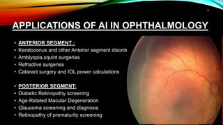
The document outlines the role of artificial intelligence (AI) in ophthalmology, detailing its history, definitions, and applications in various eye-related conditions and surgeries. It distinguishes between weak and strong AI, discusses various AI techniques such as machine learning in areas like diabetic retinopathy screening and cataract surgery, and highlights recent advancements and FDA-approved AI algorithms for eye health. The document emphasizes the potential of AI to enhance diagnostic accuracy and streamline ophthalmic practices.


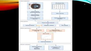
![ALGORITHM FOR IOL POWER CALCULATION
• Ladas Super Formula - Combinations of existing formulas (Hoffer Q.
Holladay-1, Holladay-l with Koch adjust - ment, Haigis, and SRK/T formulas)
and plotted into a 5-D surface
• Hill-Radial Basis Function (RBF) method - Based on Haag-Streit LENSTAR
optical biometer
• Kane formula- Cloud-based formula
Hill W. Hill-RBF Formula 3.0 [Internet]. Hill-RBF Calculator Version 3.0. https://rbfcalculator.com/. Accessed 3 Feb 2021.
Ladas JG, Siddiqui AA, Devgan U, Jun AS. A 3-D “Super Surface” combining modern intraocular lens formulas to generate a “Super Formula” and maximize accuracy.
JAMA Ophthalmol. 2015;133(12):1431–6.
Gatinel D, Debellemanière G, Saad A, Dubois M, Rampat R. Determining the theoretical effective lens position of thick intraocular lenses for machine learning based IOL
power calculation and simulation. Transl Vis Sci Technol. 2021.
18](https://image.slidesharecdn.com/artificalintelligenceinophthalmology-240525123201-a279503f/85/artifical-intelligence-in-ophthalmology-pptx-18-320.jpg)


![#AI IN DIABETIC RETINOPATHY
• In April 2018, the US Food and Drug Administration (FDA) approved an AI algorithm, developed by
IDx, used with Topcon Fundus camera (Topcon Medical) for DR identification.
• a study was done on 900-subjects in a primary-care setting (10 primary care sites) with automated
image analysis.
• Two 45-degree digital images per eye (one centered on the macula, one centered on the optic nerve)
were obtained and analyzed.
• These images were compared with the stereo, widefield fundus imaging interpreted by the
Wisconsin Fundus Photograph Reading Centre (FPRC)
Saeedi P, Petersohn I, Salpea P, Malanda B, Karuranga S, Unwin N, Colagiuri S, Guariguata L, Motala AA, Ogurtsova K, Shaw JE, Bright D, Williams R; IDF Diabetes Atlas
Committee. Global and regional diabetes prevalence estimates for 2019 and projections for 2030 and 2045: Results from the International Diabetes Federation Diabetes
Atlas, 9th edition. Diabetes Res Clin Pract. 2019 Nov;157:107843. doi: 10.1016/j.diabres.2019.107843. Epub 2019 Sep 10. PMID: 31518657.*
US Food and Drug Administration. FDA permits marketing of artificial intelligence-based device to detect certain diabetes-related eye problems. Available from:
https://www.fda.gov/NewsEvents/ Newsroom/PressAnnouncements/ucm604357.htm. Published April 11, 2018. [Last accessed on 2018 Aug 12].
30](https://image.slidesharecdn.com/artificalintelligenceinophthalmology-240525123201-a279503f/85/artifical-intelligence-in-ophthalmology-pptx-30-320.jpg)
![AI IN DIABETIC RETINOPATHY
• By autonomous comparison software provides one of the two results:*
• (1) If more than mild DR detected, refer to an eyecare professional (ECP);
• (2) If the results are negative for more than mild DR, rescreen in 12 months.
• Based on the analysis a new entity called more than minimal DR (mtmDR) was
defined- the presence of ETDRS level 35 or higher (microaneurysms plus hard
exudates, cotton wool spots, and/or mild retinal hemorrhages) and/or DME in at
least one eye**
• Sensitivity and specificity of the technology was 87.4% and 89.5% respectively for
detecting more than mild DR
• Anti-VEGF outcome prediction and dose optimization in DME.
Abramoff M. Artificial intelligence for automated detection of diabetic retinopathy in primary care. Paper presented at: Macular Society; February 22, 2018; Beverly Hills,
CA. Available from: http:// webeye.ophth.uiowa.edu/abramoff/MDA MacSocAbst 2018 02 22. Pdf [Internet]. [Last accessed on 2019 Mar 26].*
Pros and Cons of Using an AI-Based Diagnosis for Diabetic Retinopathy: Page 4 of 5 N.d. Optometry Times. Available from:
http://www.optometrytimes.com/article/pros-and-cons-using-ai-based-diagnosis-diabetic-retinopathy. [Last accessed on 2018 Oct 29**
31](https://image.slidesharecdn.com/artificalintelligenceinophthalmology-240525123201-a279503f/85/artifical-intelligence-in-ophthalmology-pptx-31-320.jpg)