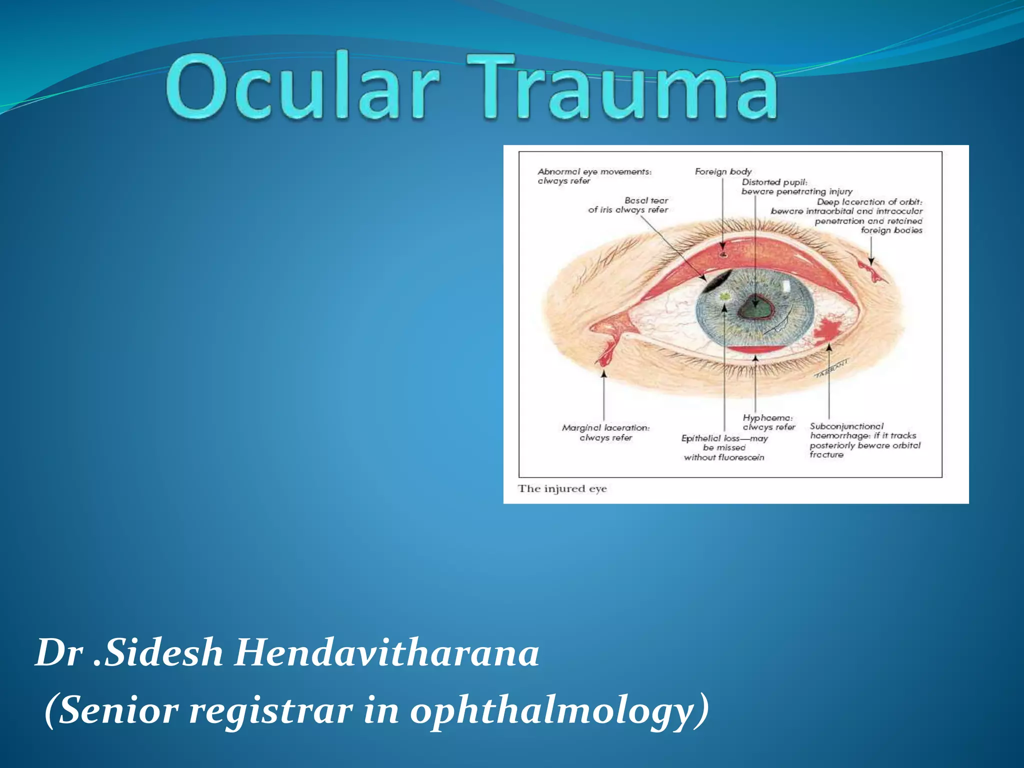
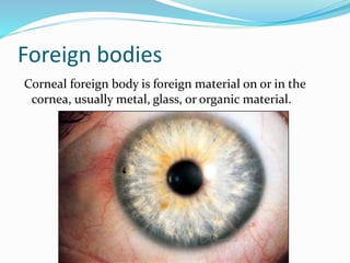
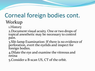
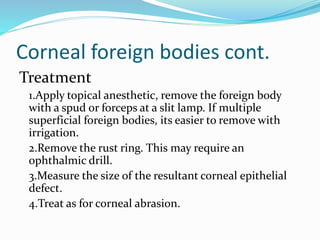
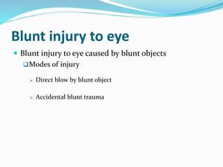
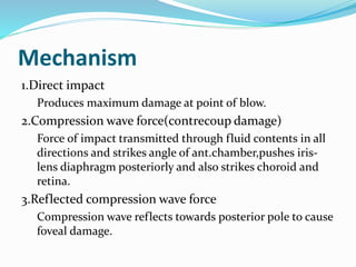
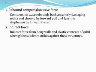
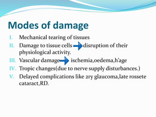
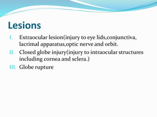
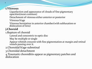
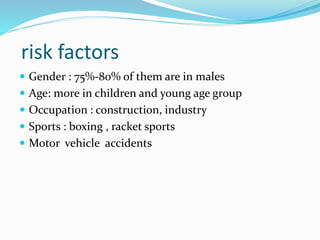
Dr .Sidesh Hendavitharana
(Senior registrar in ophthalmology)
Ocular trauma is an emergency and requires
immediate medical or surgical intervention
Classification
Direct ,
1.Mechanical injuries
 Extraocular FB
 Blunt injury(contusion)
 Penetrating and perforating injury
 Perforating injury with retained FB
2.Chemical injuries
 Acid burns
 Alkali burns
.3.Injuries due to physical agent
 Thermal injury
 Electric injury
 Radiation injury
I. Ultraviolet radiations
II. Infrared radiations
III. Ionizing radiational injuries
 Indirect ocular trauma
Foreign bodies
Corneal foreign body is foreign material on or in the
cornea, usually metal, glass, or organic material.
Corneal foreign bodies:
Symptoms
Foreign body sensation,
Tearing, History of
trauma ,photophobia ,
pain , red eye
Signs
Corneal foreign body
with or without rust ring,
edema of the lids,
conjunctiva, and cornea,
foreign body can cause
infection and/or tissue
necrosis.
Corneal foreign bodies cont.
Workup
1.History
2.Document visual acuity. One or two drops of
topical anesthetic may be necessary to control
pain.
3.Slit-lamp Examination: If there is no evidence of
perforation, evert the eyelids and inspect for
foreign bodies.
4.Dilate the eye and examine the vitreous and
retina
5.Consider a B-scan US, CT of the orbit.
Corneal foreign bodies cont.
Treatment
1.Apply topical anesthetic, remove the foreign body
with a spud or forceps at a slit lamp. If multiple
superficial foreign bodies, its easier to remove with
irrigation.
2.Remove the rust ring. This may require an
ophthalmic drill.
3.Measure the size of the resultant corneal epithelial
defect.
4.Treat as for corneal abrasion.
Blunt injury to eye
 Blunt injury to eye caused by blunt objects
Modes of injury
 Direct blow by blunt object
 Accidental blunt trauma
Mechanism
1.Direct impact
Produces maximum damage at point of blow.
2.Compression wave force(contrecoup damage)
Force of impact transmitted through fluid contents in all
directions and strikes angle of ant.chamber,pushes iris-
lens diaphragm posteriorly and also strikes choroid and
retina.
3.Reflected compression wave force
Compression wave reflects towards posterior pole to cause
foveal damage.
.
4.Rebound compression wave force
Compression wave rebounds back anteriorly damaging
retina and choroid by forward pull and lens-iris
diaphragm by forward thrust.
5.Indirect force
Indirect force from bony walls and elastic contents of orbit
when globe suddenly strikes against these structures.
Modes of damage
I. Mechanical tearing of tissues
II. Damage to tissue cells disruption of their
physiological activity.
III. Vascular damage ischemia,oedema,h’age
IV. Tropic changes(due to nerve supply disturbances.)
V. Delayed complications like 2ry glaucoma,late rossete
cataract,RD.
irri
t
irri
t
Lesions
I. Extraocular lesion(injury to eye lids,conjunctiva,
lacrimal apparatus,optic nerve and orbit.
II. Closed globe injury(injury to intraocular structures
including cornea and sclera.)
III. Globe rupture
Effects
a.Eyelids
Ecchymosis
Black eye(hematoma in loose subcutaneous tissue)
Aberasion, laceration and avulsion
Traumatic ptosis
Emphysema(due to escape of air from paranasal sinuses.
.
b.Lacrimal apparatus
Dislocation of lacrimal gland
Laceration of lacrimal passage
c.Conjunctiva
Subconjunctival h’age, chemosis, laceration
d.Cornea
1.Simple abrasion
Very painful
Detected by 2%fluorescein staining
Treatment
Antibiotic oinment with pad and bandage for 24hrs.
.
2.Recurrent corneal erosions
Follow simple aberasion caused due to fingernail trauma
Feature
Recurrent attacks of acute pain and lacrimation on
opening eye in morning(due to abnormally loose
attachment of epithelium to underlying bowman’s
membrane
Treatment
Antibiotic oinment with pad and bandage for 48hrs
Debridement of loosely attached epithelium
.
3.Partial or complete corneal tears(lamella corneal
lacerations)
4.Blood staining of cornea
Associated with hyphema and raised intraocular pressure.
Features
Reddish brown or greenish cornea
Simulates dislocation of clear lens into anterior
chamber(late stages)
Clear very slowly for periphery towards center over 2 yrs
5.Deep corneal opacity
Results from corneal stromal edema or folds in descemet’s
membrane.
.
e.Sclera
Partial thickness scleral wounds(lamella scleral
lacerations)
Rupture of eyeball(commonly at limbus or behind
insertion of recti)
f.Anterior chamber
1.Hyphema
Due to injury to iris or cilliary body vessels
2.Exudates
Following traumatic uveitis
.
g.Iris,pupil and cilliary body
1.Traumatic miosis
Due to irritation of ciliary nerves
Associated with spasm of accomodation.
2.Traumatic mydriasis(iridoplegia)permenant and
associated with traumatic cycloplegia.
3.Rupture of pupillary margin
4.Radiating tear in iris stroma (sometimes reaching upto
cilliay body)
5.Iridodialysis(detachment of iris from its root at ciliary
body)
Produces D shaped pupil and black biconvex area at
periphery.
.
6.Antiflexion of iris (rotation of detached portion of iris
such that posterior part faces anteriorly)
7.Traumatic aniridia(sinking of completely torn iris to
bottom of anterior chamber
8.Angle recession(tear longitudinal and circular muscle
fibers of ciliary body)
Deepening of anterior chamber and widening of ciliary
body
Complicated by glaucoma
Major source of bleeding
9.Inflammatory changes(traumatic iridocyclitis, post
traumatic iris atropy, and pigmentory changes
.
h.Lens
1.Vossius ring
-circular ring of stippled brown amophous pigment
granules on anterior capsule
Due to striking of contracted margin against crystalline
lens
Always smaller than pupilary size.
2.Concussion cataract
Due to inbibition of aqueous and direct mechanical effect
of injury on lens fibers
.
 Shape,
 Discrete subepithelial opacities
 Early rossete cataract(punctate)feathery lines of opacities along star shaped
suture lines in posterior cortex seen after 1-2yrs
 Traumatic zonular cataract
 Diffuse(total)concusion cataract
 Early maturation of senile cataract
 Traumatic dislocation of lens(in young children)
 Subluxation of lens
 Due to partial tear of zonules
 Lens slight displaced but still present in pupilary area.
 May be vertical(upward or downward)or lateral(nasal or temporal)
 Dislocation of lens
 Due to complete rupture of zonules
 May be intraocular dislocation into either anterior or posterior chamber or
extraocular dislocation in subconjunctival space or outside the eye ball.
 Tear in lens capsule with absorption of lens matter
 Total lens opacification
.
i.Vitreous
Liquefaction and appearance of clouds of fine pigmentary
opacities(most common)
Detachment of vitreous either anterior or posterior
Vitreous h’age
Vitreous herniation in anterior chamber(with subluxation or
dislocation of lens)
j.Choroid
1.Rupture of choroid
Lateral and concentric to optic disc
May be multiple or single
Appear whitish crescent with fine pigmentation at margin and retinal
vessels passing over it.
2.Choroidal h’age-subretinal
3.Choroidal detachment
4.Traumatic choroiditis-appear as pigmentary patches and
dislocation
.
k.Retina
1.Commotio retinae(berlin’s edema)
Milky white cloudiness at posterior pole with cherry red spot in
foveal region
May disappear or followed by pigmentory changes
2.Retinal h’age
Multiple flame shaped and preretinal subhyaloid D shaped h’age.
3.Retinal tears-common in myopic eyes and eyes with senile
degeneration
4.Traumatic proliferative retinopathy(retinitis proliferans)-2ry to
vitreous h’age forming tractional bands
5.Retinal detachment-follows retinal tears or vitreotractional bands
6.Concussion changes at macula
Traumatic macula edema pigmentory degeneration
Macular cyst rupture lamella or full thickness macular
hole.
.
l.Intraocular pressure changes
 Traumatic glaucoma(due to intense vasodilatation of ciliary
vessels,damage to trabecular meshwork and other factors
like hyphema,lens damage)
 Traumatic hypotony (due to damage of ciliary body)
m.Refractive changes
 Myopia(due to ciliary spasm or rupture of zonules or
anterior displacement of lens)
 Hypermetropia and loss of accomodation(due to damage of
ciliary body)
n.Optic nerve
 Injury associated with fracture of base of skull
 Manifest as traumatic papillitis,laceration of optic
nerve,optic nerve sheath h’age, and avulsion of optic nerve
.
o.Orbital injury
Associated with fractures of orbital walls esp.blowout #
Orbital h’age sudden proptosis
Ethmoidal sinus rupture Orbital emphysema
p.Globe rupture(full thickness wound of eye ball)
 Indirect rupture due to compresion force(more common)
 Impact momentory increase in IOP inside out injuryof
eye ball at weakest part esp.vicinity of canal of schlemm
concentric to limbus(mostly superonasal limbus)
 Associated with prolapse of uveal tissue,vitreous loss,IO
h’age and dislocation of lens
 Ultimately IOP decreases
risk factors
 Gender : 75%-80% of them are in males
 Age: more in children and young age group
 Occupation : construction, industry
 Sports : boxing , racket sports
 Motor vehicle accidents
Hyphema:
Blood in the Anterior
Chamber
Symptoms
Pain, Blurred vision, History of
blunt trauma
Signs
Blood in the Anterior Chamber.
Gross layering or clot or both,
usually visible without a slit
lamp. A total (100%) hyphema
may be black or red; when black
its called “8-ball” or “black ball”
hyphema.
Clinical classification
 Microscopic
 Grade 1(<1/3AC volume)
 Grade 2(1/3-1/2AC volume)
 Grade 3(>1/2 AC volume)
 Grade 4(total)
Problem and complication
 Rebleeding
 Depending on size of hyphema
 Grade 1 hyphema(25% will rebleed)
 Grade 3 hyphema(75% will rebleed)
 Increase IOP
 Dependent on size and rebleeding
 Corneal blood staining
 dependent on size,IOP and rebleeding
Indication for surgical treatment
 Ocular factors
 Corneal blood staining
 Total hyphema of IOP of >50mmHg for 5days(to
prevent optic nerve damage)
 hyphema that are initially total and do not resolve
below 50%at 6 days with IOP>25mmHg(to prevent
corneal blood staining)
 Hyphema that remain unresolved for 9 days (to
prevent PAS)
.
 Patient factors
 Risk of glaucoma damage(elderly,glaucoma pts,
vascular diseases)
 Risk of corneal blood staining and amblyopia
(clildren)
 Management
 Medical
 Conservative
 Bed rest , head elevation
 Topical steroids and cycloplegic agents
 Topical glaucoma medications
 Avoids aspirin
Types of surgical management
 AC paracentsis and wash out
 clot expression and limbal delivary
 Automated hyphectemy
Penetrating and perforating injury
 Three times more common in males than females.
 Common causes are assault, domestic and
occupatioanal accidents and sports.
 Extent of damage is determined by kinetic energy
caused by flying FBs.
 Risk factors include delay in primary repair, ruptured
lens capsule and a dirty wound
corneal
 Technique of primary repair depend on the extent of the
wound and associated complications such as iris
incarceration , flat anterior chamber and damage to
intraocular contents.
 Small shelving wounds with formed AC,
 May not requir suturing as they often heal spontaneously or
with the aid of a soft BCL.
 Medium sized wounds,
 Usually require suturing ,esp. the ant.chamber is shallow or
flat.
 With lens damage,
 Suture the wound and remove the lens by
phacoemulsification or with vitreous cutter +/_ IOL.
Intraocular FB
 IOFB may traumatize mechanically,introduce
infection or exert other toxic effects on the intraocular
structures
 Initial management
 Accurate history-vital to determine the origin of the FB
 Examination,
 Special attention to possible sites of entry or exit.
 Logical deduction of the probable location of a FB.
 Gonioscopy and funduscopy must be performed.
.
 CT with axial ,coronal cuts is used to detect and
localize a metallic intraocular FB
 MRI is contraindicated in the context of a metallic
IOFB.
 Technique of removal
 Magnetic removal of ferrous Fbs involves the
creation of a sclerotomy adjecent to the FB.
 Scleral bucking may be performed to reduce the risk of
retinal detachment if this is judged to be high.
 Forcep removal is used for non-magnetic FBs and
magnetic FBs that can not safely removed with a
magnet.
 Prophylaxis against infection.
siderosis
 Steel –commonest FB projected into the eye
 A ferrous IOFB undergoes dissociation resulting in the
deposition of iron in the intraocular epithelial structures
notably the lens epithelium,iris and cilliary body
epithelium and sensory retina
 Exerts toxic effect on cellular enzyme systems with
resultant cell death.
 Signs,
 Anterior capsular cataract consisting of radial iron deposits
on the anterior lens capsules and reddish brown staining of
the iris(heterochromia iridis)
 Complications include 2ry glaucoma due to trabecular
damage,pigmentory retinopathy followed by atropy of
retina and RPE.
 ERG show progressive attenuation of the b-wave over time.
chalcosis
 The ocular reaction to IOFB with a high CU content
involves a violent endophthalmitis like picture,often
progreesion to phthisis bulbi.
 Cu deposited intraocularly resulting in a picture
similar to that of wilsons disease.
 K-F ring,anterior sun flower cataract developed
 Retinal deposition results in golden plaques visible in
ophthalmoscopy.
Ocular trauma

Ocular trauma

  • 1.
    Dr .Sidesh Hendavitharana (Seniorregistrar in ophthalmology)
  • 2.
    Ocular trauma isan emergency and requires immediate medical or surgical intervention Classification Direct , 1.Mechanical injuries  Extraocular FB  Blunt injury(contusion)  Penetrating and perforating injury  Perforating injury with retained FB
  • 3.
    2.Chemical injuries  Acidburns  Alkali burns
  • 4.
    .3.Injuries due tophysical agent  Thermal injury  Electric injury  Radiation injury I. Ultraviolet radiations II. Infrared radiations III. Ionizing radiational injuries  Indirect ocular trauma
  • 5.
    Foreign bodies Corneal foreignbody is foreign material on or in the cornea, usually metal, glass, or organic material.
  • 6.
    Corneal foreign bodies: Symptoms Foreignbody sensation, Tearing, History of trauma ,photophobia , pain , red eye Signs Corneal foreign body with or without rust ring, edema of the lids, conjunctiva, and cornea, foreign body can cause infection and/or tissue necrosis.
  • 7.
    Corneal foreign bodiescont. Workup 1.History 2.Document visual acuity. One or two drops of topical anesthetic may be necessary to control pain. 3.Slit-lamp Examination: If there is no evidence of perforation, evert the eyelids and inspect for foreign bodies. 4.Dilate the eye and examine the vitreous and retina 5.Consider a B-scan US, CT of the orbit.
  • 8.
    Corneal foreign bodiescont. Treatment 1.Apply topical anesthetic, remove the foreign body with a spud or forceps at a slit lamp. If multiple superficial foreign bodies, its easier to remove with irrigation. 2.Remove the rust ring. This may require an ophthalmic drill. 3.Measure the size of the resultant corneal epithelial defect. 4.Treat as for corneal abrasion.
  • 9.
    Blunt injury toeye  Blunt injury to eye caused by blunt objects Modes of injury  Direct blow by blunt object  Accidental blunt trauma
  • 10.
    Mechanism 1.Direct impact Produces maximumdamage at point of blow. 2.Compression wave force(contrecoup damage) Force of impact transmitted through fluid contents in all directions and strikes angle of ant.chamber,pushes iris- lens diaphragm posteriorly and also strikes choroid and retina. 3.Reflected compression wave force Compression wave reflects towards posterior pole to cause foveal damage.
  • 11.
    . 4.Rebound compression waveforce Compression wave rebounds back anteriorly damaging retina and choroid by forward pull and lens-iris diaphragm by forward thrust. 5.Indirect force Indirect force from bony walls and elastic contents of orbit when globe suddenly strikes against these structures.
  • 12.
    Modes of damage I.Mechanical tearing of tissues II. Damage to tissue cells disruption of their physiological activity. III. Vascular damage ischemia,oedema,h’age IV. Tropic changes(due to nerve supply disturbances.) V. Delayed complications like 2ry glaucoma,late rossete cataract,RD. irri t irri t
  • 13.
    Lesions I. Extraocular lesion(injuryto eye lids,conjunctiva, lacrimal apparatus,optic nerve and orbit. II. Closed globe injury(injury to intraocular structures including cornea and sclera.) III. Globe rupture
  • 14.
    Effects a.Eyelids Ecchymosis Black eye(hematoma inloose subcutaneous tissue) Aberasion, laceration and avulsion Traumatic ptosis Emphysema(due to escape of air from paranasal sinuses.
  • 15.
    . b.Lacrimal apparatus Dislocation oflacrimal gland Laceration of lacrimal passage c.Conjunctiva Subconjunctival h’age, chemosis, laceration d.Cornea 1.Simple abrasion Very painful Detected by 2%fluorescein staining Treatment Antibiotic oinment with pad and bandage for 24hrs.
  • 16.
    . 2.Recurrent corneal erosions Followsimple aberasion caused due to fingernail trauma Feature Recurrent attacks of acute pain and lacrimation on opening eye in morning(due to abnormally loose attachment of epithelium to underlying bowman’s membrane Treatment Antibiotic oinment with pad and bandage for 48hrs Debridement of loosely attached epithelium
  • 17.
    . 3.Partial or completecorneal tears(lamella corneal lacerations) 4.Blood staining of cornea Associated with hyphema and raised intraocular pressure. Features Reddish brown or greenish cornea Simulates dislocation of clear lens into anterior chamber(late stages) Clear very slowly for periphery towards center over 2 yrs 5.Deep corneal opacity Results from corneal stromal edema or folds in descemet’s membrane.
  • 18.
    . e.Sclera Partial thickness scleralwounds(lamella scleral lacerations) Rupture of eyeball(commonly at limbus or behind insertion of recti) f.Anterior chamber 1.Hyphema Due to injury to iris or cilliary body vessels 2.Exudates Following traumatic uveitis
  • 19.
    . g.Iris,pupil and cilliarybody 1.Traumatic miosis Due to irritation of ciliary nerves Associated with spasm of accomodation. 2.Traumatic mydriasis(iridoplegia)permenant and associated with traumatic cycloplegia. 3.Rupture of pupillary margin 4.Radiating tear in iris stroma (sometimes reaching upto cilliay body) 5.Iridodialysis(detachment of iris from its root at ciliary body) Produces D shaped pupil and black biconvex area at periphery.
  • 20.
    . 6.Antiflexion of iris(rotation of detached portion of iris such that posterior part faces anteriorly) 7.Traumatic aniridia(sinking of completely torn iris to bottom of anterior chamber 8.Angle recession(tear longitudinal and circular muscle fibers of ciliary body) Deepening of anterior chamber and widening of ciliary body Complicated by glaucoma Major source of bleeding 9.Inflammatory changes(traumatic iridocyclitis, post traumatic iris atropy, and pigmentory changes
  • 21.
    . h.Lens 1.Vossius ring -circular ringof stippled brown amophous pigment granules on anterior capsule Due to striking of contracted margin against crystalline lens Always smaller than pupilary size. 2.Concussion cataract Due to inbibition of aqueous and direct mechanical effect of injury on lens fibers
  • 22.
    .  Shape,  Discretesubepithelial opacities  Early rossete cataract(punctate)feathery lines of opacities along star shaped suture lines in posterior cortex seen after 1-2yrs  Traumatic zonular cataract  Diffuse(total)concusion cataract  Early maturation of senile cataract  Traumatic dislocation of lens(in young children)  Subluxation of lens  Due to partial tear of zonules  Lens slight displaced but still present in pupilary area.  May be vertical(upward or downward)or lateral(nasal or temporal)  Dislocation of lens  Due to complete rupture of zonules  May be intraocular dislocation into either anterior or posterior chamber or extraocular dislocation in subconjunctival space or outside the eye ball.  Tear in lens capsule with absorption of lens matter  Total lens opacification
  • 23.
    . i.Vitreous Liquefaction and appearanceof clouds of fine pigmentary opacities(most common) Detachment of vitreous either anterior or posterior Vitreous h’age Vitreous herniation in anterior chamber(with subluxation or dislocation of lens) j.Choroid 1.Rupture of choroid Lateral and concentric to optic disc May be multiple or single Appear whitish crescent with fine pigmentation at margin and retinal vessels passing over it. 2.Choroidal h’age-subretinal 3.Choroidal detachment 4.Traumatic choroiditis-appear as pigmentary patches and dislocation
  • 24.
    . k.Retina 1.Commotio retinae(berlin’s edema) Milkywhite cloudiness at posterior pole with cherry red spot in foveal region May disappear or followed by pigmentory changes 2.Retinal h’age Multiple flame shaped and preretinal subhyaloid D shaped h’age. 3.Retinal tears-common in myopic eyes and eyes with senile degeneration 4.Traumatic proliferative retinopathy(retinitis proliferans)-2ry to vitreous h’age forming tractional bands 5.Retinal detachment-follows retinal tears or vitreotractional bands 6.Concussion changes at macula Traumatic macula edema pigmentory degeneration Macular cyst rupture lamella or full thickness macular hole.
  • 25.
    . l.Intraocular pressure changes Traumatic glaucoma(due to intense vasodilatation of ciliary vessels,damage to trabecular meshwork and other factors like hyphema,lens damage)  Traumatic hypotony (due to damage of ciliary body) m.Refractive changes  Myopia(due to ciliary spasm or rupture of zonules or anterior displacement of lens)  Hypermetropia and loss of accomodation(due to damage of ciliary body) n.Optic nerve  Injury associated with fracture of base of skull  Manifest as traumatic papillitis,laceration of optic nerve,optic nerve sheath h’age, and avulsion of optic nerve
  • 26.
    . o.Orbital injury Associated withfractures of orbital walls esp.blowout # Orbital h’age sudden proptosis Ethmoidal sinus rupture Orbital emphysema p.Globe rupture(full thickness wound of eye ball)  Indirect rupture due to compresion force(more common)  Impact momentory increase in IOP inside out injuryof eye ball at weakest part esp.vicinity of canal of schlemm concentric to limbus(mostly superonasal limbus)  Associated with prolapse of uveal tissue,vitreous loss,IO h’age and dislocation of lens  Ultimately IOP decreases
  • 27.
    risk factors  Gender: 75%-80% of them are in males  Age: more in children and young age group  Occupation : construction, industry  Sports : boxing , racket sports  Motor vehicle accidents
  • 28.
    Hyphema: Blood in theAnterior Chamber Symptoms Pain, Blurred vision, History of blunt trauma Signs Blood in the Anterior Chamber. Gross layering or clot or both, usually visible without a slit lamp. A total (100%) hyphema may be black or red; when black its called “8-ball” or “black ball” hyphema.
  • 29.
    Clinical classification  Microscopic Grade 1(<1/3AC volume)  Grade 2(1/3-1/2AC volume)  Grade 3(>1/2 AC volume)  Grade 4(total)
  • 30.
    Problem and complication Rebleeding  Depending on size of hyphema  Grade 1 hyphema(25% will rebleed)  Grade 3 hyphema(75% will rebleed)  Increase IOP  Dependent on size and rebleeding  Corneal blood staining  dependent on size,IOP and rebleeding
  • 31.
    Indication for surgicaltreatment  Ocular factors  Corneal blood staining  Total hyphema of IOP of >50mmHg for 5days(to prevent optic nerve damage)  hyphema that are initially total and do not resolve below 50%at 6 days with IOP>25mmHg(to prevent corneal blood staining)  Hyphema that remain unresolved for 9 days (to prevent PAS)
  • 32.
    .  Patient factors Risk of glaucoma damage(elderly,glaucoma pts, vascular diseases)  Risk of corneal blood staining and amblyopia (clildren)  Management  Medical  Conservative  Bed rest , head elevation  Topical steroids and cycloplegic agents  Topical glaucoma medications  Avoids aspirin
  • 33.
    Types of surgicalmanagement  AC paracentsis and wash out  clot expression and limbal delivary  Automated hyphectemy
  • 34.
    Penetrating and perforatinginjury  Three times more common in males than females.  Common causes are assault, domestic and occupatioanal accidents and sports.  Extent of damage is determined by kinetic energy caused by flying FBs.  Risk factors include delay in primary repair, ruptured lens capsule and a dirty wound
  • 35.
    corneal  Technique ofprimary repair depend on the extent of the wound and associated complications such as iris incarceration , flat anterior chamber and damage to intraocular contents.  Small shelving wounds with formed AC,  May not requir suturing as they often heal spontaneously or with the aid of a soft BCL.  Medium sized wounds,  Usually require suturing ,esp. the ant.chamber is shallow or flat.  With lens damage,  Suture the wound and remove the lens by phacoemulsification or with vitreous cutter +/_ IOL.
  • 36.
    Intraocular FB  IOFBmay traumatize mechanically,introduce infection or exert other toxic effects on the intraocular structures  Initial management  Accurate history-vital to determine the origin of the FB  Examination,  Special attention to possible sites of entry or exit.  Logical deduction of the probable location of a FB.  Gonioscopy and funduscopy must be performed.
  • 37.
    .  CT withaxial ,coronal cuts is used to detect and localize a metallic intraocular FB  MRI is contraindicated in the context of a metallic IOFB.  Technique of removal  Magnetic removal of ferrous Fbs involves the creation of a sclerotomy adjecent to the FB.  Scleral bucking may be performed to reduce the risk of retinal detachment if this is judged to be high.  Forcep removal is used for non-magnetic FBs and magnetic FBs that can not safely removed with a magnet.  Prophylaxis against infection.
  • 38.
    siderosis  Steel –commonestFB projected into the eye  A ferrous IOFB undergoes dissociation resulting in the deposition of iron in the intraocular epithelial structures notably the lens epithelium,iris and cilliary body epithelium and sensory retina  Exerts toxic effect on cellular enzyme systems with resultant cell death.  Signs,  Anterior capsular cataract consisting of radial iron deposits on the anterior lens capsules and reddish brown staining of the iris(heterochromia iridis)  Complications include 2ry glaucoma due to trabecular damage,pigmentory retinopathy followed by atropy of retina and RPE.  ERG show progressive attenuation of the b-wave over time.
  • 39.
    chalcosis  The ocularreaction to IOFB with a high CU content involves a violent endophthalmitis like picture,often progreesion to phthisis bulbi.  Cu deposited intraocularly resulting in a picture similar to that of wilsons disease.  K-F ring,anterior sun flower cataract developed  Retinal deposition results in golden plaques visible in ophthalmoscopy.