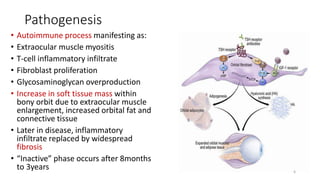
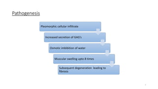
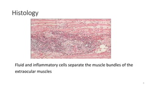
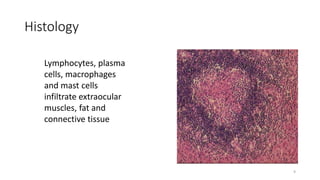
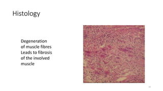
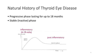
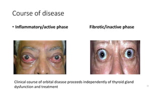
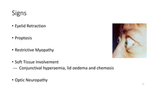
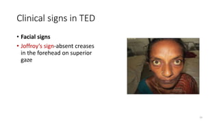
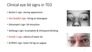
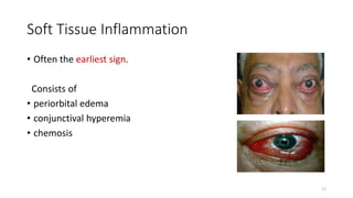
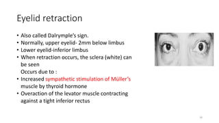
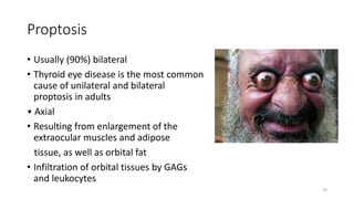
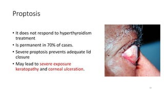
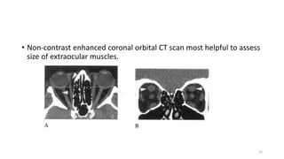
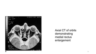
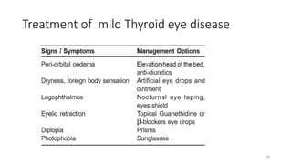
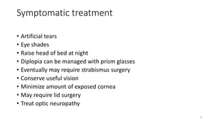
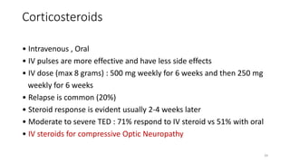
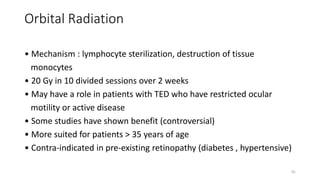
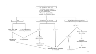
Thyroid Ophthalmopathy
1
Introduction of Thyroid eye disease
โ€ข Thyroid eye disease is an autoimmune disease producing symptoms
related to inflammation, accumulation of fluid in the orbit and also to
adipogenesis raising intra-orbital pressure.
โ€ข Synonyms
โ€ข Gravesโ€™ ophthalmopathy/orbitopathy (GO)
โ€ข Thyroid eye disease (TED)
โ€ข Thyroid associated ophthalmopathy (TAO)
โ€ข Dysthyroid ophthalmopathy
2
Epidemiology
โ€ข Prevalence of thyroid ophthalmopathy = 0.4%
Women > Men
โ€ข But severity greater in men
โ€ข Bimodal age distribution โ€“ Peak incidence in fourth and sixth decades
of life
โ€ข May be exacerbated by stress and smoking
โ€ข Most common cause of exophthalmos
โ€ข >50% of cases with Gravesโ€™ disease have eye involvement
3
Etiology
โ€ข Gravesโ€™ hyperthyroidism (90%)
โ€ข Hypothyroid Hashimotoโ€™s thyroiditis
โ€ข Euthyroid subjects with no current or past evidence of thyroid
hyper or hypofunction (so called euthyroid Gravesโ€™ disease).
โ€ข In patients with Graveโ€™s disease, eye signs may precede, coincide with
or follow the hyperthyroidism
4
Risk factors
โ€ข Smoking (strongest modifiable risk factor)
โ€ข Family history
โ€ข Monozygotic twins
5
Pathogenesis
โ€ข Autoimmune process manifesting as:
โ€ข Extraocular muscle myositis
โ€ข T-cell inflammatory infiltrate
โ€ข Fibroblast proliferation
โ€ข Glycosaminoglycan overproduction
โ€ข Increase in soft tissue mass within
bony orbit due to extraocular muscle
enlargement, increased orbital fat and
connective tissue
โ€ข Later in disease, inflammatory
infiltrate replaced by widespread
fibrosis
โ€ข โ€œInactiveโ€ phase occurs after 8months
to 3years 6
Pathogenesis
Pleomorphic cellular infiltrate
Increased secretion of GAGโ€™s
Osmotic imbibition of water
Muscular swelling upto 8 times
Subsequent degeneration leading to
fibrosis
7
Histology
Fluid and inflammatory cells separate the muscle bundles of the
extraocular muscles
8
Histology
Lymphocytes, plasma
cells, macrophages
and mast cells
infiltrate extraocular
muscles, fat and
connective tissue
9
Histology
Degeneration
of muscle fibres
Leads to fibrosis
of the involved
muscle
10
Natural History of Thyroid Eye Disease
โ€ข Progressive phase lasting for up to 18 months
โ€ข Stable (inactive) phase
11
Course of disease
โ€ข Inflammatory/active phase Fibrotic/inactive phase
Clinical course of orbital disease proceeds independently of thyroid gland
dysfunction and treatment 12
Symptoms
โ€ข Foreign body sensation
โ€ข Epiphora (tearing)
โ€ข Photophobia
โ€ข Bulging of eyes
โ€ข Puffiness of eye lids
โ€ข Diplopia
โ€ข Visual loss
13
Signs
โ€ข Eyelid Retraction
โ€ข Proptosis
โ€ข Restrictive Myopathy
โ€ข Soft Tissue Involvement
--- Conjunctival hyperaemia, lid oedema and chemosis
โ€ข Optic Neuropathy
14
Clinical signs in TED
โ€ข Facial signs
โ€ข Joffroyโ€™s sign-absent creases
in the forehead on superior
gaze
15
Clinical eye lid signs in TED
โ€ข Kocherโ€™s sign- staring appearance
โ€ข Von Graefeโ€™s sign- lid lag on downgaze
โ€ข Dalrympleโ€™s sign- lid retraction
โ€ข Stellwagโ€™s sign- incomplete & infrequent blinking
โ€ข Enroth โ€™s sign- edema of lower lid
โ€ข Griffithโ€™s sign- lower lid lag on upgaze
16
Soft Tissue Inflammation
โ€ข Often the earliest sign.
Consists of
โ€ข periorbital edema
โ€ข conjunctival hyperemia
โ€ข chemosis
17
Eyelid retraction
โ€ข Also called Dalrympleโ€™s sign.
โ€ข Normally, upper eyelid- 2mm below limbus
โ€ข Lower eyelid-inferior limbus
โ€ข When retraction occurs, the sclera (white) can
be seen
Occurs due to :
โ€ข Increased sympathetic stimulation of Mรผllerโ€™s
muscle by thyroid hormone
โ€ข Overaction of the levator muscle contracting
against a tight inferior rectus
18
Proptosis
โ€ข Usually (90%) bilateral
โ€ข Thyroid eye disease is the most common
cause of unilateral and bilateral
proptosis in adults
โ€ข Axial
โ€ข Resulting from enlargement of the
extraocular muscles and adipose
tissue, as well as orbital fat
โ€ข Infiltration of orbital tissues by GAGs
and leukocytes
19
Proptosis
โ€ข It does not respond to hyperthyroidism
treatment
โ€ข Is permanent in 70% of cases.
โ€ข Severe proptosis prevents adequate lid
closure
โ€ข May lead to severe exposure
keratopathy and corneal ulceration.
20
Restrictive Myopathy
โ€ข Eye movements are restricted due to oedema in extraocular muscles
during infiltrative stage and subsequent fibrosis.
โ€ข Despite expansion of the extraocular muscles , the muscle fibres
themselves are normal.
โ€ข IR>MR>SR>LR
โ€ข Pressure exerted by a fibrotic inferior rectus muscle on the
globe may cause a spike in intraocular pressure during upgaze.
21
Dysthyroid Optic Neuropathy
(DON)
Optic neuropathy as result of optic nerve compression
from enlargement of extraocular muscles
22
WERNERยดS CLASSIFICATION - NOSPECS
โ€ข Class 0: No signs or symptoms
โ€ข Class 1: Only signs (lid retraction, stare ยฑ lid lag)
โ€ข Class 2: Soft tissue involvement
โ€ข Class 3: Proptosis
โ€ข Class 4: Extraocular muscle involvement
โ€ข Class 5: Corneal involvement
โ€ข Class 6: Sight loss (optic nerve involvement)
23
EUGOGO classification
โ€ข Mild : eyelid swelling , lid retraction, proptosis
โ€ข Moderate-Severe : Active disease (EOM dysfunction, diplopia ,
proptosis >25 mm)
โ€ข Very severe : Compressive Optic Neuropathy , Corneal exposure
(needs emergent surgery)
24
VISA classification
โ€ข V (Vision) , I (inflammation), S (Strabismus) , A (Appearance)
โ€ข Vision/CON
โ€ข Inflammation/Congestion : based on documented change of
inflammation rather than absolute value
โ€ข Strabismus/Motility : measuring ductions and alignments
โ€ข Appearance/Exposure
โ€ข Score of 5 or more โ€”> Active disease or progression (Consider
Steroids)
25
Differential Diagnosis
โ€ข Orbital tumors (primary or metastatic)
โ€ข Orbital pseudotumor
โ€ข Wegenerโ€™s granulomatosis
โ€ข Orbital infection
โ€ข Carotid-cavernous sinus fistula
26
Diagnosis
โ€ข Characteristic eye findings
โ€ข Thyroid dysfunction
โ€ข Imaging
27
Blood investigations
โ€ข Highly sensitive & specific -- T4(thyroxine) + TSH or serum TSH
โ€ข If eye findings associates with euthyroid Gravesโ€™ disease โ€“
โ€ข Thyroid peroxidase antibody
โ€ข Antibody to thyroglobulin
โ€ข Others
โ€ข Free T4 index
โ€ข Thyroid-stimulating immunoglobulin
โ€ข Antithyroid antibodies
โ€ข Serum T3
28
Radiological Evaluation
โ€ข Usually employed if cause of exophthalmos is unclear (ie. normal
thyroid lab studies, or history/physical examination inconsistent with
thyroid disease)
โ€ข Also to determine optic nerve involvement if not obvious by
fundoscopic examination.
โ€ข Distinct sparing of muscle tendons in thyroid ophthalmopathy.
29
Radiological Evaluation
โ€ข CT scan is currently the imaging study of choice.
โ€ข MRI is sensitive for showing compression of the optic nerve.
โ€ข Neuroimaging usually reveals
โ€ข Thick muscle belly with tendon sparing
โ€ข Usually IR & MR
โ€ข Bilateral muscle enlargement is the norm
โ€ข Unilateral cases usually represent asymmetric involvement rather
than normality of the less involved side
30
Axial and coronal C.T. scan in Thyroid eye disease
31
โ€ข Non-contrast enhanced coronal orbital CT scan most helpful to assess
size of extraocular muscles.
32
Axial CT of orbits
demonstrating
medial rectus
enlargement
33
Management
โ€ข T โ€“ Tobacco abstinence
โ€ข E โ€“ Euthyroidism
โ€ข A โ€“ Artificial tears
โ€ข R โ€“ Referral
โ€ข S โ€“ Self help groups
34
Medical Management of Hyperthyroidism
โ€ข Anti-thyroid drugs : thinoamides (PTU) , carbimazole , methimazole.
โ€ข Thionamides inhibit synthesis of thyroid hormones.
โ€ข Need 6-8 weeks to achieve euthyroid state
โ€ข Side effects of anti-thyroid drugs
Skin rash , urticarial , arthralgia , fever
35
Treatment of mild Thyroid eye disease
36
Symptomatic treatment
โ€ข Artificial tears
โ€ข Eye shades
โ€ข Raise head of bed at night
โ€ข Diplopia can be managed with prism glasses
โ€ข Eventually may require strabismus surgery
โ€ข Conserve useful vision
โ€ข Minimize amount of exposed cornea
โ€ข May require lid surgery
โ€ข Treat optic neuropathy
37
Selenium
โ€ข 200 microgram/day for 6 months
โ€ข For Mild disease
โ€ข Antioxidant effect
โ€ข Immunomodulatory effect : reduce thyroid autoantibodies
โ€ข Reduces severity of disease and improve quality of life
38
Corticosteroids
โ€ข Intravenous , Oral
โ€ข IV pulses are more effective and have less side effects
โ€ข IV dose (max 8 grams) : 500 mg weekly for 6 weeks and then 250 mg
weekly for 6 weeks
โ€ข Relapse is common (20%)
โ€ข Steroid response is evident usually 2-4 weeks later
โ€ข Moderate to severe TED : 71% respond to IV steroid vs 51% with oral
โ€ข IV steroids for compressive Optic Neuropathy
39
Rituximab
โ€ข Chimeric mono-clonal antibody targets CD20
โ€ข CD20 is expressed on more than 95% of B cells and plasma cells
โ€ข RTX depletes 95% of mature B cells , blocks Ab production , and
decreases inflammatory cytokine release
โ€ข For steroid-refractory disease
โ€ข Side effects : Allergic reaction (mild) PML (severe)
40
Orbital Radiation
โ€ข Mechanism : lymphocyte sterilization, destruction of tissue
monocytes
โ€ข 20 Gy in 10 divided sessions over 2 weeks
โ€ข May have a role in patients with TED who have restricted ocular
motility or active disease
โ€ข Some studies have shown benefit (controversial)
โ€ข More suited for patients > 35 years of age
โ€ข Contra-indicated in pre-existing retinopathy (diabetes , hypertensive)
41
Botulinum Toxin
โ€ข Neurotoxin , inhibits acetylcholine release
โ€ข For upper lid retraction (transconjunctival , transcutaneous route)
โ€ข Effect on Mullerโ€™s muscle and LPS
โ€ข Side effects of Botox : bruising , ptosis and diplopia
42
Orbital Decompression for TED
โ€ข Decompression usually in stable phase of disease.
โ€ข Indications
โ€ข compressive optic neuropathy
โ€ข severe exposure keratopathy
โ€ข Post-operative complications (diplopia, vision loss)
โ€ข Outcome is variable : degree of fibrosis , fat expansion , bone
available, duration of optic neuropathy.
โ€ข Decompression โ€”> Muscle Surgery โ€”> Lid surgery
43
Strabismus Surgery for TED
โ€ข In the stable phase with stable alignments for 6 months
โ€ข Aim is single binocular vision in primary and reading position
โ€ข Typically involves release of the restricted muscle by recession rather
than resection
โ€ข Conjunctival dissection is challenging
โ€ข Use of adjustable sutures is strongly recommended due to the
variability in fibrosis, resulting in unpredictable results.
โ€ข Oblique surgery can increase area of single binocular vision
44
Eye lid surgery
โ€ข The most common indication for lid surgery is upper lid retraction.
โ€ข Graded Mullerโ€™s and levator aponeurosis weakening.
โ€ข Lower lid lengthening is indicated in lower lid retraction.
45
Psychological Impact of TED
โ€ข Disfigurement/altered facial appearance
โ€ข Misinterpretation as hostile or angry
โ€ข Almost 50% of TED suffer depression and/or anxiety
โ€ข 90% of TED have appearance concerns (young females)
โ€ข 44% have self-confidence issues
โ€ข Multidisciplinary approach (psychiatric included)
โ€ข Support groups
46
47
Conclusion
โ€ข Activation of thyrotropin receptor on orbital fibroblast by circulating
autoantibodies plays a primary role in development of thyroid
ophthalmopathy.
โ€ข Management is based on accurate assessment of both severity and
activity of disease.
โ€ข Immunosuppressive therapy is reserved for patients with clinically
active moderate to severe disease
48
Thank You
49

TED.pptx

  • 1.
  • 2.
    Introduction of Thyroideye disease โ€ข Thyroid eye disease is an autoimmune disease producing symptoms related to inflammation, accumulation of fluid in the orbit and also to adipogenesis raising intra-orbital pressure. โ€ข Synonyms โ€ข Gravesโ€™ ophthalmopathy/orbitopathy (GO) โ€ข Thyroid eye disease (TED) โ€ข Thyroid associated ophthalmopathy (TAO) โ€ข Dysthyroid ophthalmopathy 2
  • 3.
    Epidemiology โ€ข Prevalence ofthyroid ophthalmopathy = 0.4% Women > Men โ€ข But severity greater in men โ€ข Bimodal age distribution โ€“ Peak incidence in fourth and sixth decades of life โ€ข May be exacerbated by stress and smoking โ€ข Most common cause of exophthalmos โ€ข >50% of cases with Gravesโ€™ disease have eye involvement 3
  • 4.
    Etiology โ€ข Gravesโ€™ hyperthyroidism(90%) โ€ข Hypothyroid Hashimotoโ€™s thyroiditis โ€ข Euthyroid subjects with no current or past evidence of thyroid hyper or hypofunction (so called euthyroid Gravesโ€™ disease). โ€ข In patients with Graveโ€™s disease, eye signs may precede, coincide with or follow the hyperthyroidism 4
  • 5.
    Risk factors โ€ข Smoking(strongest modifiable risk factor) โ€ข Family history โ€ข Monozygotic twins 5
  • 6.
    Pathogenesis โ€ข Autoimmune processmanifesting as: โ€ข Extraocular muscle myositis โ€ข T-cell inflammatory infiltrate โ€ข Fibroblast proliferation โ€ข Glycosaminoglycan overproduction โ€ข Increase in soft tissue mass within bony orbit due to extraocular muscle enlargement, increased orbital fat and connective tissue โ€ข Later in disease, inflammatory infiltrate replaced by widespread fibrosis โ€ข โ€œInactiveโ€ phase occurs after 8months to 3years 6
  • 7.
    Pathogenesis Pleomorphic cellular infiltrate Increasedsecretion of GAGโ€™s Osmotic imbibition of water Muscular swelling upto 8 times Subsequent degeneration leading to fibrosis 7
  • 8.
    Histology Fluid and inflammatorycells separate the muscle bundles of the extraocular muscles 8
  • 9.
    Histology Lymphocytes, plasma cells, macrophages andmast cells infiltrate extraocular muscles, fat and connective tissue 9
  • 10.
    Histology Degeneration of muscle fibres Leadsto fibrosis of the involved muscle 10
  • 11.
    Natural History ofThyroid Eye Disease โ€ข Progressive phase lasting for up to 18 months โ€ข Stable (inactive) phase 11
  • 12.
    Course of disease โ€ขInflammatory/active phase Fibrotic/inactive phase Clinical course of orbital disease proceeds independently of thyroid gland dysfunction and treatment 12
  • 13.
    Symptoms โ€ข Foreign bodysensation โ€ข Epiphora (tearing) โ€ข Photophobia โ€ข Bulging of eyes โ€ข Puffiness of eye lids โ€ข Diplopia โ€ข Visual loss 13
  • 14.
    Signs โ€ข Eyelid Retraction โ€ขProptosis โ€ข Restrictive Myopathy โ€ข Soft Tissue Involvement --- Conjunctival hyperaemia, lid oedema and chemosis โ€ข Optic Neuropathy 14
  • 15.
    Clinical signs inTED โ€ข Facial signs โ€ข Joffroyโ€™s sign-absent creases in the forehead on superior gaze 15
  • 16.
    Clinical eye lidsigns in TED โ€ข Kocherโ€™s sign- staring appearance โ€ข Von Graefeโ€™s sign- lid lag on downgaze โ€ข Dalrympleโ€™s sign- lid retraction โ€ข Stellwagโ€™s sign- incomplete & infrequent blinking โ€ข Enroth โ€™s sign- edema of lower lid โ€ข Griffithโ€™s sign- lower lid lag on upgaze 16
  • 17.
    Soft Tissue Inflammation โ€ขOften the earliest sign. Consists of โ€ข periorbital edema โ€ข conjunctival hyperemia โ€ข chemosis 17
  • 18.
    Eyelid retraction โ€ข Alsocalled Dalrympleโ€™s sign. โ€ข Normally, upper eyelid- 2mm below limbus โ€ข Lower eyelid-inferior limbus โ€ข When retraction occurs, the sclera (white) can be seen Occurs due to : โ€ข Increased sympathetic stimulation of Mรผllerโ€™s muscle by thyroid hormone โ€ข Overaction of the levator muscle contracting against a tight inferior rectus 18
  • 19.
    Proptosis โ€ข Usually (90%)bilateral โ€ข Thyroid eye disease is the most common cause of unilateral and bilateral proptosis in adults โ€ข Axial โ€ข Resulting from enlargement of the extraocular muscles and adipose tissue, as well as orbital fat โ€ข Infiltration of orbital tissues by GAGs and leukocytes 19
  • 20.
    Proptosis โ€ข It doesnot respond to hyperthyroidism treatment โ€ข Is permanent in 70% of cases. โ€ข Severe proptosis prevents adequate lid closure โ€ข May lead to severe exposure keratopathy and corneal ulceration. 20
  • 21.
    Restrictive Myopathy โ€ข Eyemovements are restricted due to oedema in extraocular muscles during infiltrative stage and subsequent fibrosis. โ€ข Despite expansion of the extraocular muscles , the muscle fibres themselves are normal. โ€ข IR>MR>SR>LR โ€ข Pressure exerted by a fibrotic inferior rectus muscle on the globe may cause a spike in intraocular pressure during upgaze. 21
  • 22.
    Dysthyroid Optic Neuropathy (DON) Opticneuropathy as result of optic nerve compression from enlargement of extraocular muscles 22
  • 23.
    WERNERยดS CLASSIFICATION -NOSPECS โ€ข Class 0: No signs or symptoms โ€ข Class 1: Only signs (lid retraction, stare ยฑ lid lag) โ€ข Class 2: Soft tissue involvement โ€ข Class 3: Proptosis โ€ข Class 4: Extraocular muscle involvement โ€ข Class 5: Corneal involvement โ€ข Class 6: Sight loss (optic nerve involvement) 23
  • 24.
    EUGOGO classification โ€ข Mild: eyelid swelling , lid retraction, proptosis โ€ข Moderate-Severe : Active disease (EOM dysfunction, diplopia , proptosis >25 mm) โ€ข Very severe : Compressive Optic Neuropathy , Corneal exposure (needs emergent surgery) 24
  • 25.
    VISA classification โ€ข V(Vision) , I (inflammation), S (Strabismus) , A (Appearance) โ€ข Vision/CON โ€ข Inflammation/Congestion : based on documented change of inflammation rather than absolute value โ€ข Strabismus/Motility : measuring ductions and alignments โ€ข Appearance/Exposure โ€ข Score of 5 or more โ€”> Active disease or progression (Consider Steroids) 25
  • 26.
    Differential Diagnosis โ€ข Orbitaltumors (primary or metastatic) โ€ข Orbital pseudotumor โ€ข Wegenerโ€™s granulomatosis โ€ข Orbital infection โ€ข Carotid-cavernous sinus fistula 26
  • 27.
    Diagnosis โ€ข Characteristic eyefindings โ€ข Thyroid dysfunction โ€ข Imaging 27
  • 28.
    Blood investigations โ€ข Highlysensitive & specific -- T4(thyroxine) + TSH or serum TSH โ€ข If eye findings associates with euthyroid Gravesโ€™ disease โ€“ โ€ข Thyroid peroxidase antibody โ€ข Antibody to thyroglobulin โ€ข Others โ€ข Free T4 index โ€ข Thyroid-stimulating immunoglobulin โ€ข Antithyroid antibodies โ€ข Serum T3 28
  • 29.
    Radiological Evaluation โ€ข Usuallyemployed if cause of exophthalmos is unclear (ie. normal thyroid lab studies, or history/physical examination inconsistent with thyroid disease) โ€ข Also to determine optic nerve involvement if not obvious by fundoscopic examination. โ€ข Distinct sparing of muscle tendons in thyroid ophthalmopathy. 29
  • 30.
    Radiological Evaluation โ€ข CTscan is currently the imaging study of choice. โ€ข MRI is sensitive for showing compression of the optic nerve. โ€ข Neuroimaging usually reveals โ€ข Thick muscle belly with tendon sparing โ€ข Usually IR & MR โ€ข Bilateral muscle enlargement is the norm โ€ข Unilateral cases usually represent asymmetric involvement rather than normality of the less involved side 30
  • 31.
    Axial and coronalC.T. scan in Thyroid eye disease 31
  • 32.
    โ€ข Non-contrast enhancedcoronal orbital CT scan most helpful to assess size of extraocular muscles. 32
  • 33.
    Axial CT oforbits demonstrating medial rectus enlargement 33
  • 34.
    Management โ€ข T โ€“Tobacco abstinence โ€ข E โ€“ Euthyroidism โ€ข A โ€“ Artificial tears โ€ข R โ€“ Referral โ€ข S โ€“ Self help groups 34
  • 35.
    Medical Management ofHyperthyroidism โ€ข Anti-thyroid drugs : thinoamides (PTU) , carbimazole , methimazole. โ€ข Thionamides inhibit synthesis of thyroid hormones. โ€ข Need 6-8 weeks to achieve euthyroid state โ€ข Side effects of anti-thyroid drugs Skin rash , urticarial , arthralgia , fever 35
  • 36.
    Treatment of mildThyroid eye disease 36
  • 37.
    Symptomatic treatment โ€ข Artificialtears โ€ข Eye shades โ€ข Raise head of bed at night โ€ข Diplopia can be managed with prism glasses โ€ข Eventually may require strabismus surgery โ€ข Conserve useful vision โ€ข Minimize amount of exposed cornea โ€ข May require lid surgery โ€ข Treat optic neuropathy 37
  • 38.
    Selenium โ€ข 200 microgram/dayfor 6 months โ€ข For Mild disease โ€ข Antioxidant effect โ€ข Immunomodulatory effect : reduce thyroid autoantibodies โ€ข Reduces severity of disease and improve quality of life 38
  • 39.
    Corticosteroids โ€ข Intravenous ,Oral โ€ข IV pulses are more effective and have less side effects โ€ข IV dose (max 8 grams) : 500 mg weekly for 6 weeks and then 250 mg weekly for 6 weeks โ€ข Relapse is common (20%) โ€ข Steroid response is evident usually 2-4 weeks later โ€ข Moderate to severe TED : 71% respond to IV steroid vs 51% with oral โ€ข IV steroids for compressive Optic Neuropathy 39
  • 40.
    Rituximab โ€ข Chimeric mono-clonalantibody targets CD20 โ€ข CD20 is expressed on more than 95% of B cells and plasma cells โ€ข RTX depletes 95% of mature B cells , blocks Ab production , and decreases inflammatory cytokine release โ€ข For steroid-refractory disease โ€ข Side effects : Allergic reaction (mild) PML (severe) 40
  • 41.
    Orbital Radiation โ€ข Mechanism: lymphocyte sterilization, destruction of tissue monocytes โ€ข 20 Gy in 10 divided sessions over 2 weeks โ€ข May have a role in patients with TED who have restricted ocular motility or active disease โ€ข Some studies have shown benefit (controversial) โ€ข More suited for patients > 35 years of age โ€ข Contra-indicated in pre-existing retinopathy (diabetes , hypertensive) 41
  • 42.
    Botulinum Toxin โ€ข Neurotoxin, inhibits acetylcholine release โ€ข For upper lid retraction (transconjunctival , transcutaneous route) โ€ข Effect on Mullerโ€™s muscle and LPS โ€ข Side effects of Botox : bruising , ptosis and diplopia 42
  • 43.
    Orbital Decompression forTED โ€ข Decompression usually in stable phase of disease. โ€ข Indications โ€ข compressive optic neuropathy โ€ข severe exposure keratopathy โ€ข Post-operative complications (diplopia, vision loss) โ€ข Outcome is variable : degree of fibrosis , fat expansion , bone available, duration of optic neuropathy. โ€ข Decompression โ€”> Muscle Surgery โ€”> Lid surgery 43
  • 44.
    Strabismus Surgery forTED โ€ข In the stable phase with stable alignments for 6 months โ€ข Aim is single binocular vision in primary and reading position โ€ข Typically involves release of the restricted muscle by recession rather than resection โ€ข Conjunctival dissection is challenging โ€ข Use of adjustable sutures is strongly recommended due to the variability in fibrosis, resulting in unpredictable results. โ€ข Oblique surgery can increase area of single binocular vision 44
  • 45.
    Eye lid surgery โ€ขThe most common indication for lid surgery is upper lid retraction. โ€ข Graded Mullerโ€™s and levator aponeurosis weakening. โ€ข Lower lid lengthening is indicated in lower lid retraction. 45
  • 46.
    Psychological Impact ofTED โ€ข Disfigurement/altered facial appearance โ€ข Misinterpretation as hostile or angry โ€ข Almost 50% of TED suffer depression and/or anxiety โ€ข 90% of TED have appearance concerns (young females) โ€ข 44% have self-confidence issues โ€ข Multidisciplinary approach (psychiatric included) โ€ข Support groups 46
  • 47.
  • 48.
    Conclusion โ€ข Activation ofthyrotropin receptor on orbital fibroblast by circulating autoantibodies plays a primary role in development of thyroid ophthalmopathy. โ€ข Management is based on accurate assessment of both severity and activity of disease. โ€ข Immunosuppressive therapy is reserved for patients with clinically active moderate to severe disease 48
  • 49.