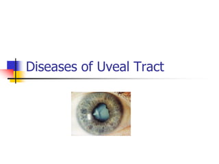
3. UVEA
• Constitutes- middle vascular coat of the eyeball
• 3 parts-
a)Iris
b)Ciliary body
c)Choroid
• Developmentally,structurally and functionally-
indivisible
• Color varies from light blue to dark brown
4. IRIS
• Anterior most part
• Avg diameter- 12mm, thickness- 0.5mm
• In centre an aperture of 3-4mm- PUPIL
• Thinnest at its root- tears away easily on blunt
trauma- IRIDODIALYSIS
• Divides space into anterior and posterior
chamber
5. MACROSCOPIC
APPEARANCE
CILIARY ZONE-
Radial streaks
Crypts- peripheral-
near the iris
central- near
collarette
Contraction furrows-
faints lines outside
collarette
PUPILLARY ZONE-
Between collarette
and pigmented frill
Pigmented frill-
black pigment at
pupillary margin
-represents ant end
of optic cup
8. FUNCTIONS OF IRIS
• CONTROLS AMOUNT OF LIGHT
ENTERING THE EYE THROUGH PUPIL
• DEFINES EYE COLOUR
• CONTROL DEPTH OF FIELD
• SOURCE OF BLOOD OCULAR TISSUES
9. CILIARY BODY
• Forward continuation of
choroid at ora serrata
• Two parts-
a) Anterior part- ciliary
processes (pars
plicata) 2-2.5mm
b) Posterior part- smooth
(pars plana) 5mm
wide temporally &
3mm nasally
11. FUNCTIONS OF CILIARY
BODY
• Site of aqueous humour production
• Maintenance of IOP
• Constitutes blood aqueous barrier
• Accommodation
• Eicosanoids are synthesised in ciliary
body
12. CHOROID
• Posterior most part
• Extension- optic disc to ora serrata
• Inner surface- smooth, brown and in
contact with RPE
• Outer surface-rough and in contact with
sclera
• Thickness- posteriorly 0.22mm
- anteriorly 0.10mm
15. FUNCTIONS OF CHOROID
• BLOOD SUPPLY TO OUTER FOUR LAYERS OF
RETINA
• MODULATION OF VASCULARISATION
• REGULATE RETINAL HEAT
• ASSIST IN THE CONTROL OF INTRAOCULAR
PRESSURE
• PIGMENT ABSORBS EXCESS LIGHT SO
AVOIDING REFLECTION
16. BLOOD SUPPLY UVEAL
TRACT
1.SHORT POSTERIOR CILIARY ARTERIES
• Branches of ophthalmic artery
• Divides into 10-20 branches, pierce sclera around optic nerve
• Supply choroid in segmental manner
2) LONG POSTERIOR CILIARY ARTERIES
• Anastomose with anterior ciliary arteries- form major arterial circle supply
ciliary body
3)ANTERIOR CILIARY ARTERIES
• From muscular arteries
• 2 each SR,IR, MR and 1 from LR
Anastomse with LPCA
• Circulous arterious major and minor
17.
18. VENOUS DRAINAGE
1. Anterior ciliary veins- tributaries of
muscular veins
2. Smaller veins from sclera- carry blood
only from sclera and not from choroid
3. Vena verticosae- 4 in no.
Drain whole of choroid
21. A. Anatomical Classification –
(IUSG) International Uveitis Study
Group
1) Anterior Uveitis – Inflammation of iris and
anterior part of ciliary body
2) Intermediate Uveitis – Involvement of posterior
part of ciliary body and extreme periphery of retina.
(Pars planitis)
3) Posterior uveitis – Retinochoroiditis, choroiditis,
retinitis, chorioretinitis
4) Diffuse or pan uveitis – Involvement of entire
uveal tract
22. B. Clinical Classification -
1) Acute – sudden symptomatic onset.
Persists for 3 weeks or less
2) Chronic – Frequently insidious and
asymptomatic. Persists for months or years
3) Recurrent
23. C. Etiological Classification
1) Exogenous-
Introduction of organism into the eye through
a perforating wound or ulcer.
2) Secondary infection-
Due to direct spread from adjoining structures-
Cornea
Sclera
Retina
24. 3) Endogenous
4) Allergic inflammation: Result of an antigen-
antibody reaction occurring in the eye due to
previous sensitization of uveal tissue to some
allergen. The allergen is a foreign protein.
Most of the cases of iridocyclitis do not have
any specific cause and are probably allergic in
nature.
26. Granulomatous Non-
granulomatous
1. Aetiology Organismal
invasion
Antigen-antibody
reaction
2. Course
a) Onset Insidious Acute
b) Duration Chronic Short
c) Inflammation Moderate Severe
D. Pathological Classification
27. Granulomatous Non-
granulomatous
3. Pathology
a) Lesion Circumscribed Diffuse
b) Iris Focal reaction Diffuse reaction
c) Keratic
precipitates
Mutton fat Fine plenty
d) Iris adhesions Coarse, few,
thick
Fine, plenty, thin
4.
Investigations
May be positive Negative
28. PATHOLOGY AND
CLINICAL SIGNS-
Inflammation of iris and ciliary body
Dilatation of blood vessels
Iris stromal edema.
SIGNS - Iris pattern altered.Iris colour
altered. Iris thickened.Also
accompanied by, ciliary congestion,
conjunctival hyperaemia and chemosis
of conjunctiva.
29. SIGNS –
Iris pattern and colour altered.
Iris thickened accompanied by, ciliary
congestion, conjunctival hyperaemia
and chemosis of conjunctiva.
30. Exudation of fibrin-rich fluid and
inflammatory cells in the tissues
Exudates escape into anterior chamber
Plasmoid aqueous
SIGNS - Aqueous flare (like the beam
of projector in smokey theatre)
31. Nutrition of corneal endothelium is
affected due to toxins
Corneal endothelium becomes sticky
and edematous
Cells desquamated at places
32. SIGN – Keratic precipitates
Inflammatory cells stick to endothelial
layer as cellular deposits .
33. In very intense cases, polymorphs pour
out to sink to bottom of anterior
chamber
SIGN – Hypopyon
34. Exudates cover the iris as a thin film and
spread over pupillary area
SIGN – Irritation of iris musculature
constrictor being more powerful than
dilator, spasm results in miosis.
If exudate is profuse
SIGN – Plastic iritis
Blockage of pupil
SIGN – impairment of sight.
35. In early stages, there is adhesion of iris to lens capsule
(Atropine may free the iris)
SIGN – Spots of exudate or pigment derived from posterior
layer of iris left permanently upon anterior capsule of
lens (valuable evidence of previous iritis)
Later on, the organization of the adhesion leads to formation of
fibrous bands between pupillary margin of iris and lens capsule
(atropine cannot rupture them)
SIGN – Posterior synechiae (more in lower part of pupil
due to effect of gravity)
36. When adhesions are localized and a
mydriatic is instilled, it causes
intervening portions of circle of pupil to
dilate.
SIGN– Festooned pupil
(due to irregular dilatation
and is a sign of present or
past iritis.)
37. Pigment epithelium on
posterior surface is pulled
around pupillary margin so
that patches of pigment on
anterior surface of iris are
seen.
SIGN – Ectropion of
uveal pigment (due to
contraction of
organizing exudates
upon iris)
38. With recurrent attacks or severe cases, the
whole circle of pupillary margin gets tied
to lens capsule.
SIGNS – Annular or ring synechiae or
Seclusio pupillae
39. Collection of aqueous behind iris since
aqueous drainage is hampered.
Iris is hence bowed forwards like sail.
SIGN – Iris Bombe (anterior chamber is
funnel shaped i.e. deepest in centre,
shallowest at periphery)
40. As iris bulges forward and comes into contact with
cornea
Adhesions of iris to cornea at periphery develop
SIGNS – Peripheral anterior synechiae
Obliteration of filtration angle (Hypertensive
iridocyclitis)
SIGNS – Rise in IOP (secondary glaucoma)
41. When exudate is more extensive
Organization of exudate across entire pupillary
area
Film of opaque fibrous tissue in pupillary area
SIGNS – Occlusio pupillae or Blocked
pupil
Exudates fill up posterior chamber if there is
much of cyclitis
When these adhesions organize, the iris
adheres to lens capsule.
SIGNS – Total posterior synechiae
42. When these adhesions organize, the iris
adheres to lens capsule.
SIGNS – Total posterior synechiae
Retraction of peripheral part of iris
Anterior chamber is abnormally deep at
periphery
In worst cases of plastic iridocyclitis
43. Cyclitic membrane formed
behind lens
Finally, degenerative
changes in ciliary body
Vitreous becomes fluid
Nutrition of lens impaired
SIGNS – Complicated
cataract
Phthisis bulbi will be the
eventuality.
44. In final stages, there is
interference with
secretion of aqueous
Fall in IOP
Eye shrinks (development
of soft eye is an
ominous sign)
SIGNS – Phthisis bulbi
45. Clinical Features
SYMPTOMS
Pain
Diminished vision
Redness of eye
lacrimation
photophobia
SIGNS
Signs of vascular
congestion
Signs of exudation
Signs of pupillary
changes
46. Clinical Features
SIGNS
Lid oedema
Circumcorneal congestion
Corneal signs
Anterior chamber signs
Iris signs
Pupillary signs
Lenticular changes
Changes in the vitreous
47. Clinical Features
SIGNS
Corneal signs
Corneal oedema
Keratic precipitates (KPs)
Mutton fat, granular, red & old KPs
Posterior corneal opacity
48. Clinical Features
SIGNS
Anterior chamber signs
1. Aqueous cells. It is an early feature of
iridocyclitis.
– = 0 cells,
± = 1–5 cells,
+1 = 6–10 cells,
+2 = 11-20 cells,
+3 = 21–50 cells, and
+4 = over 50 cells
49. Clinical Features
2. Aqueous flare. It is due to leakage of protein
particles into the aqueous humour from damaged
blood vessels. It is demonstrated on the slit lamp
examination by a point beam of light passed obliquely to
the plane of iris.
Grade :
0 = no aqueous flare,
+1 = just detectable;
+2 = moderate flare with clear iris details;
+3 = marked flare (iris details not clear);
+4 = intense flare (fixed coagulated aqueous
with considerable fibrin).
51. Clinical Features
SIGNS
Anterior chamber signs
3. Hypopyon. When exudates are heavy and thick,
they settle down in lower part of the anterior chamber
as hypopyon (sterile pus in the anterior chamber)
4. Hyphaema (blood in the anterior chamber): It may
be seen in haemorrhagic type of uveitis.
53. Clinical Features
SIGNS
Iris signs
1. Loss of normal pattern.
2. Changes in iris colour.
3. Iris nodules
4. Posterior synechiae.
5. Neovascularsation of iris
55. Clinical Features
SIGNS
Lenticular signs
1. Pigment dispersal over anterior lens capsule
2. Exudates
3. Complicated cataract
Change in the vitreous
Anterior vitreous may show exudates and
inflammatory cells after an attack of acute
iridocyclitis.
57. Character Conjunctivitis Iridocyclitis Glaucoma
Media Clear Sometimes
pupil
opaque
Corneal
oedema
Tension Normal Usually
normal
High
Pain Mild Moderate
with first
division of
trigeminal
Severe and
entire
trigeminal
58. Character Conjunctivitis Iridocyclitis Glaucoma
Tenderness Absent Marked Marked
Vision Good Fair Poor
Onset Gradual Usually
gradual
Sudden
Systemic
complications
Absent Little Prostration
and
vomiting
59. Complications of Uveitis
Hypertensive uveitis – Secondary glaucoma
Endothelial opacities in cornea due to formation of keratic
precipitates
Hypopyon and hyphaema
Suppurative uveitis may progress to end-ophthalmitis or
pan-ophthalmitis
Toxic matter goes into lens – complicated cataract.
Post inflammatory atrophy of zonules – subluxation of
lens
Vitreous – opacification of vitreous, liquification of gel,
shrinkage of gel, retinal detachment
Contd..
66. Acts in following ways
By keeping the iris and ciliary body at rest
By diminishing hyperaemia -reduces exudation
By preventing formation of posterior
synechiae and breaking down any already
formed.
Increases blood supply to anterior uvea
67. Mydriasis -the sub-conjunctival injection of 0.3
ml. of mydricaine, a mixture of atropine,
procaine and adrenaline
To avoid relapse-Atropine, or its equivalent -
continued for at least 10 days to a fortnight
after the eye appears to be quiet.
71. Corticosteroids
Indication- intractable anterior uveitis
Prednisolone-1 mg/kg body wt and
taper gradually according to response
Side effects- glaucoma & cataract
72. Aspirin (NSAIDS)
Used when steroids are C/I
Is very useful in relieving pain but if it
is intense, stronger preparation are
required.
74. Broad spectrum antibiotic
- In case of suppurative uveitis.
Specific Chemotherapy for
Tuberculosis, syphilis, gonorrhoea.
Increasing body resistance by multi-
vitamins.
75. Treatment of complications
and sequelae-
Inflammatory glaucoma- timolol 0.5%
BD & tab acetazolamide 250mg BD
C/I-latanoprost and pilocarpine
Post-inflammatory glaucoma (d/t ring
synechiae)- Laser iridotomy
Complicated cataract-cataract sx after 3
months of quiet period
Retinal detachment- anterior vitrectomy
Phthisis bulbi- enucleation
76. Hypopyon and Hyphaema may need
evacuation and A.C. Wash.
End-ophthalmitis – intravitreal injection of
Decadron and Gentamicin
Pan ophthalmitis – Evisceration
Iris Bombe
Medical – 1. Atropine 2. Diamox
Surgical – 4-dot Iridotomy
using von Graefe’s knife
YAG Laser for breaking posterior synechiae