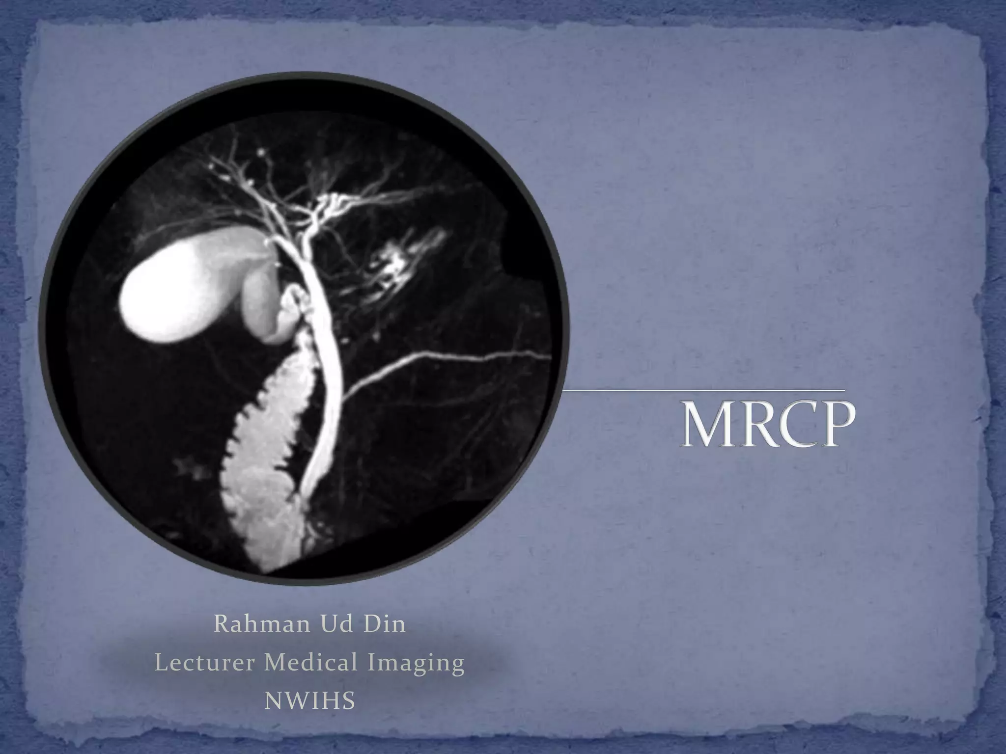
Rahman Ud Din
Lecturer Medical Imaging
NWIHS
 Wide spread clinical acceptance
 Replaced ERCP
 To visualise biliary and pancreatic tree
 Non-invasive
 No contrast injection
 No radiation
 Heavily T2-w images
 To visualise static fluid or bile in the PB-tree
 Longer TE 600-1200 ms
 Long TE, only fluid or tissues with high T2 relaxation
time will retain signal
 Background tissues with shorter TE do not retain
sufficient signal at longer TEs and are suppressed
 Seqs; used are 3D FSE and single-shot FSE
 Other seqs; include balanced SSFP and contrast
enhanced T1-w GRE seq
 3D FSE sequence
 High TE
 Respiratory triggering by tying bellows over abdomen
 MIP technique is used for 3D data
 Takes 4-5 minutes
 Limitations include respiration
 Single-shot FSE (SSFE/HASTE)
 Slab of 2-5 cm thickness
 Radial coronal slabs
 Acquired with breath hold
 No need for MIP
 Suppressed background tissues shows ducts
 Balanced SSFP (TrueFISP/FIESTA/bTFE)
 Breath hold
 Motion insensitive imaging
 Good quality imaging
 Shows ducts without motion artifacts
 Contrast-enhanced T1-w GRE sequences
(THRIVE/VBE/LAVA)
 IV injection
 Specific contrast gadobenate (Multihance), gadoxetate
(Eovist/primovist) and mangafodifir trisodium (Mn-
DPDP, Teslascan)
 Excreted through bile opacifying the bile ducts on T1-w
image
 Contrast-enhanced MRCP
 Detection of bile leak
 Visualisation of small ducts
 Secretin MRCP
 Secretin a hormone secreted by duodenal mucosa in
response to acid stimulation
 It increases secretion of water and bicarbonate by
pancreas
 IV (1 unit/kg) and T2-w images are acquired every 30
seconds for 10 minutes
 Distends pancreatic duct up to 3mm diameter
 Peak response occur at about 3-5 minutes after injection
and response completely vanish after 10 minutes
 S-MRCP improves visualisation of branches of pancreas
to diagnose chronic pancreatitis
 Main limitation is high cost of secretin
 Patient preparation
 NPO 8-12 hours
 No food in upper GIT
 Blue-berry juice and barium can empty GIT if any food
 NPO also dilate GB and bile ducts
 Examination
 Routine T2-w axial seq for planning MRCP
 3D FSE applied later (takes 4-5 minutes)
 Single-shot seq; applied
 Thin contagious section (3-4 mm) single-shot seq; in
coronal and axial plane (TE=200-300 ms)
 Coronal balanced-SSFP and axial T1-w fat sat seq;
 Cystic disease of bile duct
 Choledochal cyst
 Choledochocele
 Caroli’s disease
 Congenital anomalies
 Pancreas divisum
 Cystic duct insertion
 Medial cystic duct insertion
 Parallel course of the cystic and hepatic duct
 Aberrant right hepatic duct
 These variations are important to know in order to avoid
any complications during cholecystectomy especially
the laproscopic
Pancreas divisum
 Choledocholithiasis
 Accurate diagnosis of stone in CBD
 More accurate modality
 Compared to USG and CT
 Primary Sclerosing Cholangistis
 Characterized by multiple irregular strictures and
saccular dilatation of intraheptic and extrahepatic bile
ducts producing beaded appearance
 Good in diagnosis and in follow-up in such conditions
 ERCP may result in progression of cholestasis and may
not show ducts proximal to severe stenosis
 Postsurgical complications
 Benign strictures, retained stones, biliary leaks and
fistula
 Patency of biliary-enteric anastomosis can be seen by
MRCP
 Chronic pancreatitis
 It is characterised by pancreatic duct dilatation,
narrowing or stricture and irregularity
 Alcoholic chronic pancreatitis is usually heterogeneous
and characterised by side-branch dilatation and ductal
calcifications
 Whereas obstructive pancreatitis is more homogenous,
lack calcifications and is associated more often with
main duct dilatation
 MRCP is useful in such detection
 Identification of a surgically or endoscopically
correctable lesion
 Neoplasmic Lesions
 MRCP can show duct proximal to the obstruction
 Cause by i.e.
 Cholangiocarcinoma
 Pancreatic head carcinoma
 Fat saturated postcontrast T1-w images for the
evaluation of extent and spread of the lesion.

Magnetic Resonance Cholangiopancreatography (MRCP)

  • 1.
    Rahman Ud Din LecturerMedical Imaging NWIHS
  • 2.
     Wide spreadclinical acceptance  Replaced ERCP  To visualise biliary and pancreatic tree  Non-invasive  No contrast injection  No radiation
  • 3.
     Heavily T2-wimages  To visualise static fluid or bile in the PB-tree  Longer TE 600-1200 ms  Long TE, only fluid or tissues with high T2 relaxation time will retain signal  Background tissues with shorter TE do not retain sufficient signal at longer TEs and are suppressed
  • 4.
     Seqs; usedare 3D FSE and single-shot FSE  Other seqs; include balanced SSFP and contrast enhanced T1-w GRE seq  3D FSE sequence  High TE  Respiratory triggering by tying bellows over abdomen  MIP technique is used for 3D data  Takes 4-5 minutes  Limitations include respiration
  • 5.
     Single-shot FSE(SSFE/HASTE)  Slab of 2-5 cm thickness  Radial coronal slabs  Acquired with breath hold  No need for MIP  Suppressed background tissues shows ducts  Balanced SSFP (TrueFISP/FIESTA/bTFE)  Breath hold  Motion insensitive imaging  Good quality imaging  Shows ducts without motion artifacts
  • 6.
     Contrast-enhanced T1-wGRE sequences (THRIVE/VBE/LAVA)  IV injection  Specific contrast gadobenate (Multihance), gadoxetate (Eovist/primovist) and mangafodifir trisodium (Mn- DPDP, Teslascan)  Excreted through bile opacifying the bile ducts on T1-w image  Contrast-enhanced MRCP  Detection of bile leak  Visualisation of small ducts
  • 7.
     Secretin MRCP Secretin a hormone secreted by duodenal mucosa in response to acid stimulation  It increases secretion of water and bicarbonate by pancreas  IV (1 unit/kg) and T2-w images are acquired every 30 seconds for 10 minutes  Distends pancreatic duct up to 3mm diameter  Peak response occur at about 3-5 minutes after injection and response completely vanish after 10 minutes  S-MRCP improves visualisation of branches of pancreas to diagnose chronic pancreatitis  Main limitation is high cost of secretin
  • 8.
     Patient preparation NPO 8-12 hours  No food in upper GIT  Blue-berry juice and barium can empty GIT if any food  NPO also dilate GB and bile ducts  Examination  Routine T2-w axial seq for planning MRCP  3D FSE applied later (takes 4-5 minutes)  Single-shot seq; applied  Thin contagious section (3-4 mm) single-shot seq; in coronal and axial plane (TE=200-300 ms)  Coronal balanced-SSFP and axial T1-w fat sat seq;
  • 9.
     Cystic diseaseof bile duct  Choledochal cyst  Choledochocele  Caroli’s disease
  • 10.
     Congenital anomalies Pancreas divisum  Cystic duct insertion  Medial cystic duct insertion  Parallel course of the cystic and hepatic duct  Aberrant right hepatic duct  These variations are important to know in order to avoid any complications during cholecystectomy especially the laproscopic Pancreas divisum
  • 11.
     Choledocholithiasis  Accuratediagnosis of stone in CBD  More accurate modality  Compared to USG and CT  Primary Sclerosing Cholangistis  Characterized by multiple irregular strictures and saccular dilatation of intraheptic and extrahepatic bile ducts producing beaded appearance  Good in diagnosis and in follow-up in such conditions  ERCP may result in progression of cholestasis and may not show ducts proximal to severe stenosis
  • 12.
     Postsurgical complications Benign strictures, retained stones, biliary leaks and fistula  Patency of biliary-enteric anastomosis can be seen by MRCP
  • 13.
     Chronic pancreatitis It is characterised by pancreatic duct dilatation, narrowing or stricture and irregularity  Alcoholic chronic pancreatitis is usually heterogeneous and characterised by side-branch dilatation and ductal calcifications  Whereas obstructive pancreatitis is more homogenous, lack calcifications and is associated more often with main duct dilatation  MRCP is useful in such detection  Identification of a surgically or endoscopically correctable lesion
  • 14.
     Neoplasmic Lesions MRCP can show duct proximal to the obstruction  Cause by i.e.  Cholangiocarcinoma  Pancreatic head carcinoma  Fat saturated postcontrast T1-w images for the evaluation of extent and spread of the lesion.