

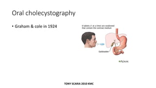
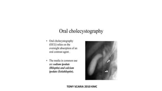
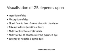
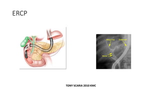
![OCG
• Now has been replaced by USG
• Contrast media used ]
• Ipanoic acid (tele[aque)
• Biloptin
• Solubiloptin
• 2 doses of 3mg dye is given
TONY SCARIA 2010 KMC](https://image.slidesharecdn.com/hepatobiliarysystem-180405015902/85/Hepatobiliary-system-radiology-revision-notes-10-320.jpg)
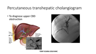
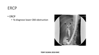
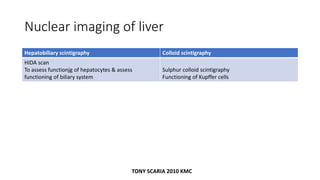


The document provides an overview of diagnostic procedures and imaging techniques related to the hepatobiliary and pancreatic systems, including oral cholecystography, ERCP, HIDA scans, and MRCP. It discusses the radiological features of different pathologies such as gallstones, cholecystitis, hepatic cysts, and pancreatic disorders. Additionally, it covers specific signs seen in imaging studies, such as the double duct sign and band-like appearance in certain conditions.


