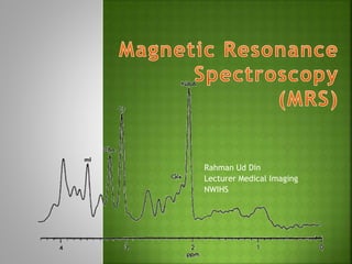
MRS is a non-invasive MRI technique that detects biochemicals in tissues and provides information to diagnose and monitor diseases. It detects small metabolites that are suppressed in standard MRI. Different metabolites including NAA, Cr, Cho, ml, Lac, Glx and lipids provide information about neuronal integrity, cell membrane turnover, hypoxia and other disease states when their levels are abnormal. MRS acquisition and interpretation requires high magnetic field homogeneity and specialized pulse sequences like STEAM and PRESS. Metabolite levels are assessed relative to Cr and compared to normal values.
Everything regarding the physics of MRA is given along with flow charts and images. Also have covered new advances and refrences taken from MR made easy and some articles related to MRI
Physicians have used palpation to detect differences in tissue stiffness as an aid to diagnosis based on the fact that the mechanical properties of tissues are often dramatically affected by the presence of disease processes such as cancer, inflammation, and fibrosis. Elastography depends on the same differences in mechanical properties between healthy and abnormal tissues using imaging to detect these differences at depths not reachable by manual palpation and presents data in color-coded display, can be performed with ultrasound, using manual pressure or low frequency sonic waves, or by MR Elastography imaging.
Magnetic Resonance Angiography and VenographyAnjan Dangal
Introduction to MR Angiography and Venography Procedure of Brain . Includes Indication, MRI protocol, planning and anatomy as well as brief intoduction to physics behind MRA and MRV principle.
differentiating different brain lesion using MR Spectroscopy which is a newer MR technique to quantify different metabolites present within the lesion and adjacent parenchyma
APPROACH TO A NEUROLOGICAL EMERGENCY CASE STARTS WITH THE BASIC TRIAGE APPROACH AS IN ANY OTHER EMERGENCY CASE. A NEUROLOGICAL ASSESSMENT IS ONLY DONE AFTER THE STABILIZATION OF THE PATIENT. THERE CAN BE MANY DIFFERENT APPROACHES BUT ALL BASICALLY AIM AT FIRST CONFIRMING IF AT ALL IT IS A NEURO CASE AND IF YES, WHERE IS THE LESION..IS IT IN THE CRANIUM OR BRAINSTEM OR THE SPINAL CORD? LESION LOCALISATION WILL NOT ONLY HELP TO UNDERSTAND BETTER THE TYPE OF THERAPY TO BE CHOSEN BUT WILL ALSO HELP TO TELL ABOUT THE PROGNOSIS OF THE CASE. MOST COMMONLY WE GET STATUS EPILEPTICUS, TRAUMATIC BRAIN INJURY, POISONING, SPINAL CORD INJURIES AND ACUTE VESTIBULAR DISEASES AS THOUGHT TO LINKED WITH NEUROLOGICAL EMERGENCY SITUATIONS. AN EMERGENCY MUST BE FAMILIAR WITH WITH THE RELEVANT HISTORY OF THE PATIENT. HE SHOULD ALSO BE KEEP A TEAM READY WHO CAN HELP HIM PUT THE IV ACCESS AND SEDATIONS WHILE HE CAN COLLECT THE BLOOD FOR BASIC ROUTINE BLOOD ANALYSIS. COUNTERING THE ONGOING STAGE OF SHOCK TO BRING IT TO NORMAL, CHECKING THE SYSTEMIC BLOOD PRESSURE, RECTAL TEMPERATURE AND OXYGEN CONCENTRATION ARE FEW OF THE MOST IMPORTANT FACTORS A CLINICIAN HAS TO DO WHILE ADMINISTERING THE MEDICS. SCORING SYSTEMS LIKE MODIFIED GLASSGOW COMA SCALE AS SUGGESTED BY DR PLATT ARE REALLY HELPFUL TO GIVE A PROGNOSTIC IDEA IN CASES LIKE CRANIO-CERBRAL INJURIES. RECENT TREATMENT UPDATES ARE REALLY HELPFUL TO KEEP HAVING BETTER OPTIONS IN CASE THE ROUTINE PROTOCOL FOR STABILIZING A SEIZURE PATIENT IS NOT WORKING.
Brief description of various neuroimaging modalities used in psychiatry which help in early detection, diagnosis and treatment of various neuropsychiatric disorders.
Title: Sense of Smell
Presenter: Dr. Faiza, Assistant Professor of Physiology
Qualifications:
MBBS (Best Graduate, AIMC Lahore)
FCPS Physiology
ICMT, CHPE, DHPE (STMU)
MPH (GC University, Faisalabad)
MBA (Virtual University of Pakistan)
Learning Objectives:
Describe the primary categories of smells and the concept of odor blindness.
Explain the structure and location of the olfactory membrane and mucosa, including the types and roles of cells involved in olfaction.
Describe the pathway and mechanisms of olfactory signal transmission from the olfactory receptors to the brain.
Illustrate the biochemical cascade triggered by odorant binding to olfactory receptors, including the role of G-proteins and second messengers in generating an action potential.
Identify different types of olfactory disorders such as anosmia, hyposmia, hyperosmia, and dysosmia, including their potential causes.
Key Topics:
Olfactory Genes:
3% of the human genome accounts for olfactory genes.
400 genes for odorant receptors.
Olfactory Membrane:
Located in the superior part of the nasal cavity.
Medially: Folds downward along the superior septum.
Laterally: Folds over the superior turbinate and upper surface of the middle turbinate.
Total surface area: 5-10 square centimeters.
Olfactory Mucosa:
Olfactory Cells: Bipolar nerve cells derived from the CNS (100 million), with 4-25 olfactory cilia per cell.
Sustentacular Cells: Produce mucus and maintain ionic and molecular environment.
Basal Cells: Replace worn-out olfactory cells with an average lifespan of 1-2 months.
Bowman’s Gland: Secretes mucus.
Stimulation of Olfactory Cells:
Odorant dissolves in mucus and attaches to receptors on olfactory cilia.
Involves a cascade effect through G-proteins and second messengers, leading to depolarization and action potential generation in the olfactory nerve.
Quality of a Good Odorant:
Small (3-20 Carbon atoms), volatile, water-soluble, and lipid-soluble.
Facilitated by odorant-binding proteins in mucus.
Membrane Potential and Action Potential:
Resting membrane potential: -55mV.
Action potential frequency in the olfactory nerve increases with odorant strength.
Adaptation Towards the Sense of Smell:
Rapid adaptation within the first second, with further slow adaptation.
Psychological adaptation greater than receptor adaptation, involving feedback inhibition from the central nervous system.
Primary Sensations of Smell:
Camphoraceous, Musky, Floral, Pepperminty, Ethereal, Pungent, Putrid.
Odor Detection Threshold:
Examples: Hydrogen sulfide (0.0005 ppm), Methyl-mercaptan (0.002 ppm).
Some toxic substances are odorless at lethal concentrations.
Characteristics of Smell:
Odor blindness for single substances due to lack of appropriate receptor protein.
Behavioral and emotional influences of smell.
Transmission of Olfactory Signals:
From olfactory cells to glomeruli in the olfactory bulb, involving lateral inhibition.
Primitive, less old, and new olfactory systems with different path
Couples presenting to the infertility clinic- Do they really have infertility...Sujoy Dasgupta
Dr Sujoy Dasgupta presented the study on "Couples presenting to the infertility clinic- Do they really have infertility? – The unexplored stories of non-consummation" in the 13th Congress of the Asia Pacific Initiative on Reproduction (ASPIRE 2024) at Manila on 24 May, 2024.
These simplified slides by Dr. Sidra Arshad present an overview of the non-respiratory functions of the respiratory tract.
Learning objectives:
1. Enlist the non-respiratory functions of the respiratory tract
2. Briefly explain how these functions are carried out
3. Discuss the significance of dead space
4. Differentiate between minute ventilation and alveolar ventilation
5. Describe the cough and sneeze reflexes
Study Resources:
1. Chapter 39, Guyton and Hall Textbook of Medical Physiology, 14th edition
2. Chapter 34, Ganong’s Review of Medical Physiology, 26th edition
3. Chapter 17, Human Physiology by Lauralee Sherwood, 9th edition
4. Non-respiratory functions of the lungs https://academic.oup.com/bjaed/article/13/3/98/278874
Ozempic: Preoperative Management of Patients on GLP-1 Receptor Agonists Saeid Safari
Preoperative Management of Patients on GLP-1 Receptor Agonists like Ozempic and Semiglutide
ASA GUIDELINE
NYSORA Guideline
2 Case Reports of Gastric Ultrasound
Prix Galien International 2024 Forum ProgramLevi Shapiro
June 20, 2024, Prix Galien International and Jerusalem Ethics Forum in ROME. Detailed agenda including panels:
- ADVANCES IN CARDIOLOGY: A NEW PARADIGM IS COMING
- WOMEN’S HEALTH: FERTILITY PRESERVATION
- WHAT’S NEW IN THE TREATMENT OF INFECTIOUS,
ONCOLOGICAL AND INFLAMMATORY SKIN DISEASES?
- ARTIFICIAL INTELLIGENCE AND ETHICS
- GENE THERAPY
- BEYOND BORDERS: GLOBAL INITIATIVES FOR DEMOCRATIZING LIFE SCIENCE TECHNOLOGIES AND PROMOTING ACCESS TO HEALTHCARE
- ETHICAL CHALLENGES IN LIFE SCIENCES
- Prix Galien International Awards Ceremony
Anti ulcer drugs and their Advance pharmacology ||
Anti-ulcer drugs are medications used to prevent and treat ulcers in the stomach and upper part of the small intestine (duodenal ulcers). These ulcers are often caused by an imbalance between stomach acid and the mucosal lining, which protects the stomach lining.
||Scope: Overview of various classes of anti-ulcer drugs, their mechanisms of action, indications, side effects, and clinical considerations.
The prostate is an exocrine gland of the male mammalian reproductive system
It is a walnut-sized gland that forms part of the male reproductive system and is located in front of the rectum and just below the urinary bladder
Function is to store and secrete a clear, slightly alkaline fluid that constitutes 10-30% of the volume of the seminal fluid that along with the spermatozoa, constitutes semen
A healthy human prostate measures (4cm-vertical, by 3cm-horizontal, 2cm ant-post ).
It surrounds the urethra just below the urinary bladder. It has anterior, median, posterior and two lateral lobes
It’s work is regulated by androgens which are responsible for male sex characteristics
Generalised disease of the prostate due to hormonal derangement which leads to non malignant enlargement of the gland (increase in the number of epithelial cells and stromal tissue)to cause compression of the urethra leading to symptoms (LUTS
Ethanol (CH3CH2OH), or beverage alcohol, is a two-carbon alcohol
that is rapidly distributed in the body and brain. Ethanol alters many
neurochemical systems and has rewarding and addictive properties. It
is the oldest recreational drug and likely contributes to more morbidity,
mortality, and public health costs than all illicit drugs combined. The
5th edition of the Diagnostic and Statistical Manual of Mental Disorders
(DSM-5) integrates alcohol abuse and alcohol dependence into a single
disorder called alcohol use disorder (AUD), with mild, moderate,
and severe subclassifications (American Psychiatric Association, 2013).
In the DSM-5, all types of substance abuse and dependence have been
combined into a single substance use disorder (SUD) on a continuum
from mild to severe. A diagnosis of AUD requires that at least two of
the 11 DSM-5 behaviors be present within a 12-month period (mild
AUD: 2–3 criteria; moderate AUD: 4–5 criteria; severe AUD: 6–11 criteria).
The four main behavioral effects of AUD are impaired control over
drinking, negative social consequences, risky use, and altered physiological
effects (tolerance, withdrawal). This chapter presents an overview
of the prevalence and harmful consequences of AUD in the U.S.,
the systemic nature of the disease, neurocircuitry and stages of AUD,
comorbidities, fetal alcohol spectrum disorders, genetic risk factors, and
pharmacotherapies for AUD.
Report Back from SGO 2024: What’s the Latest in Cervical Cancer?bkling
Are you curious about what’s new in cervical cancer research or unsure what the findings mean? Join Dr. Emily Ko, a gynecologic oncologist at Penn Medicine, to learn about the latest updates from the Society of Gynecologic Oncology (SGO) 2024 Annual Meeting on Women’s Cancer. Dr. Ko will discuss what the research presented at the conference means for you and answer your questions about the new developments.
2. MRS an exciting application of MRI
Non-invasive technique
Used to assess various metabolites or
biochemicals from the body tissues
This metabolites information used to
diagnose, monitor and follow-up diseases
1H, 13C, 19F, 23Na and 31P (theoretical MRS
can be done with)
1H & 31P (mostly clinical used MRS)
We will discuss 1H spectroscopy
3. For the brain in particular,
MRS has been a powerful research tool
and provide additional clinical information
for several disease
such as brain tumors,
metabolic disorders, and
systemic diseases
4. Principles are same as MRI
But few differences are
In MRI, images are reconstructed from all protons
in the tissue, water and fats dominate only
Contrary, MRS detect small metabolites of
clinical interest (resonate of frequency b/w
water and fats)
Small metabolites are detected when large signal
are suppressed
5. How are small metabolites from the tissue
detected?
Chemical shift
Chemical environment or electron cloud around
protons
Protons in water, fat and other compound precess at
different rate
Such change in frequency is called chemical shift
Frequency of p+ in metabolites=chemical shift=position of
metabolites peaks
Measured in ppm
Chemical shift is proportional to B°, need high Tesla
MRI
Can be performed on 0.5T or above
1.5T or above required for spectral separation & ↑SNR
6. Magnetic Field Homogeneity
MR field must be homogeneous
MRS require more homogeneous field than MRI
because smaller chemical shift needs to detect
In inhomogeneous field, smaller chemical shifts
can be misinterpreted and incorrect
concentration may be recorded
Homogeneity required for MRI is about 0.5 ppm
where for MRS it is about 0.1 ppm
The process of making the magnetic field
homogeneous is called ???!!!!!
Shimming
7. No Frequency Encoding Gradient in MRS
Similar to MRI, localisation is done by slice
selection and phase encoding gradients
The frequency encoding gradient is not used in
MRS to preserve optimal homogeneity and
chemical shift information
The spin-spin or J-coupling
Spins (p+) with a small difference of precessional
frequency, for example spins within a molecule,
interact with each other
This is facilitated by electrons around a nuclei
This spin-spin interaction modifies the resonant
frequency of the spins involved in it
J-coupling causes fusion of peaks on spectral map
e.g. doublet of lactate at 1.3ppm
8.
9.
10. In early days, localisation done on surface coil
Area covered by surface coil was VOI
From VOI metabolites information obtained
In current practice, four methods used
STEAM
PRESS
ISIS and
CSI (MRSI)
First three are single voxel MRS tech:
CSI used for multivoxel MRS technique
11. STEAM
Stimulated Echo Acquisition Method
VOI excited by 90 degree pulses in three
orthogonal planes
Echo stimulated signal is weak
STEAM used for short TE(20-30 ms) spectroscopy
PRESS
Point Resolved Spectroscopy
One 90 degree pulse and two 180 degree pulses
are applied along three orthogonal planes
The signal is strong with better SNR, PRESS used
for long TE (135, 270 ms) spectroscopy
12. ISIS
Image Selected In vivo Spectroscopy
Three frequency selective inversion pulses
applied
In the presence of the orthogonal gradients
Fourth non-invasive pulse is used for the
observation of signal
ISIS is used in 31P spectroscopy
CSI Chemical Shift Imaging
Multivoxel Spectroscopy
Large area divided into multiple voxels
Also called MRSI (imaging and spectroscopy)
Localisation is done by phase encoding in one,
two or three directions (1D, 2D or 3D spectro..)
13. Patient positioning
Global Shimming
Acquisition of MR images for localisation
Selection of MRS measurement and parameters
Improved SNR at long TR
TEs 20-30 ms, 135-145 ms and 270 ms
TEs longer than 135 ms, peaks of major metabolites
i.e. Ch, Cr, NAA and lactate visible
Other metabolites peaks are suppressed (short T2)
Selection of VOI
SVS (single voxel spectro) local or diffuse diseases
CSI used large lesion
14. Local Shimming
Optimisation of homogeneity over selected VOI
Good shim results in narrower metabolite peaks
Better spectral resolution
Good SNR
Full width at half height of H2O used as an
indicator of shim
A local shim of 4-10 Hz is desirable
Water suppression
Water peak is suppressed so that smaller
metabolite peaks are visible
Water peak suppression is done by CHESS
(chemical shift selective spectroscopy) tech
15. MRS data collection
Modern machine in use, SVS takes 3-6 minutes and CSI
takes up to 12 minutes for data acquisition
Data processing and display
Acquired data is processed to get spectrum and spectral
maps
Zero point of spectrum is set in the software itself by
frequency of a particular compound called
Tetramethysilane (TMS)
Interpretation
Area under peak of any metabolite is directly
proportional to the number of spins
Absolute values for each varies with age and population
Interpretation based on ratios of metabolites and
comparison with normal side
16.
17. NAA: N-Acetylaspartate
Peak position: 2.02 ppm
There is some contribution from NAAG and
glutamate to the NAA peak
It is a neuronal marker and any insult to the brain
causing neuronal loss or degeneration causes
reduction in NAA
It is absent in tissues/lesions with no neurons e.g.
metastasis and meningioma
NAA is reduced in: hypoxia, infraction,
Alzheimer’s, herpes, encephalitis, hydrocephalus,
Alexander’s disease, epilepsy, some neoplasms,
stroke, NPH, close head trauma,
NAA increased in: Canavan’s disease
18. Cr: Creatine
Peak position: 3.0 ppm. Second peak at 3.94 ppm
Cr peak creatine, CrPO4, GABA (Gamma-
aminobutyric acid), Lysine and glutathione
Cr serves as high energy phosphate and as a buffer
in ATP/ADP reservoir. Increase in amount with age
Cr increased in: hypometabolic states and in trauma
Cr reduced in: hypermetabolic states, hypoxia,
stroke and some tumor
Cr remain stable in many disease hence serves as
reference or control peak for the comparison
19. Cho: Choline
Peak position: 3.22 ppm
Choline is phospholipids of the cell membrane
Precursor of acetyl choline and phosphatidylcholine
Indicator of cell membrane integrity
Cho increases with increased cell membrane
synthesis and increased cell turnover
Cho is increased in: chronic hypoxia, epilepsy,
Alzheimer’s, gliomas and some other tumors,
trauma, infraction, hyperosmolar states, diabetes
mellitus
Cho is reduced in: hepatic encephalopathy and
stroke
20. ml: Myo-inositol
Peak position: 3.56 ppm, second peak at 4.1 ppm
MI acts as an osmolyte and is marker of gliosis
Involved in hormone sensitive neuroreception and is
precursor of glucuronic acid
It is the dominent peak in newborn babies and
decrease with age
MI is increased in: Alzheimer’s, frontal lobe
dementias, diabetes and hyperosmolar states
MI decreased in: hepatic and hypoxic
encephalopathy, stroke, tumor, osmotic pontine
myelinolysis and hyponatremia
21. Lac: Lactate
Peak position: 1.3 pm
Doublet
Inverted at TE of 135 ms with PRESS, upright at
other TEs on PRESS and at all the TEs with STEAM
sequences
Not seen in normal brain spectrum
Elevated in hypoxia, tumor, mitochondrial
encephalopathy, intracranial hemorrahage, stroke,
hypoventilation, Canavan’s disease, Alexander’s
disease and hydrocephalus
22. Glx: Glutamate (Glu) and Glutamine (Gln)
Peak position: 2-2.45 ppm for beta and gamma Glx
Second peak of alpha Glx at 3.6-3.8 ppm
Glu is excitatory neurotransmitter and GABA is
product of Glu
It has role in detoxification and regulation of
neurotransmitter activities
Glx peak is elevated in head injury, hepatic
encephalopathy and hypoxia
23. Lipids
Peak position: 0.9, 1.3, 1.5 ppm
Not seen in normal brain spectrum
Seen in acute destruction of myelin
Increased in high grade tumors (reflects necrosis),
stroke and MS lesions
Aminoacids
Alanine (at 1.3-1.4 ppm), Valine (0.9 ppm) and
leucine (3.6 ppm)
Usually multiplets visualised at short TE
Inverted at 135 ms
Alanine is seen in the meningioma whereas valine
and leucine are marker of abcess
24. Glucose
When present, seen next to Cho peak on its left
side
May increases in diabetes, parenteral feeding and
hepatic encephalopathy
GABA
Peak position: 1.9 and 2.3 ppm
Used for monitoring of vigabatrin therapy given in
children with myoclonic jerks