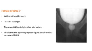
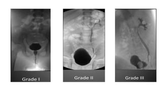
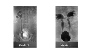
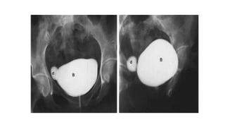
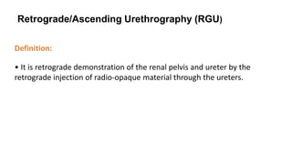
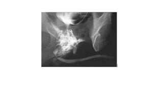
SlideShare a Scribd company logo
Micturating Cystourethrography (MCU)
&
Retrograde Urethrography (RGU)
By
Dr. J K PATIL
D.Y. PATIL MEDICAL COLLEGE AND HOSPITAL
RADIODIAGNOSIS DEPARTMENT
ANATOMY OF URETHRA
• MALE URETHRA IS 18-20 cms LONG
• EXTENDS FROM BLADDER NECK TILL THE MEATAL OPENING AT PENIS
• It has four named regions:
 Prostatic urethra:
• Is approximately 3 cm in length.
• Passes through the prostate gland.
 Membranous urethra:
• Is approximately 1 cm in length.
• Passes through the urogenital diaphragm.
 Bulbar urethra
From inferior aspect of urogenital diaphragm to
penoscrotal junction.
 Spongy (penile) urethra:
Passes through the length of the penis.
PARTS OF URETHRA
 ANTERIOR URETHRA
-PENILE URETHRA
-BULBAR URETHRA
 POSTERIOR URETHRA
- MEMBRANOUS URETHRA
- PROSTATIC URETHRA
SPHINCTERS OF URETHRA
INTERNAL URETHRAL SPHINCTER -
• involuntary in nature
• Supplied by sympathetic nerves
• It controls the neck of the bladder & prostatic
urethra above the opening of ejaculatory ducts
EXTERNAL URETHRAL SPHINCTER-
• voluntary in nature
• Supplied by perineal branch of the pudendal nerve (s2-s4)
• Controls the membranous urethra & is responsible for voluntary holding of urine
Female urethra :-
• Widest at bladder neck.
• 4-5cms in length
• Narrowest & least distensible at meatus.
• This forms the Spinning top configuration of urethra
on normal MCU.
Urethrography is of 2 types:
1. Ascending/Retrograde urethrography - Contrast is retrogradely injected
in the ureter with the urethral orifice occluded to prevent reflux of contrast.
2. Descending /MCU- Bladder is filled with contrast via suprapubic or
retrograde catheterization and the urethra is assessed during voiding.
• Voiding cystourethrogram/ Micturating cystourethrogram demonstrates the lower
urinary tract
• Helps in detection of:
 vesico-ureteral reflux
 bladder pathology
congenital or acquired anomalies of bladder outflow tract.
Micturating Cystourethrogram (MCU)
INDICATIONS
Children:
1. UTI
2. Voiding difficulties like dysuria, thin stream, dribbling, frequency, urgency.
3. Vesico ureteric reflux.
4. Other congenital anomalies : Meningomyelocele, Sacral agenesis,Rectal anomalies.
5. For post operative evaluation of ureteric abnormalities.
6. Pelvic Trauma.
7. In renal failure to exclude reflux.
8. Boys with hematuria-MCU can demonstrate posterior uretheral valve or polyp.
Adults
Main indications:
1. Trauma to urethra.
2. Urethral stricture.
3. Suspected urethral diverticula.
CONTRAST MEDIA
• Water soluble constrast media like Urograffin 60% are used which is diluted with
normal saline in 1 :3 ratio.
• The estimated volume of contrast medium to be given:
Less than one year
Weight (kg) x 7 = capacity (ml)
Less than two years
(2 x age in years + 2) × 30 = capacity (ml)
More than two years
(Age in years/2 + 6) × 30 = capacity (ml)
Procedure
• Using a sterile technique, a catheter is introduced into the bladder.
• A 5f feeding tube with side holes are used for children and in older children or
adults 8f or 10 f catheters are used.
• In girls after initial inspection of perineum to identify any local genitilia
abnormalities (cystoceles or labial fusion ) the catheter is introduced.
• When it enters the bladder a varying amount of urine will flow through it .if no
flow a catheter is introduced till urine is obtained.
• Suprapubic pressure Is sometimes helpful.
• In males, foreskin is retracted and catheter is introduced.
• The catheter should be lubricated with anaesthetic jelly and inserted slowly and
gently into the urthera holding the penis is vertical position.
• The normal bladder capacity in children is estimated to be 1 ounce i.e 29 cc.
• For newborns -30 to 35 cc can be instilled.
• For upto 3 yrs - 200 to 250 cc
• Adequate capacity is reached when the child becomes uncomfortable and begins
voiding around the catheter.
Filming
In children
• In children up to the age of 2 yrs bladder is filled by hand injection.
For older children contrast medium is instilled from a bottle elevated one metre above examination table.
• During filling, fluoroscopic screening is performed at short intervals to see if vesicoureteral reflux, diverticuli
or other abnormalities are present. The child is turned oblique on both sides to ensure that minimal reflux
is not overlooked.
• If reflux appears, films are taken in the appropriate oblique projection. If the bladder appears normal, one
film is taken in the frontal projection at the end of filling.
• Voiding starts in infants the moment the catheter is removed.
• At the end of voiding, a frontal film is made of the entire abdomen including the kidney region in order to
prevent overlooking the vesicoureteral reflux which is apparent only on termination of voiding and may
reach the upper collecting system.
In adult male :
• Bladder is filled in the usual way as in older child and voiding filming is done in both oblique
projection views.
• The voiding study in male adults can be modified by getting the patient to void against resistance
i.e. by compression of distal part of penis or using penile clamp thus enhancing the visualization
of urethra by artificial distention.This is known as CHOKE CYSTOURETHROGRAPHY
In adult female:
• The procedure is essentially the same
• In addition to the standard exposures, a double exposed film taken at rest and during straining
demonstrates the degree of bladder descent if any.
Filming Technique:
Scout film:
• The first image that is taken while performing the MCU and VCUG is the image of KUB that
is called scout film. We evaluate the spine, pelvis, and soft tissues on the scout film.
• After several seconds of the contrast media begins to flow, the image of minimally filled
bladder is taken in Antero-posterior (AP) projection.
• During early filling a ureterocele or tumor can be detected
and it may obscure as more contrast material enters
into the bladder.
Voiding phase
• The image taken during voiding may demonstrate the urethral strictures or obstructions.
• They will also give the details of the presence or absence of vesicoureteral reflux.
• Voiding film necessary because gives the determination of reflux because reflux may
only happen with the pressure generated by voiding.
Post-voiding film
• A post-voiding film may demonstrate the reflux or extravasation of
urine from the bladder or urethra.
• A normal post-void film has no reflux and no residual urine.
COMPLICATIONS
• Contrast reaction.
• Contrast induced cystitis.
• UTI.
• Catheter trauma.
• Bladder perforation - overfilling.
• Retention of a foley catheter.
• Catheterisation of vagina / ectopic ureter.
• Radiation exposure
• Autonomic dysreflexia- in paraplegic patients due to spinal cord injury at or above t6
level, forceful injection of contrast causes severe headache ,sweating,hypertension
with bradycardia due to forceful opening of bladder neck
ALTERNATE TECHNIQUES
1) SUPRAPUBIC BLADDER PUNCTURE.
Sometimes in PUV & pelvic trauma - not possible to catheterize.
2) EXCRETION MCU (MCU followed by IVU)
• This method makes use of contrast media accumulated in the urinary bladdder during ivp
Advantages : avoidance of physical and psychological trauma of catherization
• Avoidance of infection
Disadvatanges : visualization is not usually adequate Takes longer time
• Vu reflux visualised poorly.
.
Retrograde/Ascending Urethrography (RGU)
Definition:
• It is retrograde demonstration of the renal pelvis and ureter by the
retrograde injection of radio-opaque material through the ureters.
INDICATIONS:-
• Stricture
• Urethral Trauma
• Fistulae or false passages
• Congenital abnormalities
 Periurethral / prostatic abscess
CONTRAINDICATIONS:-
• Acute UTI
• Recent instrumentation
CONTRAST MEDIUM :-
• Iopamidol (LOCM)
EQUIPMENT :-
Tilting radiography table.
• Fluoroscopy/spot film device.
• Foley's catheter, Syringe, Gloves
PREPARATION :-
• Patient is asked to micturate prior to the procedure
TECHINIQUE :-
Preliminary film - coned supine PA view of bladder base and urethra
• Patient is made to lie in supine position and slightly tilted
with legs position as shown in the image.
• Using aseptic conditions, the tip of the Foley's catheter is
inserted in the urethra after applying lignocaine jelly
for 2 to 4 cm length.
• Pressure is applied over the glans penis to avoid expulsion of the catheter and also to
straighten the penis over the ipsilateral leg and prevent urethral overlap.
• Contrast medium is injected slowly under fluoroscopic control.
IMAGING:-
• Supine PA before injecting contrast medium.
• 30º left anterior oblique
• 30º right anterior oblique
AFTER CARE:-
1. Observation.
2. Prophylatic antibiotics may be used
COMPLICATIONS :-
• Contrast reaction (due to absorption through bladder mucosa)
• UTI
• Urethral trauma.
• Extravasation of contrast - due to use of excessive pressure in
stricture.
Advantages of MR - MCU and RGU
1. These studies are most valuable to detect congenital anomalies, posterior urethral
injuries, and with urethral and periurethral tumours.
2. It is a better imaging modality for assessing the post traumatic pelvic anatomy &
non-invasive method for measuring stricture length.
3. It clearly shows the extent of scar tissue as well as prostatic displacement.
4. MR uretherography is more accurate in estimating the length of obliterative
urethral stricture than RGU combined with Voiding cystouretherography
Limitations of conventional RGU combined with voiding
cystouretherography:
1. It does not provide accurate length of the defect because of poor prostatic
urethral filling.
2. It does not provide information regarding extent of fibrosis of corpora spongiosa
or prostatic displacement.
3. The stricture length is overestimated if bladder neck does not relax.
Advantages of CT urethrography
1. C.T. voiding uretherography is more comfortable to the patient
because it requires adaptation only in one position.
2. Less time consuming; takes only few seconds
3. Comparison of lurninal size & stricture length for follow up is possible.
4. Extralurninal pathology can be detected
5. Good patient compliance
6. Ability to survey whole urinary tract from kidney to urethra
RGU/ASU vs VCUG/MCU
• RGU/ASU is carried out to visualise anterior urethral abnormalities
• VCUG/MCU for posterior urethral abnormalities.
• Additionally, although the bladder is not generally the main target of the exam, as with a
cystogram, a VCUG/MCU may be useful in detection of bladder abnormalities and vesico-
ureteric reflux (VUR).
• In a trauma situation, an RGU/ASU should be performed first. A VCUG/MCU should not be
performed first because blindly trying to introduce a Foley catheter into the bladder in a
trauma setting may lead to creating additional urethral damage with the catheter
THANK YOU

More Related Content

What's hot

Mcu
McuMcu
Mcu
McuMcu
Excretory urography
Excretory urographyExcretory urography
Excretory urographyairwave12
 
Rgu & mcu final presentation
Rgu & mcu final presentationRgu & mcu final presentation
Rgu & mcu final presentation
Parth Nathwani
 
Hypotonic duodenography
Hypotonic duodenographyHypotonic duodenography
Hypotonic duodenography
Maajid Mohi ud din
 
Enteroclysis( small bowel enema)
Enteroclysis( small bowel enema)Enteroclysis( small bowel enema)
Enteroclysis( small bowel enema)
Ankit Mishra
 
IVP Best presnetation
IVP Best presnetationIVP Best presnetation
IVP Best presnetation
DR SACHIN SURA
 
Micturating Cystourethrogram(MCU) ppt.pptx
Micturating Cystourethrogram(MCU) ppt.pptxMicturating Cystourethrogram(MCU) ppt.pptx
Micturating Cystourethrogram(MCU) ppt.pptx
Abhishek Singh
 
CT urography
CT urography CT urography
CT urography
SabitaMandal1
 
RGU (Retrograde urethrogram), MCU (Micturating cystourethrogram) and its inte...
RGU (Retrograde urethrogram), MCU (Micturating cystourethrogram) and its inte...RGU (Retrograde urethrogram), MCU (Micturating cystourethrogram) and its inte...
RGU (Retrograde urethrogram), MCU (Micturating cystourethrogram) and its inte...
Milan Silwal
 
Barium follow through and small bowel enema sahara mahato
Barium follow through and small bowel enema  sahara mahatoBarium follow through and small bowel enema  sahara mahato
Barium follow through and small bowel enema sahara mahato
sahara mahato
 
CT Urography
CT UrographyCT Urography
Retrograde Pyelography
Retrograde PyelographyRetrograde Pyelography
Retrograde Pyelography
Dr. Yash Kumar Achantani
 
Barium follow through & small bowel enema ranju
Barium follow through & small bowel enema   ranjuBarium follow through & small bowel enema   ranju
Barium follow through & small bowel enema ranju
RABIN PAUDEL
 
Ct & mr enterography
Ct & mr enterographyCt & mr enterography
Ct & mr enterography
Rakesh Ca
 
Intravenous urography
Intravenous urographyIntravenous urography
Intravenous urography
Milan Silwal
 
Barium enema
Barium enemaBarium enema
Barium enema
Athul Nampoothiri
 
CT Enteroclysis
CT EnteroclysisCT Enteroclysis
CT Enteroclysis
Dr. Yash Kumar Achantani
 
Imaging of biliary tract
Imaging of biliary tractImaging of biliary tract
Imaging of biliary tract
Maajid Mohi ud din
 

What's hot (20)

Mcu
McuMcu
Mcu
 
Mcu
McuMcu
Mcu
 
Excretory urography
Excretory urographyExcretory urography
Excretory urography
 
T Tube
T TubeT Tube
T Tube
 
Rgu & mcu final presentation
Rgu & mcu final presentationRgu & mcu final presentation
Rgu & mcu final presentation
 
Hypotonic duodenography
Hypotonic duodenographyHypotonic duodenography
Hypotonic duodenography
 
Enteroclysis( small bowel enema)
Enteroclysis( small bowel enema)Enteroclysis( small bowel enema)
Enteroclysis( small bowel enema)
 
IVP Best presnetation
IVP Best presnetationIVP Best presnetation
IVP Best presnetation
 
Micturating Cystourethrogram(MCU) ppt.pptx
Micturating Cystourethrogram(MCU) ppt.pptxMicturating Cystourethrogram(MCU) ppt.pptx
Micturating Cystourethrogram(MCU) ppt.pptx
 
CT urography
CT urography CT urography
CT urography
 
RGU (Retrograde urethrogram), MCU (Micturating cystourethrogram) and its inte...
RGU (Retrograde urethrogram), MCU (Micturating cystourethrogram) and its inte...RGU (Retrograde urethrogram), MCU (Micturating cystourethrogram) and its inte...
RGU (Retrograde urethrogram), MCU (Micturating cystourethrogram) and its inte...
 
Barium follow through and small bowel enema sahara mahato
Barium follow through and small bowel enema  sahara mahatoBarium follow through and small bowel enema  sahara mahato
Barium follow through and small bowel enema sahara mahato
 
CT Urography
CT UrographyCT Urography
CT Urography
 
Retrograde Pyelography
Retrograde PyelographyRetrograde Pyelography
Retrograde Pyelography
 
Barium follow through & small bowel enema ranju
Barium follow through & small bowel enema   ranjuBarium follow through & small bowel enema   ranju
Barium follow through & small bowel enema ranju
 
Ct & mr enterography
Ct & mr enterographyCt & mr enterography
Ct & mr enterography
 
Intravenous urography
Intravenous urographyIntravenous urography
Intravenous urography
 
Barium enema
Barium enemaBarium enema
Barium enema
 
CT Enteroclysis
CT EnteroclysisCT Enteroclysis
CT Enteroclysis
 
Imaging of biliary tract
Imaging of biliary tractImaging of biliary tract
Imaging of biliary tract
 

Similar to MCU AND RGU

RGU and MCU by capt alauddin, MD phase A.pptx
RGU and MCU by capt alauddin, MD phase A.pptxRGU and MCU by capt alauddin, MD phase A.pptx
RGU and MCU by capt alauddin, MD phase A.pptx
Alauddin Md
 
ASU & MCU --- RADIOLOGY IMAGING PROCEDURE
ASU & MCU --- RADIOLOGY IMAGING PROCEDUREASU & MCU --- RADIOLOGY IMAGING PROCEDURE
ASU & MCU --- RADIOLOGY IMAGING PROCEDURE
Sayantan Biswas
 
Mcu by armaan lone
Mcu by   armaan loneMcu by   armaan lone
Mcu by armaan lone
Armaan LONE
 
Vcu ppt
Vcu pptVcu ppt
Vcu ppt
dypradio
 
Mcu
McuMcu
Intravenous urography
Intravenous urographyIntravenous urography
Intravenous urography
rajen ray
 
INTRAVENOUS UROGRAM (IVU)
INTRAVENOUS UROGRAM (IVU)INTRAVENOUS UROGRAM (IVU)
INTRAVENOUS UROGRAM (IVU)
Jai Kumar
 
Radiographic round on mcug
Radiographic round on mcugRadiographic round on mcug
Radiographic round on mcug
SanzzuTimilsina
 
Retrograde pyeloureterography
Retrograde pyeloureterographyRetrograde pyeloureterography
Retrograde pyeloureterography
dypradio
 
Imaging
ImagingImaging
MCU and AUG - CYSTOURETHEROGRAM Urology.pptx
MCU and AUG - CYSTOURETHEROGRAM Urology.pptxMCU and AUG - CYSTOURETHEROGRAM Urology.pptx
MCU and AUG - CYSTOURETHEROGRAM Urology.pptx
Kuppan Thenappan
 
urethrogram.pptx
urethrogram.pptxurethrogram.pptx
urethrogram.pptx
AliyaIshak1
 
Uro procedure
Uro procedureUro procedure
Uro procedure
Dr-Girish Gunari
 
Management of genitourinary fistula
Management of genitourinary fistulaManagement of genitourinary fistula
Management of genitourinary fistuladrmcbansal
 
Methods of imaging of the urinary tract using contrast
Methods of imaging of the urinary tract using contrastMethods of imaging of the urinary tract using contrast
Methods of imaging of the urinary tract using contrast
Dr Abdalla M. Gamal
 
URETHRAL STRICTURES.pdf
URETHRAL STRICTURES.pdfURETHRAL STRICTURES.pdf
URETHRAL STRICTURES.pdf
Shapi. MD
 
INTRAVENOUS UROGRAPHY INDICATIONS CONTRAINDICATIONS
INTRAVENOUS UROGRAPHY INDICATIONS CONTRAINDICATIONSINTRAVENOUS UROGRAPHY INDICATIONS CONTRAINDICATIONS
INTRAVENOUS UROGRAPHY INDICATIONS CONTRAINDICATIONS
obscmch
 
RGU MCU and its interpretation in pathology of Urinary Bladder & Urethra
RGU MCU and its interpretation in pathology of Urinary Bladder & UrethraRGU MCU and its interpretation in pathology of Urinary Bladder & Urethra
RGU MCU and its interpretation in pathology of Urinary Bladder & Urethradbc9427
 
Antegrade & retrograde urethrogram
Antegrade & retrograde urethrogramAntegrade & retrograde urethrogram
Antegrade & retrograde urethrogram
Amlendu Kumar
 
OPPOSING URETHROGRAM (OUG)
OPPOSING URETHROGRAM (OUG)OPPOSING URETHROGRAM (OUG)
OPPOSING URETHROGRAM (OUG)
Jai Kumar
 

Similar to MCU AND RGU (20)

RGU and MCU by capt alauddin, MD phase A.pptx
RGU and MCU by capt alauddin, MD phase A.pptxRGU and MCU by capt alauddin, MD phase A.pptx
RGU and MCU by capt alauddin, MD phase A.pptx
 
ASU & MCU --- RADIOLOGY IMAGING PROCEDURE
ASU & MCU --- RADIOLOGY IMAGING PROCEDUREASU & MCU --- RADIOLOGY IMAGING PROCEDURE
ASU & MCU --- RADIOLOGY IMAGING PROCEDURE
 
Mcu by armaan lone
Mcu by   armaan loneMcu by   armaan lone
Mcu by armaan lone
 
Vcu ppt
Vcu pptVcu ppt
Vcu ppt
 
Mcu
McuMcu
Mcu
 
Intravenous urography
Intravenous urographyIntravenous urography
Intravenous urography
 
INTRAVENOUS UROGRAM (IVU)
INTRAVENOUS UROGRAM (IVU)INTRAVENOUS UROGRAM (IVU)
INTRAVENOUS UROGRAM (IVU)
 
Radiographic round on mcug
Radiographic round on mcugRadiographic round on mcug
Radiographic round on mcug
 
Retrograde pyeloureterography
Retrograde pyeloureterographyRetrograde pyeloureterography
Retrograde pyeloureterography
 
Imaging
ImagingImaging
Imaging
 
MCU and AUG - CYSTOURETHEROGRAM Urology.pptx
MCU and AUG - CYSTOURETHEROGRAM Urology.pptxMCU and AUG - CYSTOURETHEROGRAM Urology.pptx
MCU and AUG - CYSTOURETHEROGRAM Urology.pptx
 
urethrogram.pptx
urethrogram.pptxurethrogram.pptx
urethrogram.pptx
 
Uro procedure
Uro procedureUro procedure
Uro procedure
 
Management of genitourinary fistula
Management of genitourinary fistulaManagement of genitourinary fistula
Management of genitourinary fistula
 
Methods of imaging of the urinary tract using contrast
Methods of imaging of the urinary tract using contrastMethods of imaging of the urinary tract using contrast
Methods of imaging of the urinary tract using contrast
 
URETHRAL STRICTURES.pdf
URETHRAL STRICTURES.pdfURETHRAL STRICTURES.pdf
URETHRAL STRICTURES.pdf
 
INTRAVENOUS UROGRAPHY INDICATIONS CONTRAINDICATIONS
INTRAVENOUS UROGRAPHY INDICATIONS CONTRAINDICATIONSINTRAVENOUS UROGRAPHY INDICATIONS CONTRAINDICATIONS
INTRAVENOUS UROGRAPHY INDICATIONS CONTRAINDICATIONS
 
RGU MCU and its interpretation in pathology of Urinary Bladder & Urethra
RGU MCU and its interpretation in pathology of Urinary Bladder & UrethraRGU MCU and its interpretation in pathology of Urinary Bladder & Urethra
RGU MCU and its interpretation in pathology of Urinary Bladder & Urethra
 
Antegrade & retrograde urethrogram
Antegrade & retrograde urethrogramAntegrade & retrograde urethrogram
Antegrade & retrograde urethrogram
 
OPPOSING URETHROGRAM (OUG)
OPPOSING URETHROGRAM (OUG)OPPOSING URETHROGRAM (OUG)
OPPOSING URETHROGRAM (OUG)
 

More from dypradio

X ray Generator physics behind x ray generation.ppt
X ray Generator physics behind x ray generation.pptX ray Generator physics behind x ray generation.ppt
X ray Generator physics behind x ray generation.ppt
dypradio
 
X RAY ATTENUATION AND DIFFERENT TYPES OF FILTERS
X RAY ATTENUATION AND DIFFERENT TYPES OF FILTERS  X RAY ATTENUATION AND DIFFERENT TYPES OF FILTERS
X RAY ATTENUATION AND DIFFERENT TYPES OF FILTERS
dypradio
 
Mammography -A ppt bt J K PATIL, Prof,dept of radiology
Mammography -A ppt bt J K PATIL, Prof,dept of radiologyMammography -A ppt bt J K PATIL, Prof,dept of radiology
Mammography -A ppt bt J K PATIL, Prof,dept of radiology
dypradio
 
Ultrasound Features of Fetal Syndromes Part 1.pptx
Ultrasound Features of Fetal Syndromes Part 1.pptxUltrasound Features of Fetal Syndromes Part 1.pptx
Ultrasound Features of Fetal Syndromes Part 1.pptx
dypradio
 
USG Fetal Gut .pptx
USG Fetal Gut .pptxUSG Fetal Gut .pptx
USG Fetal Gut .pptx
dypradio
 
Anatomy and Anomalies of Aorta .pptx
Anatomy and Anomalies of Aorta .pptxAnatomy and Anomalies of Aorta .pptx
Anatomy and Anomalies of Aorta .pptx
dypradio
 
Fetal cns ppt.pptx
Fetal cns ppt.pptxFetal cns ppt.pptx
Fetal cns ppt.pptx
dypradio
 
CAROTID DOPPLER BY DR NITIN WADHWANI
CAROTID DOPPLER BY DR NITIN WADHWANICAROTID DOPPLER BY DR NITIN WADHWANI
CAROTID DOPPLER BY DR NITIN WADHWANI
dypradio
 
ASPERGILLOSIS.pptx
ASPERGILLOSIS.pptxASPERGILLOSIS.pptx
ASPERGILLOSIS.pptx
dypradio
 
evaluation of fetal anatomy in 1st trimester.pptx
evaluation of fetal anatomy in 1st trimester.pptxevaluation of fetal anatomy in 1st trimester.pptx
evaluation of fetal anatomy in 1st trimester.pptx
dypradio
 
ULTRASOUND EVALUATION OF ANEUPLOIDY IN FIRST AND SECOND TRIMESTER
ULTRASOUND EVALUATION OF ANEUPLOIDY IN FIRST AND SECOND TRIMESTERULTRASOUND EVALUATION OF ANEUPLOIDY IN FIRST AND SECOND TRIMESTER
ULTRASOUND EVALUATION OF ANEUPLOIDY IN FIRST AND SECOND TRIMESTER
dypradio
 
chromosomal anomalies.pptx
chromosomal anomalies.pptxchromosomal anomalies.pptx
chromosomal anomalies.pptx
dypradio
 
scrotal doppler
scrotal doppler  scrotal doppler
scrotal doppler
dypradio
 
mri diffusion .pptx
mri diffusion .pptxmri diffusion .pptx
mri diffusion .pptx
dypradio
 
imaging of scrotum [Repaired] [Repaired].pptx
imaging of scrotum [Repaired] [Repaired].pptximaging of scrotum [Repaired] [Repaired].pptx
imaging of scrotum [Repaired] [Repaired].pptx
dypradio
 
Radiological anatomy of brain.pptx
Radiological anatomy of brain.pptxRadiological anatomy of brain.pptx
Radiological anatomy of brain.pptx
dypradio
 
Female infertility.pptx
Female infertility.pptxFemale infertility.pptx
Female infertility.pptx
dypradio
 
radiation protection and personnel monitoring in radiology.pptx
radiation protection and personnel monitoring in radiology.pptxradiation protection and personnel monitoring in radiology.pptx
radiation protection and personnel monitoring in radiology.pptx
dypradio
 
ANATOMY AND IMAGING OF LYMPHATIC SYSTEM
ANATOMY AND IMAGING OF LYMPHATIC SYSTEMANATOMY AND IMAGING OF LYMPHATIC SYSTEM
ANATOMY AND IMAGING OF LYMPHATIC SYSTEM
dypradio
 
radiological procedures presentation.pptx
radiological procedures presentation.pptxradiological procedures presentation.pptx
radiological procedures presentation.pptx
dypradio
 

More from dypradio (20)

X ray Generator physics behind x ray generation.ppt
X ray Generator physics behind x ray generation.pptX ray Generator physics behind x ray generation.ppt
X ray Generator physics behind x ray generation.ppt
 
X RAY ATTENUATION AND DIFFERENT TYPES OF FILTERS
X RAY ATTENUATION AND DIFFERENT TYPES OF FILTERS  X RAY ATTENUATION AND DIFFERENT TYPES OF FILTERS
X RAY ATTENUATION AND DIFFERENT TYPES OF FILTERS
 
Mammography -A ppt bt J K PATIL, Prof,dept of radiology
Mammography -A ppt bt J K PATIL, Prof,dept of radiologyMammography -A ppt bt J K PATIL, Prof,dept of radiology
Mammography -A ppt bt J K PATIL, Prof,dept of radiology
 
Ultrasound Features of Fetal Syndromes Part 1.pptx
Ultrasound Features of Fetal Syndromes Part 1.pptxUltrasound Features of Fetal Syndromes Part 1.pptx
Ultrasound Features of Fetal Syndromes Part 1.pptx
 
USG Fetal Gut .pptx
USG Fetal Gut .pptxUSG Fetal Gut .pptx
USG Fetal Gut .pptx
 
Anatomy and Anomalies of Aorta .pptx
Anatomy and Anomalies of Aorta .pptxAnatomy and Anomalies of Aorta .pptx
Anatomy and Anomalies of Aorta .pptx
 
Fetal cns ppt.pptx
Fetal cns ppt.pptxFetal cns ppt.pptx
Fetal cns ppt.pptx
 
CAROTID DOPPLER BY DR NITIN WADHWANI
CAROTID DOPPLER BY DR NITIN WADHWANICAROTID DOPPLER BY DR NITIN WADHWANI
CAROTID DOPPLER BY DR NITIN WADHWANI
 
ASPERGILLOSIS.pptx
ASPERGILLOSIS.pptxASPERGILLOSIS.pptx
ASPERGILLOSIS.pptx
 
evaluation of fetal anatomy in 1st trimester.pptx
evaluation of fetal anatomy in 1st trimester.pptxevaluation of fetal anatomy in 1st trimester.pptx
evaluation of fetal anatomy in 1st trimester.pptx
 
ULTRASOUND EVALUATION OF ANEUPLOIDY IN FIRST AND SECOND TRIMESTER
ULTRASOUND EVALUATION OF ANEUPLOIDY IN FIRST AND SECOND TRIMESTERULTRASOUND EVALUATION OF ANEUPLOIDY IN FIRST AND SECOND TRIMESTER
ULTRASOUND EVALUATION OF ANEUPLOIDY IN FIRST AND SECOND TRIMESTER
 
chromosomal anomalies.pptx
chromosomal anomalies.pptxchromosomal anomalies.pptx
chromosomal anomalies.pptx
 
scrotal doppler
scrotal doppler  scrotal doppler
scrotal doppler
 
mri diffusion .pptx
mri diffusion .pptxmri diffusion .pptx
mri diffusion .pptx
 
imaging of scrotum [Repaired] [Repaired].pptx
imaging of scrotum [Repaired] [Repaired].pptximaging of scrotum [Repaired] [Repaired].pptx
imaging of scrotum [Repaired] [Repaired].pptx
 
Radiological anatomy of brain.pptx
Radiological anatomy of brain.pptxRadiological anatomy of brain.pptx
Radiological anatomy of brain.pptx
 
Female infertility.pptx
Female infertility.pptxFemale infertility.pptx
Female infertility.pptx
 
radiation protection and personnel monitoring in radiology.pptx
radiation protection and personnel monitoring in radiology.pptxradiation protection and personnel monitoring in radiology.pptx
radiation protection and personnel monitoring in radiology.pptx
 
ANATOMY AND IMAGING OF LYMPHATIC SYSTEM
ANATOMY AND IMAGING OF LYMPHATIC SYSTEMANATOMY AND IMAGING OF LYMPHATIC SYSTEM
ANATOMY AND IMAGING OF LYMPHATIC SYSTEM
 
radiological procedures presentation.pptx
radiological procedures presentation.pptxradiological procedures presentation.pptx
radiological procedures presentation.pptx
 

Recently uploaded

basicmodesofventilation2022-220313203758.pdf
basicmodesofventilation2022-220313203758.pdfbasicmodesofventilation2022-220313203758.pdf
basicmodesofventilation2022-220313203758.pdf
aljamhori teaching hospital
 
263778731218 Abortion Clinic /Pills In Harare ,
263778731218 Abortion Clinic /Pills In Harare ,263778731218 Abortion Clinic /Pills In Harare ,
263778731218 Abortion Clinic /Pills In Harare ,
sisternakatoto
 
Prix Galien International 2024 Forum Program
Prix Galien International 2024 Forum ProgramPrix Galien International 2024 Forum Program
Prix Galien International 2024 Forum Program
Levi Shapiro
 
HOT NEW PRODUCT! BIG SALES FAST SHIPPING NOW FROM CHINA!! EU KU DB BK substit...
HOT NEW PRODUCT! BIG SALES FAST SHIPPING NOW FROM CHINA!! EU KU DB BK substit...HOT NEW PRODUCT! BIG SALES FAST SHIPPING NOW FROM CHINA!! EU KU DB BK substit...
HOT NEW PRODUCT! BIG SALES FAST SHIPPING NOW FROM CHINA!! EU KU DB BK substit...
GL Anaacs
 
Lung Cancer: Artificial Intelligence, Synergetics, Complex System Analysis, S...
Lung Cancer: Artificial Intelligence, Synergetics, Complex System Analysis, S...Lung Cancer: Artificial Intelligence, Synergetics, Complex System Analysis, S...
Lung Cancer: Artificial Intelligence, Synergetics, Complex System Analysis, S...
Oleg Kshivets
 
For Better Surat #ℂall #Girl Service ❤85270-49040❤ Surat #ℂall #Girls
For Better Surat #ℂall #Girl Service ❤85270-49040❤ Surat #ℂall #GirlsFor Better Surat #ℂall #Girl Service ❤85270-49040❤ Surat #ℂall #Girls
For Better Surat #ℂall #Girl Service ❤85270-49040❤ Surat #ℂall #Girls
Savita Shen $i11
 
Tom Selleck Health: A Comprehensive Look at the Iconic Actor’s Wellness Journey
Tom Selleck Health: A Comprehensive Look at the Iconic Actor’s Wellness JourneyTom Selleck Health: A Comprehensive Look at the Iconic Actor’s Wellness Journey
Tom Selleck Health: A Comprehensive Look at the Iconic Actor’s Wellness Journey
greendigital
 
How STIs Influence the Development of Pelvic Inflammatory Disease.pptx
How STIs Influence the Development of Pelvic Inflammatory Disease.pptxHow STIs Influence the Development of Pelvic Inflammatory Disease.pptx
How STIs Influence the Development of Pelvic Inflammatory Disease.pptx
FFragrant
 
Ozempic: Preoperative Management of Patients on GLP-1 Receptor Agonists
Ozempic: Preoperative Management of Patients on GLP-1 Receptor Agonists  Ozempic: Preoperative Management of Patients on GLP-1 Receptor Agonists
Ozempic: Preoperative Management of Patients on GLP-1 Receptor Agonists
Saeid Safari
 
Flu Vaccine Alert in Bangalore Karnataka
Flu Vaccine Alert in Bangalore KarnatakaFlu Vaccine Alert in Bangalore Karnataka
Flu Vaccine Alert in Bangalore Karnataka
addon Scans
 
Ophthalmology Clinical Tests for OSCE exam
Ophthalmology Clinical Tests for OSCE examOphthalmology Clinical Tests for OSCE exam
Ophthalmology Clinical Tests for OSCE exam
KafrELShiekh University
 
Pulmonary Thromboembolism - etilogy, types, medical- Surgical and nursing man...
Pulmonary Thromboembolism - etilogy, types, medical- Surgical and nursing man...Pulmonary Thromboembolism - etilogy, types, medical- Surgical and nursing man...
Pulmonary Thromboembolism - etilogy, types, medical- Surgical and nursing man...
VarunMahajani
 
Hemodialysis: Chapter 3, Dialysis Water Unit - Dr.Gawad
Hemodialysis: Chapter 3, Dialysis Water Unit - Dr.GawadHemodialysis: Chapter 3, Dialysis Water Unit - Dr.Gawad
Hemodialysis: Chapter 3, Dialysis Water Unit - Dr.Gawad
NephroTube - Dr.Gawad
 
Pharynx and Clinical Correlations BY Dr.Rabia Inam Gandapore.pptx
Pharynx and Clinical Correlations BY Dr.Rabia Inam Gandapore.pptxPharynx and Clinical Correlations BY Dr.Rabia Inam Gandapore.pptx
Pharynx and Clinical Correlations BY Dr.Rabia Inam Gandapore.pptx
Dr. Rabia Inam Gandapore
 
KDIGO 2024 guidelines for diabetologists
KDIGO 2024 guidelines for diabetologistsKDIGO 2024 guidelines for diabetologists
KDIGO 2024 guidelines for diabetologists
د.محمود نجيب
 
Cervical & Brachial Plexus By Dr. RIG.pptx
Cervical & Brachial Plexus By Dr. RIG.pptxCervical & Brachial Plexus By Dr. RIG.pptx
Cervical & Brachial Plexus By Dr. RIG.pptx
Dr. Rabia Inam Gandapore
 
Surat @ℂall @Girls ꧁❤8527049040❤꧂@ℂall @Girls Service Vip Top Model Safe
Surat @ℂall @Girls ꧁❤8527049040❤꧂@ℂall @Girls Service Vip Top Model SafeSurat @ℂall @Girls ꧁❤8527049040❤꧂@ℂall @Girls Service Vip Top Model Safe
Surat @ℂall @Girls ꧁❤8527049040❤꧂@ℂall @Girls Service Vip Top Model Safe
Savita Shen $i11
 
The Normal Electrocardiogram - Part I of II
The Normal Electrocardiogram - Part I of IIThe Normal Electrocardiogram - Part I of II
The Normal Electrocardiogram - Part I of II
MedicoseAcademics
 
ANATOMY AND PHYSIOLOGY OF URINARY SYSTEM.pptx
ANATOMY AND PHYSIOLOGY OF URINARY SYSTEM.pptxANATOMY AND PHYSIOLOGY OF URINARY SYSTEM.pptx
ANATOMY AND PHYSIOLOGY OF URINARY SYSTEM.pptx
Swetaba Besh
 
Non-respiratory Functions of the Lungs.pdf
Non-respiratory Functions of the Lungs.pdfNon-respiratory Functions of the Lungs.pdf
Non-respiratory Functions of the Lungs.pdf
MedicoseAcademics
 

Recently uploaded (20)

basicmodesofventilation2022-220313203758.pdf
basicmodesofventilation2022-220313203758.pdfbasicmodesofventilation2022-220313203758.pdf
basicmodesofventilation2022-220313203758.pdf
 
263778731218 Abortion Clinic /Pills In Harare ,
263778731218 Abortion Clinic /Pills In Harare ,263778731218 Abortion Clinic /Pills In Harare ,
263778731218 Abortion Clinic /Pills In Harare ,
 
Prix Galien International 2024 Forum Program
Prix Galien International 2024 Forum ProgramPrix Galien International 2024 Forum Program
Prix Galien International 2024 Forum Program
 
HOT NEW PRODUCT! BIG SALES FAST SHIPPING NOW FROM CHINA!! EU KU DB BK substit...
HOT NEW PRODUCT! BIG SALES FAST SHIPPING NOW FROM CHINA!! EU KU DB BK substit...HOT NEW PRODUCT! BIG SALES FAST SHIPPING NOW FROM CHINA!! EU KU DB BK substit...
HOT NEW PRODUCT! BIG SALES FAST SHIPPING NOW FROM CHINA!! EU KU DB BK substit...
 
Lung Cancer: Artificial Intelligence, Synergetics, Complex System Analysis, S...
Lung Cancer: Artificial Intelligence, Synergetics, Complex System Analysis, S...Lung Cancer: Artificial Intelligence, Synergetics, Complex System Analysis, S...
Lung Cancer: Artificial Intelligence, Synergetics, Complex System Analysis, S...
 
For Better Surat #ℂall #Girl Service ❤85270-49040❤ Surat #ℂall #Girls
For Better Surat #ℂall #Girl Service ❤85270-49040❤ Surat #ℂall #GirlsFor Better Surat #ℂall #Girl Service ❤85270-49040❤ Surat #ℂall #Girls
For Better Surat #ℂall #Girl Service ❤85270-49040❤ Surat #ℂall #Girls
 
Tom Selleck Health: A Comprehensive Look at the Iconic Actor’s Wellness Journey
Tom Selleck Health: A Comprehensive Look at the Iconic Actor’s Wellness JourneyTom Selleck Health: A Comprehensive Look at the Iconic Actor’s Wellness Journey
Tom Selleck Health: A Comprehensive Look at the Iconic Actor’s Wellness Journey
 
How STIs Influence the Development of Pelvic Inflammatory Disease.pptx
How STIs Influence the Development of Pelvic Inflammatory Disease.pptxHow STIs Influence the Development of Pelvic Inflammatory Disease.pptx
How STIs Influence the Development of Pelvic Inflammatory Disease.pptx
 
Ozempic: Preoperative Management of Patients on GLP-1 Receptor Agonists
Ozempic: Preoperative Management of Patients on GLP-1 Receptor Agonists  Ozempic: Preoperative Management of Patients on GLP-1 Receptor Agonists
Ozempic: Preoperative Management of Patients on GLP-1 Receptor Agonists
 
Flu Vaccine Alert in Bangalore Karnataka
Flu Vaccine Alert in Bangalore KarnatakaFlu Vaccine Alert in Bangalore Karnataka
Flu Vaccine Alert in Bangalore Karnataka
 
Ophthalmology Clinical Tests for OSCE exam
Ophthalmology Clinical Tests for OSCE examOphthalmology Clinical Tests for OSCE exam
Ophthalmology Clinical Tests for OSCE exam
 
Pulmonary Thromboembolism - etilogy, types, medical- Surgical and nursing man...
Pulmonary Thromboembolism - etilogy, types, medical- Surgical and nursing man...Pulmonary Thromboembolism - etilogy, types, medical- Surgical and nursing man...
Pulmonary Thromboembolism - etilogy, types, medical- Surgical and nursing man...
 
Hemodialysis: Chapter 3, Dialysis Water Unit - Dr.Gawad
Hemodialysis: Chapter 3, Dialysis Water Unit - Dr.GawadHemodialysis: Chapter 3, Dialysis Water Unit - Dr.Gawad
Hemodialysis: Chapter 3, Dialysis Water Unit - Dr.Gawad
 
Pharynx and Clinical Correlations BY Dr.Rabia Inam Gandapore.pptx
Pharynx and Clinical Correlations BY Dr.Rabia Inam Gandapore.pptxPharynx and Clinical Correlations BY Dr.Rabia Inam Gandapore.pptx
Pharynx and Clinical Correlations BY Dr.Rabia Inam Gandapore.pptx
 
KDIGO 2024 guidelines for diabetologists
KDIGO 2024 guidelines for diabetologistsKDIGO 2024 guidelines for diabetologists
KDIGO 2024 guidelines for diabetologists
 
Cervical & Brachial Plexus By Dr. RIG.pptx
Cervical & Brachial Plexus By Dr. RIG.pptxCervical & Brachial Plexus By Dr. RIG.pptx
Cervical & Brachial Plexus By Dr. RIG.pptx
 
Surat @ℂall @Girls ꧁❤8527049040❤꧂@ℂall @Girls Service Vip Top Model Safe
Surat @ℂall @Girls ꧁❤8527049040❤꧂@ℂall @Girls Service Vip Top Model SafeSurat @ℂall @Girls ꧁❤8527049040❤꧂@ℂall @Girls Service Vip Top Model Safe
Surat @ℂall @Girls ꧁❤8527049040❤꧂@ℂall @Girls Service Vip Top Model Safe
 
The Normal Electrocardiogram - Part I of II
The Normal Electrocardiogram - Part I of IIThe Normal Electrocardiogram - Part I of II
The Normal Electrocardiogram - Part I of II
 
ANATOMY AND PHYSIOLOGY OF URINARY SYSTEM.pptx
ANATOMY AND PHYSIOLOGY OF URINARY SYSTEM.pptxANATOMY AND PHYSIOLOGY OF URINARY SYSTEM.pptx
ANATOMY AND PHYSIOLOGY OF URINARY SYSTEM.pptx
 
Non-respiratory Functions of the Lungs.pdf
Non-respiratory Functions of the Lungs.pdfNon-respiratory Functions of the Lungs.pdf
Non-respiratory Functions of the Lungs.pdf
 

MCU AND RGU

  • 1. Micturating Cystourethrography (MCU) & Retrograde Urethrography (RGU) By Dr. J K PATIL D.Y. PATIL MEDICAL COLLEGE AND HOSPITAL RADIODIAGNOSIS DEPARTMENT
  • 2. ANATOMY OF URETHRA • MALE URETHRA IS 18-20 cms LONG • EXTENDS FROM BLADDER NECK TILL THE MEATAL OPENING AT PENIS • It has four named regions:  Prostatic urethra: • Is approximately 3 cm in length. • Passes through the prostate gland.  Membranous urethra: • Is approximately 1 cm in length. • Passes through the urogenital diaphragm.
  • 3.  Bulbar urethra From inferior aspect of urogenital diaphragm to penoscrotal junction.  Spongy (penile) urethra: Passes through the length of the penis.
  • 4. PARTS OF URETHRA  ANTERIOR URETHRA -PENILE URETHRA -BULBAR URETHRA  POSTERIOR URETHRA - MEMBRANOUS URETHRA - PROSTATIC URETHRA
  • 5. SPHINCTERS OF URETHRA INTERNAL URETHRAL SPHINCTER - • involuntary in nature • Supplied by sympathetic nerves • It controls the neck of the bladder & prostatic urethra above the opening of ejaculatory ducts EXTERNAL URETHRAL SPHINCTER- • voluntary in nature • Supplied by perineal branch of the pudendal nerve (s2-s4) • Controls the membranous urethra & is responsible for voluntary holding of urine
  • 6. Female urethra :- • Widest at bladder neck. • 4-5cms in length • Narrowest & least distensible at meatus. • This forms the Spinning top configuration of urethra on normal MCU.
  • 7. Urethrography is of 2 types: 1. Ascending/Retrograde urethrography - Contrast is retrogradely injected in the ureter with the urethral orifice occluded to prevent reflux of contrast. 2. Descending /MCU- Bladder is filled with contrast via suprapubic or retrograde catheterization and the urethra is assessed during voiding.
  • 8. • Voiding cystourethrogram/ Micturating cystourethrogram demonstrates the lower urinary tract • Helps in detection of:  vesico-ureteral reflux  bladder pathology congenital or acquired anomalies of bladder outflow tract. Micturating Cystourethrogram (MCU)
  • 9. INDICATIONS Children: 1. UTI 2. Voiding difficulties like dysuria, thin stream, dribbling, frequency, urgency. 3. Vesico ureteric reflux. 4. Other congenital anomalies : Meningomyelocele, Sacral agenesis,Rectal anomalies. 5. For post operative evaluation of ureteric abnormalities. 6. Pelvic Trauma. 7. In renal failure to exclude reflux. 8. Boys with hematuria-MCU can demonstrate posterior uretheral valve or polyp.
  • 10. Adults Main indications: 1. Trauma to urethra. 2. Urethral stricture. 3. Suspected urethral diverticula.
  • 11. CONTRAST MEDIA • Water soluble constrast media like Urograffin 60% are used which is diluted with normal saline in 1 :3 ratio. • The estimated volume of contrast medium to be given: Less than one year Weight (kg) x 7 = capacity (ml) Less than two years (2 x age in years + 2) × 30 = capacity (ml) More than two years (Age in years/2 + 6) × 30 = capacity (ml)
  • 12. Procedure • Using a sterile technique, a catheter is introduced into the bladder. • A 5f feeding tube with side holes are used for children and in older children or adults 8f or 10 f catheters are used. • In girls after initial inspection of perineum to identify any local genitilia abnormalities (cystoceles or labial fusion ) the catheter is introduced. • When it enters the bladder a varying amount of urine will flow through it .if no flow a catheter is introduced till urine is obtained.
  • 13. • Suprapubic pressure Is sometimes helpful. • In males, foreskin is retracted and catheter is introduced. • The catheter should be lubricated with anaesthetic jelly and inserted slowly and gently into the urthera holding the penis is vertical position. • The normal bladder capacity in children is estimated to be 1 ounce i.e 29 cc. • For newborns -30 to 35 cc can be instilled. • For upto 3 yrs - 200 to 250 cc • Adequate capacity is reached when the child becomes uncomfortable and begins voiding around the catheter.
  • 14. Filming In children • In children up to the age of 2 yrs bladder is filled by hand injection. For older children contrast medium is instilled from a bottle elevated one metre above examination table. • During filling, fluoroscopic screening is performed at short intervals to see if vesicoureteral reflux, diverticuli or other abnormalities are present. The child is turned oblique on both sides to ensure that minimal reflux is not overlooked. • If reflux appears, films are taken in the appropriate oblique projection. If the bladder appears normal, one film is taken in the frontal projection at the end of filling. • Voiding starts in infants the moment the catheter is removed. • At the end of voiding, a frontal film is made of the entire abdomen including the kidney region in order to prevent overlooking the vesicoureteral reflux which is apparent only on termination of voiding and may reach the upper collecting system.
  • 15. In adult male : • Bladder is filled in the usual way as in older child and voiding filming is done in both oblique projection views. • The voiding study in male adults can be modified by getting the patient to void against resistance i.e. by compression of distal part of penis or using penile clamp thus enhancing the visualization of urethra by artificial distention.This is known as CHOKE CYSTOURETHROGRAPHY In adult female: • The procedure is essentially the same • In addition to the standard exposures, a double exposed film taken at rest and during straining demonstrates the degree of bladder descent if any.
  • 16. Filming Technique: Scout film: • The first image that is taken while performing the MCU and VCUG is the image of KUB that is called scout film. We evaluate the spine, pelvis, and soft tissues on the scout film. • After several seconds of the contrast media begins to flow, the image of minimally filled bladder is taken in Antero-posterior (AP) projection. • During early filling a ureterocele or tumor can be detected and it may obscure as more contrast material enters into the bladder.
  • 17. Voiding phase • The image taken during voiding may demonstrate the urethral strictures or obstructions. • They will also give the details of the presence or absence of vesicoureteral reflux. • Voiding film necessary because gives the determination of reflux because reflux may only happen with the pressure generated by voiding.
  • 18. Post-voiding film • A post-voiding film may demonstrate the reflux or extravasation of urine from the bladder or urethra. • A normal post-void film has no reflux and no residual urine.
  • 19.
  • 20. COMPLICATIONS • Contrast reaction. • Contrast induced cystitis. • UTI. • Catheter trauma. • Bladder perforation - overfilling. • Retention of a foley catheter. • Catheterisation of vagina / ectopic ureter. • Radiation exposure • Autonomic dysreflexia- in paraplegic patients due to spinal cord injury at or above t6 level, forceful injection of contrast causes severe headache ,sweating,hypertension with bradycardia due to forceful opening of bladder neck
  • 21. ALTERNATE TECHNIQUES 1) SUPRAPUBIC BLADDER PUNCTURE. Sometimes in PUV & pelvic trauma - not possible to catheterize. 2) EXCRETION MCU (MCU followed by IVU) • This method makes use of contrast media accumulated in the urinary bladdder during ivp Advantages : avoidance of physical and psychological trauma of catherization • Avoidance of infection Disadvatanges : visualization is not usually adequate Takes longer time • Vu reflux visualised poorly. .
  • 22.
  • 23.
  • 24.
  • 25.
  • 26.
  • 27. Retrograde/Ascending Urethrography (RGU) Definition: • It is retrograde demonstration of the renal pelvis and ureter by the retrograde injection of radio-opaque material through the ureters.
  • 28. INDICATIONS:- • Stricture • Urethral Trauma • Fistulae or false passages • Congenital abnormalities  Periurethral / prostatic abscess CONTRAINDICATIONS:- • Acute UTI • Recent instrumentation
  • 29. CONTRAST MEDIUM :- • Iopamidol (LOCM) EQUIPMENT :- Tilting radiography table. • Fluoroscopy/spot film device. • Foley's catheter, Syringe, Gloves PREPARATION :- • Patient is asked to micturate prior to the procedure
  • 30. TECHINIQUE :- Preliminary film - coned supine PA view of bladder base and urethra • Patient is made to lie in supine position and slightly tilted with legs position as shown in the image. • Using aseptic conditions, the tip of the Foley's catheter is inserted in the urethra after applying lignocaine jelly for 2 to 4 cm length. • Pressure is applied over the glans penis to avoid expulsion of the catheter and also to straighten the penis over the ipsilateral leg and prevent urethral overlap. • Contrast medium is injected slowly under fluoroscopic control.
  • 31. IMAGING:- • Supine PA before injecting contrast medium. • 30º left anterior oblique • 30º right anterior oblique
  • 32. AFTER CARE:- 1. Observation. 2. Prophylatic antibiotics may be used COMPLICATIONS :- • Contrast reaction (due to absorption through bladder mucosa) • UTI • Urethral trauma. • Extravasation of contrast - due to use of excessive pressure in stricture.
  • 33. Advantages of MR - MCU and RGU 1. These studies are most valuable to detect congenital anomalies, posterior urethral injuries, and with urethral and periurethral tumours. 2. It is a better imaging modality for assessing the post traumatic pelvic anatomy & non-invasive method for measuring stricture length. 3. It clearly shows the extent of scar tissue as well as prostatic displacement. 4. MR uretherography is more accurate in estimating the length of obliterative urethral stricture than RGU combined with Voiding cystouretherography
  • 34. Limitations of conventional RGU combined with voiding cystouretherography: 1. It does not provide accurate length of the defect because of poor prostatic urethral filling. 2. It does not provide information regarding extent of fibrosis of corpora spongiosa or prostatic displacement. 3. The stricture length is overestimated if bladder neck does not relax.
  • 35. Advantages of CT urethrography 1. C.T. voiding uretherography is more comfortable to the patient because it requires adaptation only in one position. 2. Less time consuming; takes only few seconds 3. Comparison of lurninal size & stricture length for follow up is possible. 4. Extralurninal pathology can be detected 5. Good patient compliance 6. Ability to survey whole urinary tract from kidney to urethra
  • 36.
  • 37.
  • 38.
  • 39. RGU/ASU vs VCUG/MCU • RGU/ASU is carried out to visualise anterior urethral abnormalities • VCUG/MCU for posterior urethral abnormalities. • Additionally, although the bladder is not generally the main target of the exam, as with a cystogram, a VCUG/MCU may be useful in detection of bladder abnormalities and vesico- ureteric reflux (VUR). • In a trauma situation, an RGU/ASU should be performed first. A VCUG/MCU should not be performed first because blindly trying to introduce a Foley catheter into the bladder in a trauma setting may lead to creating additional urethral damage with the catheter

Editor's Notes

  1. Voiding cyctourethrography in AP and oblique projections sowing bladder diverticulum
  2. Retrograde Urethrogram showing extravastion of contarst at the site of posterior uretrhal injury