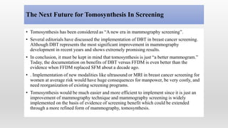
DIGITAL BREAST
TOMOSYNTHESIS
HISTORY
• The first full-field digital mammography was approved for use in January 2000. Several other
full-field digital detector systems were developed and approved for use over the next decade.
• Tomosynthesis imaging was first demonstrated by Niklason in 1997 but was not commercially
available until 2011.
• In February 2011, the first digital breast tomosynthesis (DBT) system (Hologic Selenia
Dimensions) was introduced. Two other systems have been approved since then, GE SenoClaire
(August 2014) and Siemens MAMMOMAT Inspiration (April 2015).
• Early DBT studies were viewed on dedicated workstations due to the large data sets involved
and due to the proprietary compression algorithms that were originally employed.
• The digital imaging and communication (DICOM) standard now supports DBT, allowing
viewing and reading of DBT studies on conventional picture archiving and communication
systems (PACS) workstations.
INTRODUCTION
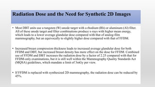
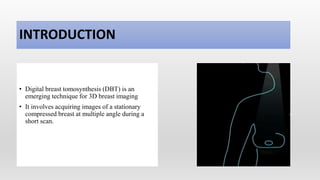
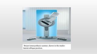
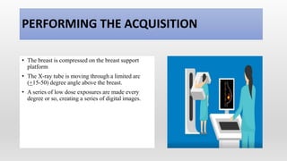
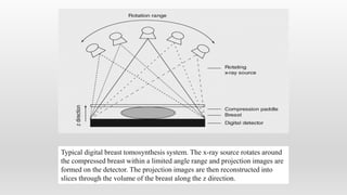
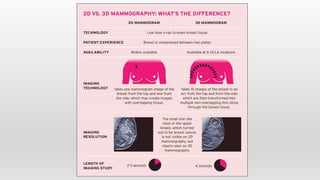
• Digital breast tomosynthesis (DBT) is an
emerging technique for 3D breast imaging
• It involves acquiring images of a stationary
compressed breast at multiple angle during a
short scan.
Breast tomosynthesis scanner, shown in the medio-
lateral-oblique position.
PATIENT PREPARETION
• Ask patient about prior surgeries, hormone use, and family or personal history of breast cancer.
• Do not schedule screening mammogram for the week before menstrual period, breasts are
usually tender during this time. The best time for a screening mammogram is one week
following your period.
• Ask patient’s pregnancy status.
• Not wear deodorant, talcum powder or lotion under arms or on breasts on the day of the exam.
These can be confused for calcium deposits on the mammogram.
• Discuss any breast symptoms or problems to the patient before performing the exam.
• Check prior mammograms. This is needed for comparison with current exam.
PERFORMING THE ACQUISITION
• The breast is compressed on the breast support
platform
• The X-ray tube is moving through a limited arc
(+15-50) degree angle above the breast.
• A series of low dose exposures are made every
degree or so, creating a series of digital images.
Typical digital breast tomosynthesis system. The x-ray source rotates around
the compressed breast within a limited angle range and projection images are
formed on the detector. The projection images are then reconstructed into
slices through the volume of the breast along the z direction.
IMAGE FORMATION
• Multiple projection images are acquired at
different angles
• A volume image is then reconstructed from the
data
• Only the structures belonging to that plane will
be focused. While other structures from different
planes will be blurred out.
TOMOSYNTHESIS IMAGES
DBT images
Tomographic planes (slices)
1mm thickness
Slabs (thicker slices)
6mm & 10mm
2D synthetic views (2D
images from DBT data)
ADVANTAGES
• Overlapping tissue structures are
resolved
• Improved visualization of lesion
margins
• Detecting subtle lesions
• Patient call back is reduced
Example of tomosynthesis. Improved visualization of architectural
distortion (circle in [b]) in the center of the right breast in tomosynthesis, as
compared to reconstructed 2D digital images. (a) Reconstructed 2D image.
(b) Tomosynthesis: single slice image.
The range of breast density, from fatty breasts (far left) to extremely dense breasts (far
right).
Manufacturer Fuji GE Hologic IMS Siemens
Anode material W (tungsten) Mo or Rh W W W
Filter material Al or Rh Mo or Rh Ag (silver) Ag Rh
Detector a-Se FPD CsI FPD a-Se FPD a-Se FPD a-Se FPD
Pixel size (μm) 150/100 (ST
mode) 100/50
(HR mode)
100 140* (*70
rebinned)
85 85
Pixel shape Hexagonal Square Square Square Square
Tube motion Continuous Step-and-
shoot
Continuous Step-and-shoot Continuous
Sweep angle (°) 15 (ST mode)
40 (HR mode)
25 15 40 50
N° of projections 15 9 15 13 25
Dose/projection Uniform Uniform Uniform Variable Uniform
Antiscatter grid No Yes No No No
Physical Characteristics Of The Five Commercial Solutions For Digital Breast
Tomosynthesis
Radiation Dose and the Need for Synthetic 2D Images
• Most DBT units use a tungsten (W) anode target with a rhodium (Rh) or aluminum (Al) filter.
All of these anode target and filter combinations produce x-rays with higher mean energy,
which leads to a lower average glandular dose compared with that of analog-film
mammography, but an equivocally to slightly higher dose compared with that of FFDM.
• Increased breast compression thickness leads to increased average glandular dose for both
FFDM and DBT, but increased breast density has more effect on the dose for FFDM. Combined
use of FFDM and DBT increases the radiation dose by a factor of 2.25 compared with that for
FFDM-only examinations, but it is still well within the Mammography Quality Standards Act
(MQSA) guidelines, which mandate a limit of 3mGy per view.
• If FFDM is replaced with synthesized 2D mammography, the radiation dose can be reduced by
45%.
The Next Future for Tomosynthesis In Screening
• Tomosynthesis has been considered as “A new era in mammography screening”.
• Several editorials have discussed the implementation of DBT in breast cancer screening.
Although DBT represents the most significant improvement in mammography
development in recent years and shows extremely promising results.
• In conclusion, it must be kept in mind that tomosynthesis is just “a better mammogram.”
Today, the documentation on benefits of DBT versus FFDM is even better than the
evidence when FFDM replaced SFM about a decade ago.
• . Implementation of new modalities like ultrasound or MRI in breast cancer screening for
women at average risk would have huge consequences for manpower, be very costly, and
need reorganization of existing screening programs.
• Tomosynthesis would be much easier and more efficient to implement since it is just an
improvement of mammography technique and mammography screening is widely
implemented on the basis of evidence of screening benefit which could be extended
through a more refined form of mammography, tomosynthesis.
THANK YOU
Ms. Mamta Panda
mpanda483@gmail.com

Digital breast tomosynthesis

  • 1.
  • 2.
    HISTORY • The firstfull-field digital mammography was approved for use in January 2000. Several other full-field digital detector systems were developed and approved for use over the next decade. • Tomosynthesis imaging was first demonstrated by Niklason in 1997 but was not commercially available until 2011. • In February 2011, the first digital breast tomosynthesis (DBT) system (Hologic Selenia Dimensions) was introduced. Two other systems have been approved since then, GE SenoClaire (August 2014) and Siemens MAMMOMAT Inspiration (April 2015). • Early DBT studies were viewed on dedicated workstations due to the large data sets involved and due to the proprietary compression algorithms that were originally employed. • The digital imaging and communication (DICOM) standard now supports DBT, allowing viewing and reading of DBT studies on conventional picture archiving and communication systems (PACS) workstations.
  • 3.
    INTRODUCTION • Digital breasttomosynthesis (DBT) is an emerging technique for 3D breast imaging • It involves acquiring images of a stationary compressed breast at multiple angle during a short scan.
  • 4.
    Breast tomosynthesis scanner,shown in the medio- lateral-oblique position.
  • 5.
    PATIENT PREPARETION • Askpatient about prior surgeries, hormone use, and family or personal history of breast cancer. • Do not schedule screening mammogram for the week before menstrual period, breasts are usually tender during this time. The best time for a screening mammogram is one week following your period. • Ask patient’s pregnancy status. • Not wear deodorant, talcum powder or lotion under arms or on breasts on the day of the exam. These can be confused for calcium deposits on the mammogram. • Discuss any breast symptoms or problems to the patient before performing the exam. • Check prior mammograms. This is needed for comparison with current exam.
  • 6.
    PERFORMING THE ACQUISITION •The breast is compressed on the breast support platform • The X-ray tube is moving through a limited arc (+15-50) degree angle above the breast. • A series of low dose exposures are made every degree or so, creating a series of digital images.
  • 7.
    Typical digital breasttomosynthesis system. The x-ray source rotates around the compressed breast within a limited angle range and projection images are formed on the detector. The projection images are then reconstructed into slices through the volume of the breast along the z direction.
  • 8.
    IMAGE FORMATION • Multipleprojection images are acquired at different angles • A volume image is then reconstructed from the data • Only the structures belonging to that plane will be focused. While other structures from different planes will be blurred out.
  • 9.
    TOMOSYNTHESIS IMAGES DBT images Tomographicplanes (slices) 1mm thickness Slabs (thicker slices) 6mm & 10mm 2D synthetic views (2D images from DBT data)
  • 11.
    ADVANTAGES • Overlapping tissuestructures are resolved • Improved visualization of lesion margins • Detecting subtle lesions • Patient call back is reduced Example of tomosynthesis. Improved visualization of architectural distortion (circle in [b]) in the center of the right breast in tomosynthesis, as compared to reconstructed 2D digital images. (a) Reconstructed 2D image. (b) Tomosynthesis: single slice image.
  • 12.
    The range ofbreast density, from fatty breasts (far left) to extremely dense breasts (far right).
  • 13.
    Manufacturer Fuji GEHologic IMS Siemens Anode material W (tungsten) Mo or Rh W W W Filter material Al or Rh Mo or Rh Ag (silver) Ag Rh Detector a-Se FPD CsI FPD a-Se FPD a-Se FPD a-Se FPD Pixel size (μm) 150/100 (ST mode) 100/50 (HR mode) 100 140* (*70 rebinned) 85 85 Pixel shape Hexagonal Square Square Square Square Tube motion Continuous Step-and- shoot Continuous Step-and-shoot Continuous Sweep angle (°) 15 (ST mode) 40 (HR mode) 25 15 40 50 N° of projections 15 9 15 13 25 Dose/projection Uniform Uniform Uniform Variable Uniform Antiscatter grid No Yes No No No Physical Characteristics Of The Five Commercial Solutions For Digital Breast Tomosynthesis
  • 14.
    Radiation Dose andthe Need for Synthetic 2D Images • Most DBT units use a tungsten (W) anode target with a rhodium (Rh) or aluminum (Al) filter. All of these anode target and filter combinations produce x-rays with higher mean energy, which leads to a lower average glandular dose compared with that of analog-film mammography, but an equivocally to slightly higher dose compared with that of FFDM. • Increased breast compression thickness leads to increased average glandular dose for both FFDM and DBT, but increased breast density has more effect on the dose for FFDM. Combined use of FFDM and DBT increases the radiation dose by a factor of 2.25 compared with that for FFDM-only examinations, but it is still well within the Mammography Quality Standards Act (MQSA) guidelines, which mandate a limit of 3mGy per view. • If FFDM is replaced with synthesized 2D mammography, the radiation dose can be reduced by 45%.
  • 15.
    The Next Futurefor Tomosynthesis In Screening • Tomosynthesis has been considered as “A new era in mammography screening”. • Several editorials have discussed the implementation of DBT in breast cancer screening. Although DBT represents the most significant improvement in mammography development in recent years and shows extremely promising results. • In conclusion, it must be kept in mind that tomosynthesis is just “a better mammogram.” Today, the documentation on benefits of DBT versus FFDM is even better than the evidence when FFDM replaced SFM about a decade ago. • . Implementation of new modalities like ultrasound or MRI in breast cancer screening for women at average risk would have huge consequences for manpower, be very costly, and need reorganization of existing screening programs. • Tomosynthesis would be much easier and more efficient to implement since it is just an improvement of mammography technique and mammography screening is widely implemented on the basis of evidence of screening benefit which could be extended through a more refined form of mammography, tomosynthesis.
  • 16.
    THANK YOU Ms. MamtaPanda mpanda483@gmail.com