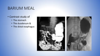
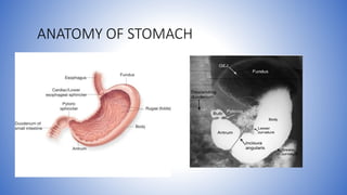
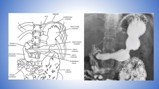
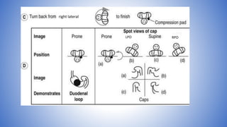
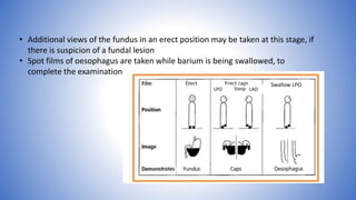
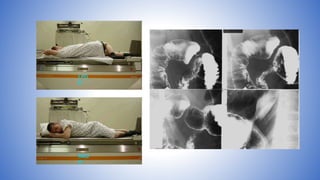
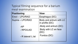
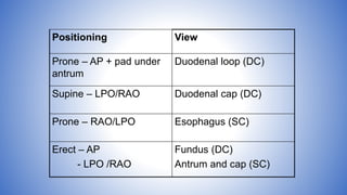
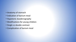
BARIUM MEAL
PRADEEP KUMAR
BARIUM MEAL
• Contrast study of
• The stomach
• The duodenum &
• The distal esophagus
ANATOMY OF STOMACH
• Surface of the gastric mucosa has a reticular pattern produced
by multiple interconnecting grooves
• Areae gastricae:
• Varies from fine reticular to coarse nodular pattern
produced by the grooves- hallmark of normal
• most prominent in the body and antrum
• 2-4mm in size
• >4mm or focal abnormalities like distortion is a sign of
gastritis or superficial infiltration by ca
• Its presence – atrophic gastritis unlikely
RADIOLOGICAL FEATURES
• Cardia :
• part of stomach where the
abdominal part of the esophagus
enters the stomach.
• Appearance is variable
• When viewed en face in LAO position,
shows a rosette of folds radiating from
the esophageal orifice, with a curved
mucosal fold forming a hood over
these emerging folds – BURNAUS SIGN
• Or, may show crescentic line
• Fundus:
• Dome-shaped and directly adjacent to the cardia, projecting superiorly.
• It is typically air-filled and is the portion of the stomach where the gastric bubble is
commonly appreciated on upright plain films.
• Mucosa of fundus (and body) is thrown into longitudinal folds or rugae.
• These are effaced to some extent when stomach is distended
• But along the greater curvature where they are most prominent, are still be seen in
distended stomach.
• Body:
• Lies inferior to the fundus and extends from the cardiac orifice to the level of
the incisura angularis, a notch in the lower part of the lesser curvature.
• The shape and size of which is quite variable due to peristalsis.
• Antrum:
• Extends from the incisura angularis to the pyloric opening.
• Pylorus:
• The most distal segment of the stomach and consists of a thickened band of
circular muscle that forms a sphincter functioning to control the rate of
gastric emptying into the duodenum.
• The length of the pyloric canal is about 1cm in adults.
ANATOMY OF DUODENUM
• Extends from pylorus to duodenojejunal flexure
• Radiographically consists of:
• Duodenal cap or duodenal bulb (1st part)
• Descending (2nd part)
• Horizontal (3rd part)
• Ascending (4th part)
• Duodenal cap:
• Symmetric, regular and triangular or conical
• Fine velvety reticular surface pattern due to presence of villi
• Sometimes spotted surface pattern due to barium caught in mucosal pits.
• Undistended: a fold pattern and a prominent fold curves around the inferior bend
between the first and second parts of the duodenum.
• Beyond the cap, mucosal folds are a normal feature and are seen as narrow
bands extending across the whole width of duodenum.
• Major papilla (of Vater):
• Projects into the lumen on the inner side of the 2nd part
• Running down from the papilla are 3 folds:
• A central longitudinal fold
• Oblique folds on either side
• Join and form an arch over the top of the papilla.
• Minor papilla (of Santorini):
• occasionally seen on the anterior wall, 2 cm proximal to
the major papilla.
INDICATIONS
• Symptoms related to upper GIT:
• Dyspepsia
• Weight loss
• Epigastric pain
• Anorexia
• Anemia
• Heart burn
• Vomiting
• Upper GI hemorrhage
• Upper abdominal masses
• Gastric or duodenal obstruction
• Malignancies of oesophago-gastric junction, stomach and duodenum
• Systemic diseases like tuberculosis affecting the upper GIT
• Motility disorders
• GI reflux
• In children to identify a cause for vomiting due to:
• gastro-esophageal reflux
• pyloric obstruction
• malrotation
• Assessment of perforation &/or post-surgery (anastomosis)
CONTRAINDICATIONS
• Suspected cases of gastro- duodenal perforation – barium peritonitis
• H/o or suspicion of aspiration – alternative contrast media
• Large bowel obstruction – barium inspissation occurs
• Fistulous communication with any organs other than parts of GIT
• Recent biopsy from GIT – barium granuloma at biopsy site
METHODS
1. Single-contrast + graded
compression (SCGC)
2. Double-contrast (DC): routine
contrast examination for
gastroduodenal disease
3. Biphasic method: Combined DC +
SCGC.
SINGLE CONTRAST STUDY
• Filling the stomach and duodenum with a low-density barium
suspension or a water-soluble contrast agent and applying graded
compression to permit the examiner to “see through” these viscera
• The stomach is compressed either manually or by positioning to allow
for adequate x-ray penetration in the evaluation of each anatomical
segment.
• Assesses thickness of the gastric folds and evaluation of gastric
emptying.
• Large luminal defects can be detected.
• The anterior gastric wall is evaluated on the prone films, an area which
may not be well demonstrated on a routine double-contrast examination
Advantages Disadvantages
Optimal for patients who are immobile or unable
to swallow gas forming tablets
Lack of sensitivity in detecting small erosion/
linear ulceration, superficial gastric carcinoma
and subtle mucosal abnormalities
Pylorospasm, fistulae and enlarged gastric rugae
are best seen
Distension can be achieved only at the expense
of increasing opacity in SC study – lesions not
caught in profile can be obscured
Filling defects due to large masses in pyloric and
duodenal region are more easily identifiable
Some parts of the anatomy such as gastric
fundus and cardia, colonic flexures, and
rectosigmoid are not easily accessible to
palpation.
Procedure of choice to examine patients with
suspected gastric or duodenal obstruction
Physical limitations to effective compression –
obesity and recent surgery
DOUBLE CONTRAST STUDY
• Combines the principles of distension, mucosal coating and proper
projection
• A small amount of high-density barium suspension is used to coat the
mucosal surface and air or CO2 gas to distend the lumen.
• Images are obtained as the patient rolls in various positions to coat the
gastric mucosa with contrast.
Advantages
• Conventional single contrast studies have several limitations, which
may result in diagnostic error.
• Double-contrast technique provides exquisite detail of the mucosal
surface of the stomach. Very useful for small mucosal lesions like
polyps, mucosal erosions and ulcers, recurrent tumors and post
operative studies.
• Gives excellent definition of the lesser and greater curvatures and the
posterior wall of the stomach.
Limitations
• Contraindicated in the search for mechanical problems such as obstruction or
fistula
• Whenever it is important to control the barium column precisely, a single
contrast study is preferred, such as in – intussusception, suspected
diverticulitis, acute obstruction and Hirschprung’s disease
• Not satisfactory for imaging anterior wall of distal stomach and duodenum
• Limited use in case of immobile/ elderly
BIPHASIC CONTRAST STUDY
• Performed with a single barium suspension that can provide excellent mucosal
coating during gaseous distension in the DC phase of the study and also sufficient
transparency to permit “see through” of the contrast agent during the SCGC
phase
• For biphasic study bubbly barium is developed.
• A medium-density barium suspension which can be administered simultaneously
with a gas-producing agent in the form of a cold, carbonated drink (“bubbly
barium”)
CONTRAST MEDIA
• Barium sulphate:
• High density, low viscosity barium sulphate for DC study.
• 250% w/v
• ~135ml
• Low density barium (100%w/v) for single contrast study.
• Medium density bubbly barium for biphasic study.
• Water soluble contrast:
• Perforation or post-operative anastomotic failure suspected
• Effervescent:
• Used for double contrast study.
• Carbex granules or tablets
• Composed of sodium bicarbonate, citrate and an antifoaming agent
(simethicone).
• When swallowed with a small amount of water, the granules or
tablets release 300–500 ml of carbon dioxide which distends the
stomach
PHARMACOLOGICAL AGENTS
• To relax the stomach and delay gastric emptying
• Buscopan 20mg i.v. or
• Glucagon 0.1 to 0.2mg i.v. (0.3 mg in case slow procedure)
PATIENT PREPARATION
• NPO x 6 hours to ensure adequate gastric emptying
• Avoid smoking
• Question the patient about:
• relevant symptoms
• previous gastric surgery
• modification of technique required
• having been NPO since midnight
• the possibility of pregnancy
• Explain the procedure to the patient.
• Describe how to perform breath-holding during spot filming
• Caution the patient not to belch after ingesting carbex granules or
drinking the “bubbly barium”.
TECHNIQUE
• If the patient is to have both a cervical/esophageal barium swallow (BS) and
UGI, start with the UGI and do the examination of the hypopharynx and
cervical esophagus at the end of the study.
• Spot-film technique:
• Phototimed 90 kVp (70-120kVp) is used for all double-contrast filming.
• 125 kVp (120-150kVp) for all single-contrast filming.
PRELIMINARY FILM
• Required
• if there is any question of perforation or
obstruction,
• history of recent contrast examination
• Raise the x-ray table to the upright position.
• The patient stands on the footrest with his
back against the table top. (If patient cannot
stand, elevate head of table 30°-45°.)
DOUBLE CONTRAST METHOD
• Turn the x-ray table into a horizontal position. Give the patient a pillow on which
to rest his head
• While the patient is lying supine on the x-ray table, slowly inject Buscopan or
Glucagon intravenously
• A gas producing agent is swallowed
• Patient then drinks barium while lying on left side, supported by the elbow.
• Prevents barium from reaching the duodenum too quickly obscuring the greater curve of the
stomach.
 Ask patient to roll toward you or rightward (counter-clockwise, as
viewed from the foot end of table) through three 360° rotations,
stopping in the steep LPO or left lateral position.
 If patient cannot turn around, he should be rolled back and forth
three times from one lateral position to the other.
 This is done to obtain good barium coating of the gastric mucosa
while the CO2 will distend the gastric lumen.
 Good coating has been achieved if the areae gastricae in the antrum
are visible
LPO RPO Right
Gastric antrum (patient LPO)
Gastric body, inferior portion (patient supine, AP)
Fundus (patient right lateral)
Gastric body, superior portion (patient RPO)
Elevate head of table 15° to keep barium from
flowing back into gastric fundus as the patient
rolls back into RPO position
• Have patient make another counter-clockwise rotation (as viewed
from foot of table) to refresh the barium coating of the gastric
mucosa. Stop in the steep LPO position.
• Take four DC spot images of the entire stomach using the largest
FOV in the following sequence:
• LPO position
• AP (supine)
• RPO
• Right lateral (Wait until duodenal C-loop is sufficiently filled with barium;
otherwise, take this film at end of study.)
LPO AP
RPO
RL
Spot films of duodenal loops
• Prone: the patient lies on a compression pad to prevent barium from
flooding into the duodenum
• An additional view to demonstrate the anterior wall of the duodenal
loop may be taken in LPO position
Spot films of duodenal cap
• Prone
• LPO (patient attains this position from the prone position by rolling first onto the
left side)
• Supine
• RPO
right lateral
LPO RPO
• Additional views of the fundus in an erect position may be taken at this stage, if
there is suspicion of a fundal lesion
• Spot films of oesophagus are taken while barium is being swallowed, to
complete the examination
LPO LAO
Swallow LPO
• Decrease kVp to 90.
• Turn patient into LPO position.
• Quickly scan the mediastinum to be sure that the esophagus is
empty of barium.
• Turn the table into horizontal position and resume fluoroscopy.
• Take two DC spots (4-on-1 film format) of duodenal bulb and two
DC spots of air-filled duodenal C-loop.
• Observe for spontaneous gastro esophageal reflux as you turn
patient towards you (counter-clockwise if viewed from foot of table)
into RAO position.
• Increase kVp to 125.
• Have the patient drink several single swallows of dilute, non-carbonated barium
through a straw.
• Observe esophageal motility and also look for anatomic lesions.
• Take one SC spot (2-on-1 format) of the barium-distended lower esophagus and
gastric cardia during breath-holding and one SC spot of the same area during
Valsalva maneuver to evaluate for a possible sliding hiatal hernia.
• Take one overhead film (14" x
17", 125 kVp) of the abdomen
with patient in prone position.
Modifications for young children
• Main indication: identify the cause for
vomiting (mainly 3 major causes – gastro-
esophageal reflux, pyloric obstruction &
malrotation)
• It is essential that the position of the
duodeno-jejunal flexure is demonstrated
• Single contrast technique using 30% w/v
BaSO4 and no paralytic agent
• A relatively small volume of BaSO4 enough to just fill the fundus, is given to the
infant in the supine position. A film of the distended oesophagus is exposed.
• Child is turned semi-prone into RAO position, film taken as barium passes
through the pylorus.
• 20-400 caudocranial angulation – pylorus is shown even better
• Once the barium enters the duodenum, the infant is returned to the supine
position, and with the child perfectly straight a 2nd film is exposed as barium
passes around the duodenojejunal flexure
• Once malrotation has been diagnosed or excluded, a further volume of barium is
administered until the stomach is reasonably full and barium lies against the GE
junction. The child is gently rotated through 1800 in an attempt to elicit GE reflux.
• In newborn infants with upper intestinal obstruction, eg. duodenal atresia, the
diagnosis may be confirmed if 20 ml air injected down the nasogastric tube.
• If the diagnosis remains in doubt, it can be replaced by a positive contrast agent
(dilute barium or LOCM if the risk of aspiration is high)
Modifications in partial gastrectomy or gastric
drainage procedure
• Start with prone swallow using high-density barium
• When barium reaches duodenum or gastroenterostomy, patient is quickly turned
supine for double contrast of these structures
• Double contrast filming of esophagus and stomach can then follow
Modifications in suspected gastroduodenal
perforation
• Water soluble contrast used
• Profile views of filled stomach are obtained
• Then the patient is turned on right to allow duodenal filling & turned through
360o. If no obvious extravasation , remain on right side for 10 mins
• If no perforation seen, but still strongly suspected clinically, delayed films may
show contrast excreted through urinary tract, since gastrograffin is absorbed from
peritoneal cavity
AFTER CARE
• Patient should be warned that bowel motions will be white and difficult to flush
for few days
• Advised to take adequate water to prevent barium impaction. Laxative may be
used if required
• If buscopan is used, the blurring of vision should be subsided before patient
leaves the department.
COMPLICATIONS
• Leakage of Ba from unsuspected perforation leading to Ba peritonitis
• Barium impaction (partial bowel obstruction to complete bowel
obstruction)
• Barium appendicitis if Ba impacted in appendix
• Barium embolisation if a bleeding ulcer is present
• Adverse effects of pharmacological agents used.
• Aspiration of stomach contents (due to buscopan)
• Acute gastric dilatation
HYPOTONIC DUODENOGRAPHY
• Is performed as a separate study if the duodenal loop is the prime area of
interest
• Tubeless method can be performed as part of routine DC barium meal or as
a specific examination of the duodenum
• The barium suspension and effervescent agent are given, and when barium is present
in the duodenum a smooth muscle relaxant is injected intravenously
• Filming is done same as in DC barium meal to demonstrate various parts of duodenum
• Tube method: Duodenal intubation (Bilbao-Dotter duodenal catheter) gives
consistently better results
• The quantity of barium and air being insufflated can be controlled and there
is no overlying barium in the gastric antrum or the jejunal loops
• INDICATIONS
Primary lesion of duodenum
Duodenitis
To demonstrate details of duodenum
Poor distension , unusual position of duodenum
To investigate obscure GI bleeding
• TECHNIQUE
The tip of the catheter is placed on the lower part of ascending
duodenum and abt. 40 ml of Ba- suspension is injected
As second part of duodenum fills ,buscopan is given I/V
During atonic state when duodenum distended with contrast, air is
injected through catheter
Spots films of duodenal loop are taken in
-SUPINE
-LPO/RAO
-PRONE
For proximal duodenum - head elevated 40 -60 degree
Narrow segment of duodenum- by single contrast Ba column
Typical filming sequence for a barium
meal examination
Positioning View
Erect – LPO/RAO Esophagus (DC)
Supine – LPO/RAO
-AP
- RPO/LAO
- R lateral/ L lat
Body and antrum with LC
in profile (DC)
-Body and antrum (DC)
-Body with LC en face
(DC)
-Fundus (DC)
Positioning View
Prone – AP + pad under
antrum
Duodenal loop (DC)
Supine – LPO/RAO Duodenal cap (DC)
Prone – RAO/LPO Esophagus (SC)
Erect – AP
- LPO /RAO
Fundus (DC)
Antrum and cap (SC)
• Anatomy of stomach
• Indication of barium meal
• Hypotonic duodenography
• Modifications for young children
• Single vs double contrast
• Complication of barium meal
Barium meal PPT Slide  PK

Barium meal PPT Slide PK

  • 1.
  • 2.
    BARIUM MEAL • Contraststudy of • The stomach • The duodenum & • The distal esophagus
  • 3.
  • 5.
    • Surface ofthe gastric mucosa has a reticular pattern produced by multiple interconnecting grooves • Areae gastricae: • Varies from fine reticular to coarse nodular pattern produced by the grooves- hallmark of normal • most prominent in the body and antrum • 2-4mm in size • >4mm or focal abnormalities like distortion is a sign of gastritis or superficial infiltration by ca • Its presence – atrophic gastritis unlikely RADIOLOGICAL FEATURES
  • 6.
    • Cardia : •part of stomach where the abdominal part of the esophagus enters the stomach. • Appearance is variable • When viewed en face in LAO position, shows a rosette of folds radiating from the esophageal orifice, with a curved mucosal fold forming a hood over these emerging folds – BURNAUS SIGN • Or, may show crescentic line
  • 7.
    • Fundus: • Dome-shapedand directly adjacent to the cardia, projecting superiorly. • It is typically air-filled and is the portion of the stomach where the gastric bubble is commonly appreciated on upright plain films. • Mucosa of fundus (and body) is thrown into longitudinal folds or rugae. • These are effaced to some extent when stomach is distended • But along the greater curvature where they are most prominent, are still be seen in distended stomach.
  • 8.
    • Body: • Liesinferior to the fundus and extends from the cardiac orifice to the level of the incisura angularis, a notch in the lower part of the lesser curvature. • The shape and size of which is quite variable due to peristalsis. • Antrum: • Extends from the incisura angularis to the pyloric opening. • Pylorus: • The most distal segment of the stomach and consists of a thickened band of circular muscle that forms a sphincter functioning to control the rate of gastric emptying into the duodenum. • The length of the pyloric canal is about 1cm in adults.
  • 9.
    ANATOMY OF DUODENUM •Extends from pylorus to duodenojejunal flexure • Radiographically consists of: • Duodenal cap or duodenal bulb (1st part) • Descending (2nd part) • Horizontal (3rd part) • Ascending (4th part)
  • 10.
    • Duodenal cap: •Symmetric, regular and triangular or conical • Fine velvety reticular surface pattern due to presence of villi • Sometimes spotted surface pattern due to barium caught in mucosal pits. • Undistended: a fold pattern and a prominent fold curves around the inferior bend between the first and second parts of the duodenum. • Beyond the cap, mucosal folds are a normal feature and are seen as narrow bands extending across the whole width of duodenum.
  • 11.
    • Major papilla(of Vater): • Projects into the lumen on the inner side of the 2nd part • Running down from the papilla are 3 folds: • A central longitudinal fold • Oblique folds on either side • Join and form an arch over the top of the papilla. • Minor papilla (of Santorini): • occasionally seen on the anterior wall, 2 cm proximal to the major papilla.
  • 12.
    INDICATIONS • Symptoms relatedto upper GIT: • Dyspepsia • Weight loss • Epigastric pain • Anorexia • Anemia • Heart burn • Vomiting • Upper GI hemorrhage • Upper abdominal masses • Gastric or duodenal obstruction • Malignancies of oesophago-gastric junction, stomach and duodenum • Systemic diseases like tuberculosis affecting the upper GIT
  • 13.
    • Motility disorders •GI reflux • In children to identify a cause for vomiting due to: • gastro-esophageal reflux • pyloric obstruction • malrotation • Assessment of perforation &/or post-surgery (anastomosis)
  • 14.
    CONTRAINDICATIONS • Suspected casesof gastro- duodenal perforation – barium peritonitis • H/o or suspicion of aspiration – alternative contrast media • Large bowel obstruction – barium inspissation occurs • Fistulous communication with any organs other than parts of GIT • Recent biopsy from GIT – barium granuloma at biopsy site
  • 15.
    METHODS 1. Single-contrast +graded compression (SCGC) 2. Double-contrast (DC): routine contrast examination for gastroduodenal disease 3. Biphasic method: Combined DC + SCGC.
  • 16.
    SINGLE CONTRAST STUDY •Filling the stomach and duodenum with a low-density barium suspension or a water-soluble contrast agent and applying graded compression to permit the examiner to “see through” these viscera • The stomach is compressed either manually or by positioning to allow for adequate x-ray penetration in the evaluation of each anatomical segment. • Assesses thickness of the gastric folds and evaluation of gastric emptying. • Large luminal defects can be detected. • The anterior gastric wall is evaluated on the prone films, an area which may not be well demonstrated on a routine double-contrast examination
  • 17.
    Advantages Disadvantages Optimal forpatients who are immobile or unable to swallow gas forming tablets Lack of sensitivity in detecting small erosion/ linear ulceration, superficial gastric carcinoma and subtle mucosal abnormalities Pylorospasm, fistulae and enlarged gastric rugae are best seen Distension can be achieved only at the expense of increasing opacity in SC study – lesions not caught in profile can be obscured Filling defects due to large masses in pyloric and duodenal region are more easily identifiable Some parts of the anatomy such as gastric fundus and cardia, colonic flexures, and rectosigmoid are not easily accessible to palpation. Procedure of choice to examine patients with suspected gastric or duodenal obstruction Physical limitations to effective compression – obesity and recent surgery
  • 18.
    DOUBLE CONTRAST STUDY •Combines the principles of distension, mucosal coating and proper projection • A small amount of high-density barium suspension is used to coat the mucosal surface and air or CO2 gas to distend the lumen. • Images are obtained as the patient rolls in various positions to coat the gastric mucosa with contrast.
  • 19.
    Advantages • Conventional singlecontrast studies have several limitations, which may result in diagnostic error. • Double-contrast technique provides exquisite detail of the mucosal surface of the stomach. Very useful for small mucosal lesions like polyps, mucosal erosions and ulcers, recurrent tumors and post operative studies. • Gives excellent definition of the lesser and greater curvatures and the posterior wall of the stomach.
  • 20.
    Limitations • Contraindicated inthe search for mechanical problems such as obstruction or fistula • Whenever it is important to control the barium column precisely, a single contrast study is preferred, such as in – intussusception, suspected diverticulitis, acute obstruction and Hirschprung’s disease • Not satisfactory for imaging anterior wall of distal stomach and duodenum • Limited use in case of immobile/ elderly
  • 21.
    BIPHASIC CONTRAST STUDY •Performed with a single barium suspension that can provide excellent mucosal coating during gaseous distension in the DC phase of the study and also sufficient transparency to permit “see through” of the contrast agent during the SCGC phase • For biphasic study bubbly barium is developed. • A medium-density barium suspension which can be administered simultaneously with a gas-producing agent in the form of a cold, carbonated drink (“bubbly barium”)
  • 22.
    CONTRAST MEDIA • Bariumsulphate: • High density, low viscosity barium sulphate for DC study. • 250% w/v • ~135ml • Low density barium (100%w/v) for single contrast study. • Medium density bubbly barium for biphasic study. • Water soluble contrast: • Perforation or post-operative anastomotic failure suspected • Effervescent: • Used for double contrast study. • Carbex granules or tablets • Composed of sodium bicarbonate, citrate and an antifoaming agent (simethicone). • When swallowed with a small amount of water, the granules or tablets release 300–500 ml of carbon dioxide which distends the stomach
  • 23.
    PHARMACOLOGICAL AGENTS • Torelax the stomach and delay gastric emptying • Buscopan 20mg i.v. or • Glucagon 0.1 to 0.2mg i.v. (0.3 mg in case slow procedure)
  • 24.
    PATIENT PREPARATION • NPOx 6 hours to ensure adequate gastric emptying • Avoid smoking • Question the patient about: • relevant symptoms • previous gastric surgery • modification of technique required • having been NPO since midnight • the possibility of pregnancy • Explain the procedure to the patient. • Describe how to perform breath-holding during spot filming • Caution the patient not to belch after ingesting carbex granules or drinking the “bubbly barium”.
  • 25.
    TECHNIQUE • If thepatient is to have both a cervical/esophageal barium swallow (BS) and UGI, start with the UGI and do the examination of the hypopharynx and cervical esophagus at the end of the study. • Spot-film technique: • Phototimed 90 kVp (70-120kVp) is used for all double-contrast filming. • 125 kVp (120-150kVp) for all single-contrast filming.
  • 26.
    PRELIMINARY FILM • Required •if there is any question of perforation or obstruction, • history of recent contrast examination • Raise the x-ray table to the upright position. • The patient stands on the footrest with his back against the table top. (If patient cannot stand, elevate head of table 30°-45°.)
  • 27.
    DOUBLE CONTRAST METHOD •Turn the x-ray table into a horizontal position. Give the patient a pillow on which to rest his head • While the patient is lying supine on the x-ray table, slowly inject Buscopan or Glucagon intravenously • A gas producing agent is swallowed • Patient then drinks barium while lying on left side, supported by the elbow. • Prevents barium from reaching the duodenum too quickly obscuring the greater curve of the stomach.
  • 28.
     Ask patientto roll toward you or rightward (counter-clockwise, as viewed from the foot end of table) through three 360° rotations, stopping in the steep LPO or left lateral position.  If patient cannot turn around, he should be rolled back and forth three times from one lateral position to the other.  This is done to obtain good barium coating of the gastric mucosa while the CO2 will distend the gastric lumen.  Good coating has been achieved if the areae gastricae in the antrum are visible
  • 29.
  • 30.
  • 31.
    Gastric body, inferiorportion (patient supine, AP)
  • 32.
  • 33.
    Gastric body, superiorportion (patient RPO) Elevate head of table 15° to keep barium from flowing back into gastric fundus as the patient rolls back into RPO position
  • 34.
    • Have patientmake another counter-clockwise rotation (as viewed from foot of table) to refresh the barium coating of the gastric mucosa. Stop in the steep LPO position. • Take four DC spot images of the entire stomach using the largest FOV in the following sequence: • LPO position • AP (supine) • RPO • Right lateral (Wait until duodenal C-loop is sufficiently filled with barium; otherwise, take this film at end of study.)
  • 35.
  • 36.
    Spot films ofduodenal loops • Prone: the patient lies on a compression pad to prevent barium from flooding into the duodenum • An additional view to demonstrate the anterior wall of the duodenal loop may be taken in LPO position
  • 37.
    Spot films ofduodenal cap • Prone • LPO (patient attains this position from the prone position by rolling first onto the left side) • Supine • RPO
  • 38.
  • 39.
    • Additional viewsof the fundus in an erect position may be taken at this stage, if there is suspicion of a fundal lesion • Spot films of oesophagus are taken while barium is being swallowed, to complete the examination LPO LAO Swallow LPO
  • 40.
    • Decrease kVpto 90. • Turn patient into LPO position. • Quickly scan the mediastinum to be sure that the esophagus is empty of barium. • Turn the table into horizontal position and resume fluoroscopy. • Take two DC spots (4-on-1 film format) of duodenal bulb and two DC spots of air-filled duodenal C-loop. • Observe for spontaneous gastro esophageal reflux as you turn patient towards you (counter-clockwise if viewed from foot of table) into RAO position.
  • 42.
    • Increase kVpto 125. • Have the patient drink several single swallows of dilute, non-carbonated barium through a straw. • Observe esophageal motility and also look for anatomic lesions. • Take one SC spot (2-on-1 format) of the barium-distended lower esophagus and gastric cardia during breath-holding and one SC spot of the same area during Valsalva maneuver to evaluate for a possible sliding hiatal hernia.
  • 43.
    • Increase kVpto 125. • Have the patient drink several single swallows of dilute, non-carbonated barium through a straw. • Observe esophageal motility and also look for anatomic lesions. • Take one SC spot (2-on-1 format) of the barium-distended lower esophagus and gastric cardia during breath-holding and one SC spot of the same area during Valsalva maneuver to evaluate for a possible sliding hiatal hernia.
  • 45.
    • Take oneoverhead film (14" x 17", 125 kVp) of the abdomen with patient in prone position.
  • 46.
    Modifications for youngchildren • Main indication: identify the cause for vomiting (mainly 3 major causes – gastro- esophageal reflux, pyloric obstruction & malrotation) • It is essential that the position of the duodeno-jejunal flexure is demonstrated • Single contrast technique using 30% w/v BaSO4 and no paralytic agent
  • 47.
    • A relativelysmall volume of BaSO4 enough to just fill the fundus, is given to the infant in the supine position. A film of the distended oesophagus is exposed. • Child is turned semi-prone into RAO position, film taken as barium passes through the pylorus. • 20-400 caudocranial angulation – pylorus is shown even better • Once the barium enters the duodenum, the infant is returned to the supine position, and with the child perfectly straight a 2nd film is exposed as barium passes around the duodenojejunal flexure • Once malrotation has been diagnosed or excluded, a further volume of barium is administered until the stomach is reasonably full and barium lies against the GE junction. The child is gently rotated through 1800 in an attempt to elicit GE reflux.
  • 48.
    • In newborninfants with upper intestinal obstruction, eg. duodenal atresia, the diagnosis may be confirmed if 20 ml air injected down the nasogastric tube. • If the diagnosis remains in doubt, it can be replaced by a positive contrast agent (dilute barium or LOCM if the risk of aspiration is high)
  • 49.
    Modifications in partialgastrectomy or gastric drainage procedure • Start with prone swallow using high-density barium • When barium reaches duodenum or gastroenterostomy, patient is quickly turned supine for double contrast of these structures • Double contrast filming of esophagus and stomach can then follow
  • 50.
    Modifications in suspectedgastroduodenal perforation • Water soluble contrast used • Profile views of filled stomach are obtained • Then the patient is turned on right to allow duodenal filling & turned through 360o. If no obvious extravasation , remain on right side for 10 mins • If no perforation seen, but still strongly suspected clinically, delayed films may show contrast excreted through urinary tract, since gastrograffin is absorbed from peritoneal cavity
  • 51.
    AFTER CARE • Patientshould be warned that bowel motions will be white and difficult to flush for few days • Advised to take adequate water to prevent barium impaction. Laxative may be used if required • If buscopan is used, the blurring of vision should be subsided before patient leaves the department.
  • 52.
    COMPLICATIONS • Leakage ofBa from unsuspected perforation leading to Ba peritonitis • Barium impaction (partial bowel obstruction to complete bowel obstruction) • Barium appendicitis if Ba impacted in appendix • Barium embolisation if a bleeding ulcer is present • Adverse effects of pharmacological agents used. • Aspiration of stomach contents (due to buscopan) • Acute gastric dilatation
  • 53.
    HYPOTONIC DUODENOGRAPHY • Isperformed as a separate study if the duodenal loop is the prime area of interest • Tubeless method can be performed as part of routine DC barium meal or as a specific examination of the duodenum • The barium suspension and effervescent agent are given, and when barium is present in the duodenum a smooth muscle relaxant is injected intravenously • Filming is done same as in DC barium meal to demonstrate various parts of duodenum • Tube method: Duodenal intubation (Bilbao-Dotter duodenal catheter) gives consistently better results • The quantity of barium and air being insufflated can be controlled and there is no overlying barium in the gastric antrum or the jejunal loops
  • 54.
    • INDICATIONS Primary lesionof duodenum Duodenitis To demonstrate details of duodenum Poor distension , unusual position of duodenum To investigate obscure GI bleeding
  • 55.
    • TECHNIQUE The tipof the catheter is placed on the lower part of ascending duodenum and abt. 40 ml of Ba- suspension is injected As second part of duodenum fills ,buscopan is given I/V During atonic state when duodenum distended with contrast, air is injected through catheter
  • 56.
    Spots films ofduodenal loop are taken in -SUPINE -LPO/RAO -PRONE For proximal duodenum - head elevated 40 -60 degree Narrow segment of duodenum- by single contrast Ba column
  • 57.
    Typical filming sequencefor a barium meal examination Positioning View Erect – LPO/RAO Esophagus (DC) Supine – LPO/RAO -AP - RPO/LAO - R lateral/ L lat Body and antrum with LC in profile (DC) -Body and antrum (DC) -Body with LC en face (DC) -Fundus (DC)
  • 58.
    Positioning View Prone –AP + pad under antrum Duodenal loop (DC) Supine – LPO/RAO Duodenal cap (DC) Prone – RAO/LPO Esophagus (SC) Erect – AP - LPO /RAO Fundus (DC) Antrum and cap (SC)
  • 59.
    • Anatomy ofstomach • Indication of barium meal • Hypotonic duodenography • Modifications for young children • Single vs double contrast • Complication of barium meal

Editor's Notes

  • #4 The lesser curvature forms the right gastric border and extends from the cardia to the pylorus. The greater curvature forms the left gastric border and extends from the cardia, over the dome of the fundus, to the pylorus.
  • #6 Non visualization- sign of diffuse atrophic gastritis
  • #11 Fleck (=spot) is a loculation of barium of any size from few mm to 2cm which strongly suggests a break in the normal mucosal structure and ulceration. There are certain locations in the duodenal bulb where fleck formation may be a normal variant and these must be differentiated from pathological variety. These are: When the pylorus closes, there may be a dimple of mucosa at the base of the bulb in which the barium may accumulate, giving rise to the appearance of a fleck. The outer periphery of the bulb occasionally acts as a groove, or sinus, in which barium may accumulate and when seen in profile, gives the appearance of fleck formation at the base of the bulb The concentration of rugae at the apex of the bulb may simulate fleck formation Peristaltic waves passing over the duodenal bulb may simulate an inconstant variety of fleck
  • #17 100 % w/v barium low density is used in single contrast for anatomy and pathology
  • #25 Secretions will prevent adequate mucosal coating and may mimic tumors
  • #35 *RPO (First, turn patient into right lateral position and elevate head of table 15° to keep barium from flowing back into gastric fundus as he rolls back into RPO position.)
  • #37 From left lateral position the patient returns to a supine position and then rolls onto the left side and over into a prone position. This sequence is required to avoid barium flooding into the duodenal loop, which would occur if the patient were to roll onto the right side to achieve a prone position.
  • #50 Needed due to quick flooding of barium into the jejunum
  • #51 However this sign is not specific for perforation, since inflamed or ischaemic mucosa can allow absorption & thus renal excretion