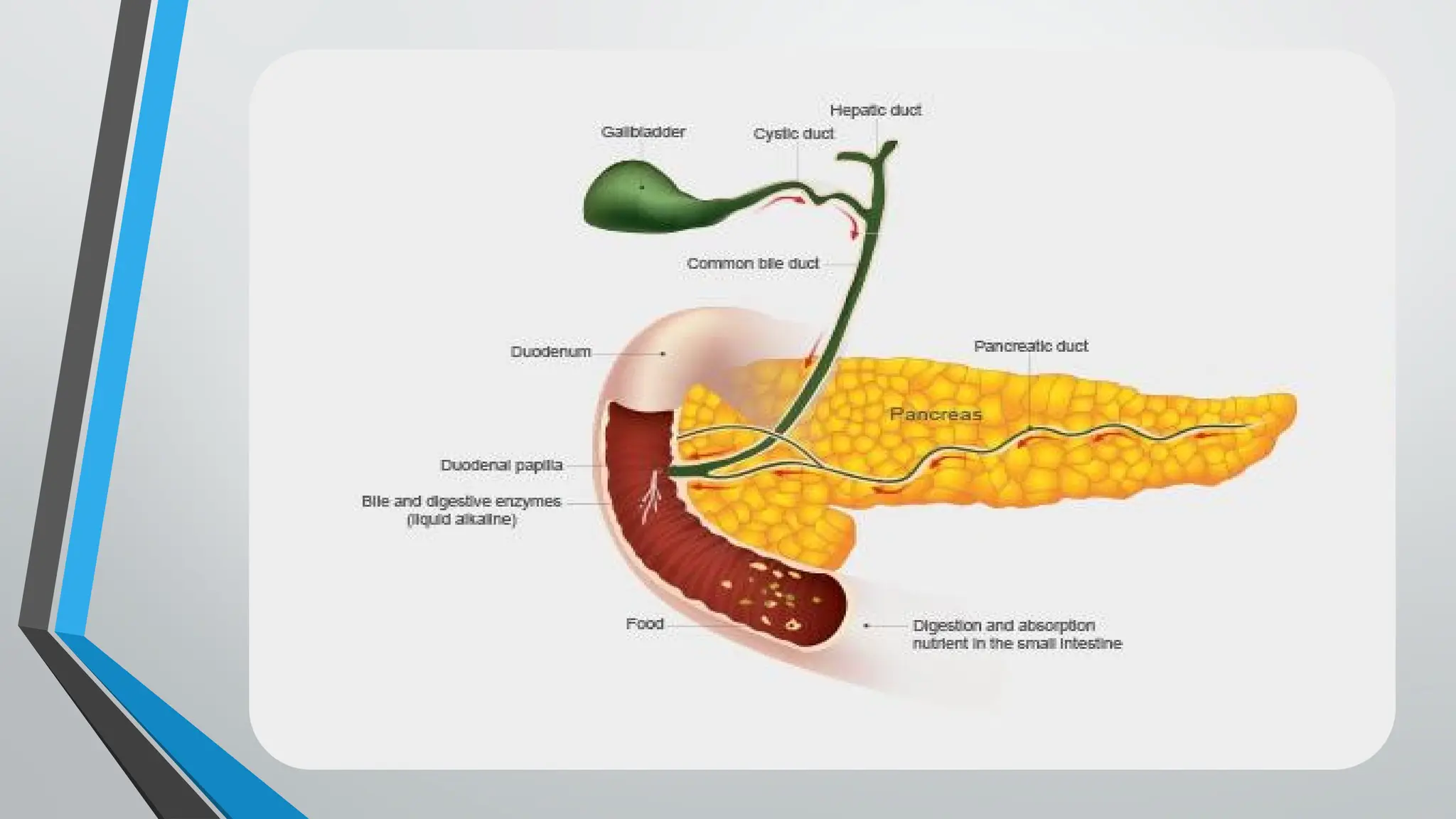
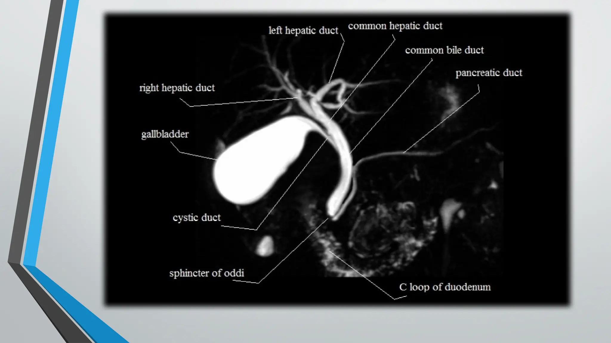
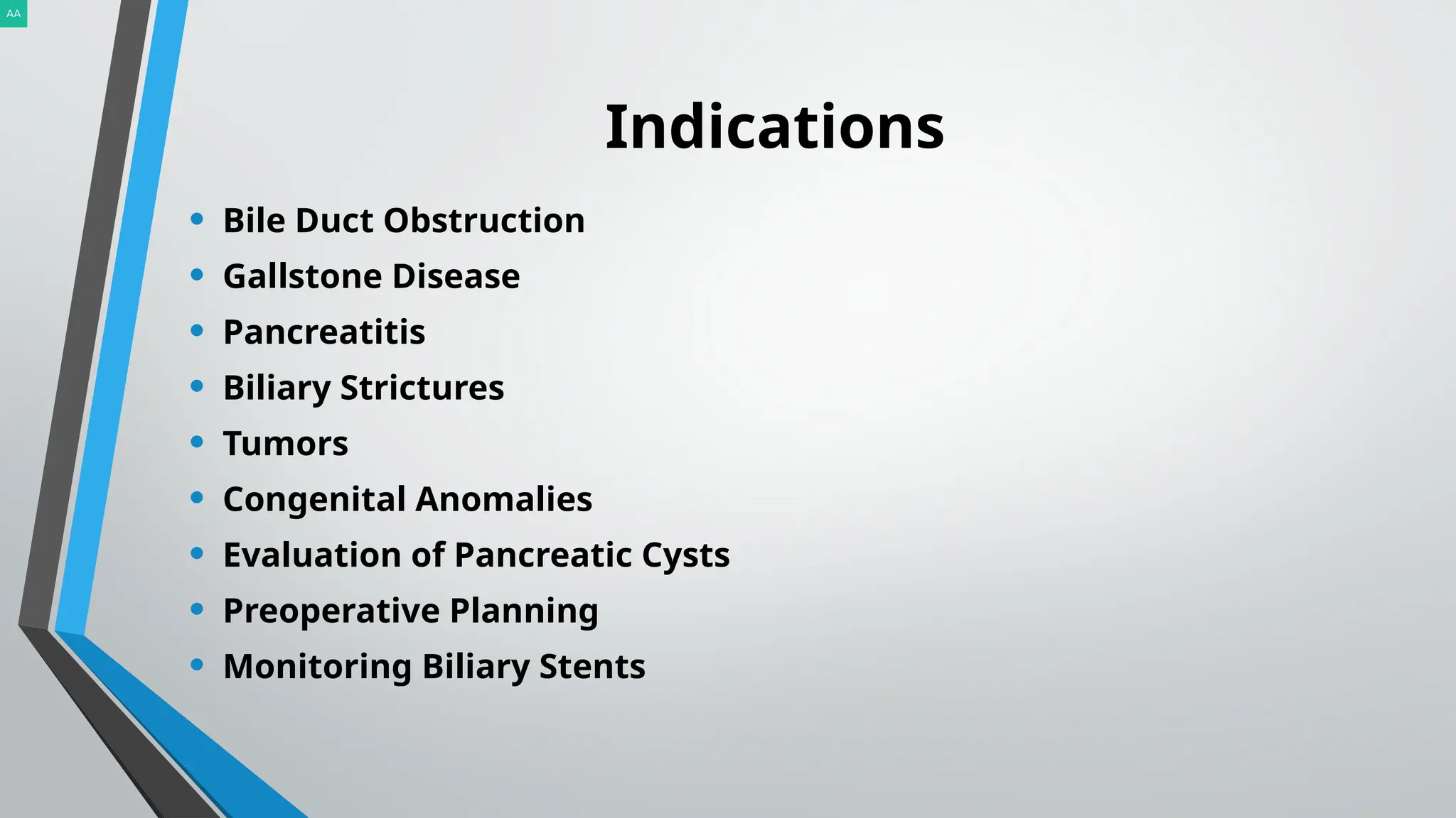
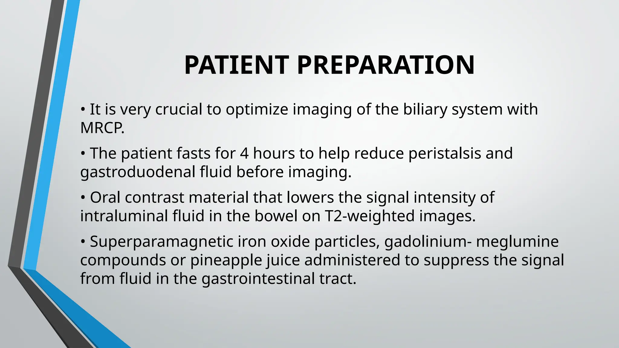
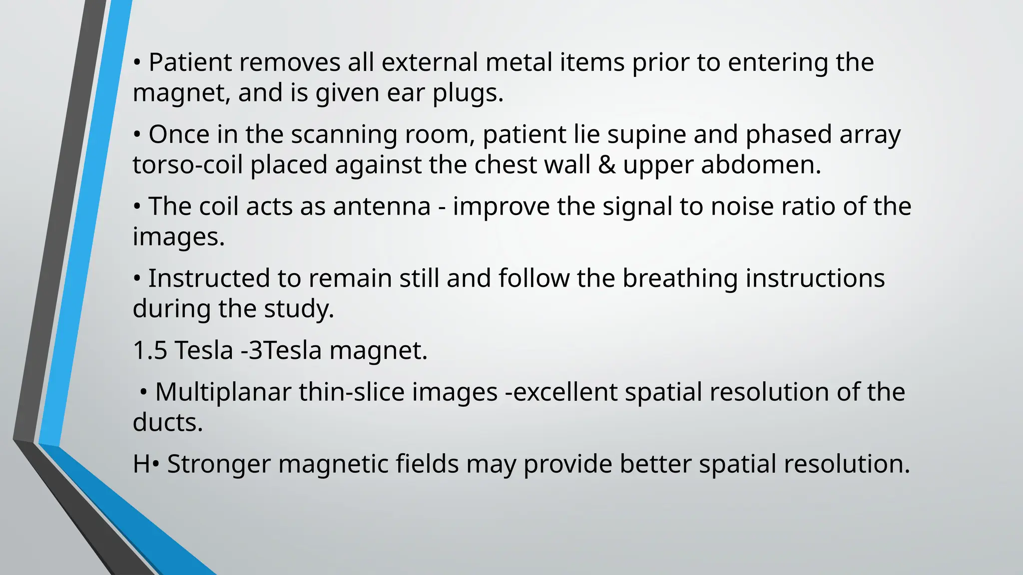
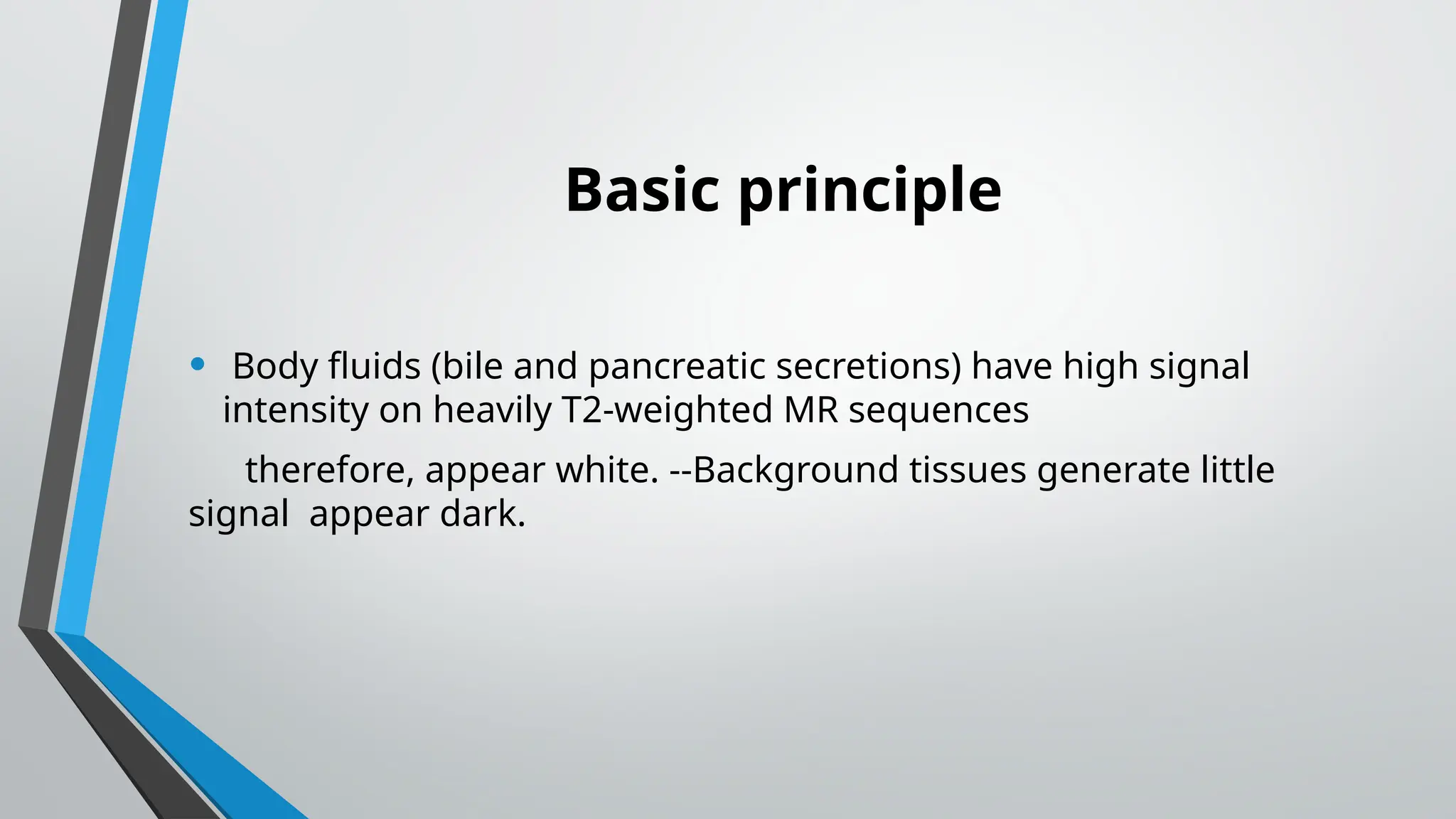
MRCP
By: AALIA ABDULLAH
ASSIST.PROF. RADIOLOGY & IMAGING
MEWAR UNIVERSITY.
MRCP
• MAGNETIC resonance cholangiopancreatography (MRCP) is a
radiologic technique that produces images of the
pancreatobiliary tree.
• It is a specialized imaging technique that uses magnetic
resonance imaging (MRI) to visualize the bile ducts and
pancreatic ducts. MRCP provides detailed images of the liver,
gallbladder, bile ducts, and pancreas without the need for
invasive procedures like endoscopic retrograde
cholangiopancreatography (ERCP).
• Introduced in 1991.
• Magnetic resonance cholangiopancreatography (MRCP) is
performed with heavily T2-weighted fast spin-echo sequences.
• Noninvasive, less costly, and sensitive alternative to diagnostic
ERCP
• The MRCP procedure typically takes about 30 to 60 minutes, but
the actual time may vary depending on the specific imaging
requirements.
• Bile Duct Obstruction
• Gallstone Disease
• Pancreatitis
• Biliary Strictures
• Tumors
• Congenital Anomalies
• Evaluation of Pancreatic Cysts
• Preoperative Planning
• Monitoring Biliary Stents
Indications
PATIENT PREPARATION
• It is very crucial to optimize imaging of the biliary system with
MRCP.
• The patient fasts for 4 hours to help reduce peristalsis and
gastroduodenal fluid before imaging.
• Oral contrast material that lowers the signal intensity of
intraluminal fluid in the bowel on T2-weighted images.
• Superparamagnetic iron oxide particles, gadolinium- meglumine
compounds or pineapple juice administered to suppress the signal
from fluid in the gastrointestinal tract.
• Patient removes all external metal items prior to entering the
magnet, and is given ear plugs.
• Once in the scanning room, patient lie supine and phased array
torso-coil placed against the chest wall & upper abdomen.
• The coil acts as antenna - improve the signal to noise ratio of the
images.
• Instructed to remain still and follow the breathing instructions
during the study.
1.5 Tesla -3Tesla magnet.
• Multiplanar thin-slice images -excellent spatial resolution of the
ducts.
H• Stronger magnetic fields may provide better spatial resolution.
Basic principle
• Body fluids (bile and pancreatic secretions) have high signal
intensity on heavily T2-weighted MR sequences
therefore, appear white. --Background tissues generate little
signal appear dark.
• Earlier sequences such as GRE & FSE provided MRCP images ,but
these long sequences often suffered from motion artifact & poor
spatial resolution.
• • Images are now acquired in one of the following sequences that
use heavy T2 weighting .
• 1) Single shot fast-spin echo (SSFSE) 2) Half Fourier single-shot
turbo spin echo (HASTE) 3) Rapid acquisition relaxation enhanced
(RARE). These ultrafast techniques obtain images rapidly with
reduced artifacts.
MRCP (Magnetic resonance cholangiopancreatography)

MRCP (Magnetic resonance cholangiopancreatography)

  • 1.
    MRCP By: AALIA ABDULLAH ASSIST.PROF.RADIOLOGY & IMAGING MEWAR UNIVERSITY.
  • 2.
    MRCP • MAGNETIC resonancecholangiopancreatography (MRCP) is a radiologic technique that produces images of the pancreatobiliary tree. • It is a specialized imaging technique that uses magnetic resonance imaging (MRI) to visualize the bile ducts and pancreatic ducts. MRCP provides detailed images of the liver, gallbladder, bile ducts, and pancreas without the need for invasive procedures like endoscopic retrograde cholangiopancreatography (ERCP).
  • 3.
    • Introduced in1991. • Magnetic resonance cholangiopancreatography (MRCP) is performed with heavily T2-weighted fast spin-echo sequences. • Noninvasive, less costly, and sensitive alternative to diagnostic ERCP • The MRCP procedure typically takes about 30 to 60 minutes, but the actual time may vary depending on the specific imaging requirements.
  • 6.
    • Bile DuctObstruction • Gallstone Disease • Pancreatitis • Biliary Strictures • Tumors • Congenital Anomalies • Evaluation of Pancreatic Cysts • Preoperative Planning • Monitoring Biliary Stents Indications
  • 7.
    PATIENT PREPARATION • Itis very crucial to optimize imaging of the biliary system with MRCP. • The patient fasts for 4 hours to help reduce peristalsis and gastroduodenal fluid before imaging. • Oral contrast material that lowers the signal intensity of intraluminal fluid in the bowel on T2-weighted images. • Superparamagnetic iron oxide particles, gadolinium- meglumine compounds or pineapple juice administered to suppress the signal from fluid in the gastrointestinal tract.
  • 8.
    • Patient removesall external metal items prior to entering the magnet, and is given ear plugs. • Once in the scanning room, patient lie supine and phased array torso-coil placed against the chest wall & upper abdomen. • The coil acts as antenna - improve the signal to noise ratio of the images. • Instructed to remain still and follow the breathing instructions during the study. 1.5 Tesla -3Tesla magnet. • Multiplanar thin-slice images -excellent spatial resolution of the ducts. H• Stronger magnetic fields may provide better spatial resolution.
  • 9.
    Basic principle • Bodyfluids (bile and pancreatic secretions) have high signal intensity on heavily T2-weighted MR sequences therefore, appear white. --Background tissues generate little signal appear dark.
  • 10.
    • Earlier sequencessuch as GRE & FSE provided MRCP images ,but these long sequences often suffered from motion artifact & poor spatial resolution. • • Images are now acquired in one of the following sequences that use heavy T2 weighting . • 1) Single shot fast-spin echo (SSFSE) 2) Half Fourier single-shot turbo spin echo (HASTE) 3) Rapid acquisition relaxation enhanced (RARE). These ultrafast techniques obtain images rapidly with reduced artifacts.