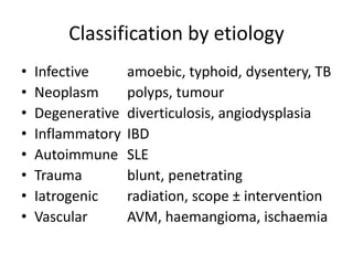
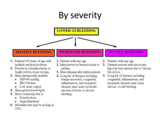
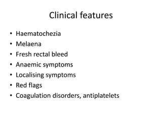
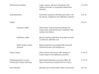
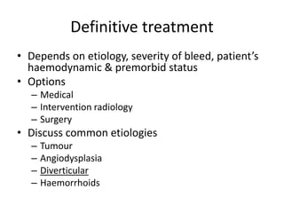
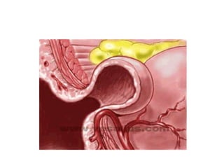
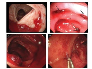
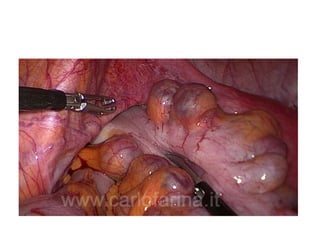
Lower GI bleed
Chea Chan Hooi
Surgeon
Sibu Hospital
Content
• Introduction
• Definition
• Classification
• Clinical features
• Principles of management
• Special discussion – Diverticular bleed
Introduction
• 20 – 33% of GI bleed cases
• Wide spectrum of presentation
• Potentially life-threatening
Definition
• Bleeding within GI tract distal to ligament of
Trietz
Classification by etiology
• Infective amoebic, typhoid, dysentery, TB
• Neoplasm polyps, tumour
• Degenerative diverticulosis, angiodysplasia
• Inflammatory IBD
• Autoimmune SLE
• Trauma blunt, penetrating
• Iatrogenic radiation, scope ± intervention
• Vascular AVM, haemangioma, ischaemia
By severity
Clinical features
• Haematochezia
• Melaena
• Fresh rectal bleed
• Anaemic symptoms
• Localising symptoms
• Red flags
• Coagulation disorders, antiplatelets
Principles of management
• Resuscitation
– IVF challenge
– Blood product transfusion
– Majority stop spontaneously
• Identify cause of bleed + haemostasis
• Definitive treatment
Investigations
• Endoscopy
– Anoscopy
– Proctoscopy
– Sigmoidoscopy
– Colonoscopy
• Initial diagnostic method of choice
• IDs cause up to 90%
– Enteroscopy
– Capsule endoscopy
• Imaging
– Angiography
• Bleed rate ≥1.0ml/min
• Pros : therapeutic in same setting
• Cons : invasive, high dose radiation
– CT angiography of mesentery
• Bleed rate ≥0.5ml/min
• Pros : anatomically detailed, relatively easily available
• Cons : non-therapeutic
– RBC-tagged scintigraphy
• Bleed rate ≥0.1ml/min
• Pros : detect minute amount of bleeding
• Cons : poor anatomical correlation, time consuming, now widely
available
Enteroscopy
• Push enteroscopy
– An extra-long upper endoscope with an average reach of
proximal 70cm of small bowel
• Balloon –assisted enteroscopy
– Single & double balloon
– Inflatable balloons to grip the intestine to facilitate deep
enteroscopy
– Largest body of evidence on enteroscopy is based on
double-balloon enteroscopy
• Spiral enteroscopy
– A specialised overtube with a compliant spiral located at
the distal tip
Definitive treatment
• Depends on etiology, severity of bleed, patient’s
haemodynamic & premorbid status
• Options
– Medical
– Intervention radiology
– Surgery
• Discuss common etiologies
– Tumour
– Angiodysplasia
– Diverticular
– Haemorrhoids
Diverticular bleed
Introduction
• Most common cause of LGIB
• Bleeding complicates 5 – 15% of patients with
diverticulosis
• 80% will cease spontaneously
• 30 – 40% will have recurrent bleed
• Bleeding tends to complicate right sided
diverticuli
Pathogenesis of colonic diverticular
bleed
• At sites of weaknesses in the colonic wall –
where vasa recta penetrates
• Mucosa herniates thru  pseudodiverticulum
• Vasa recta draped over the dome of the
diverticulum  susceptible to trauma and
disruption
• Intimal damages  eccentric thickening 
wall weakening  rupture
Other contributory factors
• Atherosclerosis
• NSAIDs
• Hyperuricaemia  oxidised LDL  oxygen
free radicals
• Faecoliths
Principles of management
• Resuscitation
• Endoscopy for localisation & haemostasis
• Angioembolisation
• Surgery
TA!
Q&A?

Lower gi bleed

  • 1.
    Lower GI bleed CheaChan Hooi Surgeon Sibu Hospital
  • 2.
    Content • Introduction • Definition •Classification • Clinical features • Principles of management • Special discussion – Diverticular bleed
  • 3.
    Introduction • 20 –33% of GI bleed cases • Wide spectrum of presentation • Potentially life-threatening
  • 4.
    Definition • Bleeding withinGI tract distal to ligament of Trietz
  • 5.
    Classification by etiology •Infective amoebic, typhoid, dysentery, TB • Neoplasm polyps, tumour • Degenerative diverticulosis, angiodysplasia • Inflammatory IBD • Autoimmune SLE • Trauma blunt, penetrating • Iatrogenic radiation, scope ± intervention • Vascular AVM, haemangioma, ischaemia
  • 6.
  • 7.
    Clinical features • Haematochezia •Melaena • Fresh rectal bleed • Anaemic symptoms • Localising symptoms • Red flags • Coagulation disorders, antiplatelets
  • 9.
    Principles of management •Resuscitation – IVF challenge – Blood product transfusion – Majority stop spontaneously • Identify cause of bleed + haemostasis • Definitive treatment
  • 10.
    Investigations • Endoscopy – Anoscopy –Proctoscopy – Sigmoidoscopy – Colonoscopy • Initial diagnostic method of choice • IDs cause up to 90% – Enteroscopy – Capsule endoscopy
  • 11.
    • Imaging – Angiography •Bleed rate ≥1.0ml/min • Pros : therapeutic in same setting • Cons : invasive, high dose radiation – CT angiography of mesentery • Bleed rate ≥0.5ml/min • Pros : anatomically detailed, relatively easily available • Cons : non-therapeutic – RBC-tagged scintigraphy • Bleed rate ≥0.1ml/min • Pros : detect minute amount of bleeding • Cons : poor anatomical correlation, time consuming, now widely available
  • 13.
    Enteroscopy • Push enteroscopy –An extra-long upper endoscope with an average reach of proximal 70cm of small bowel • Balloon –assisted enteroscopy – Single & double balloon – Inflatable balloons to grip the intestine to facilitate deep enteroscopy – Largest body of evidence on enteroscopy is based on double-balloon enteroscopy • Spiral enteroscopy – A specialised overtube with a compliant spiral located at the distal tip
  • 19.
    Definitive treatment • Dependson etiology, severity of bleed, patient’s haemodynamic & premorbid status • Options – Medical – Intervention radiology – Surgery • Discuss common etiologies – Tumour – Angiodysplasia – Diverticular – Haemorrhoids
  • 20.
  • 21.
    Introduction • Most commoncause of LGIB • Bleeding complicates 5 – 15% of patients with diverticulosis • 80% will cease spontaneously • 30 – 40% will have recurrent bleed • Bleeding tends to complicate right sided diverticuli
  • 22.
    Pathogenesis of colonicdiverticular bleed • At sites of weaknesses in the colonic wall – where vasa recta penetrates • Mucosa herniates thru  pseudodiverticulum • Vasa recta draped over the dome of the diverticulum  susceptible to trauma and disruption • Intimal damages  eccentric thickening  wall weakening  rupture
  • 23.
    Other contributory factors •Atherosclerosis • NSAIDs • Hyperuricaemia  oxidised LDL  oxygen free radicals • Faecoliths
  • 26.
    Principles of management •Resuscitation • Endoscopy for localisation & haemostasis • Angioembolisation • Surgery
  • 30.