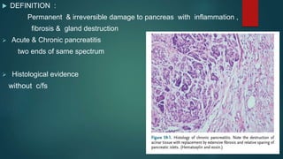
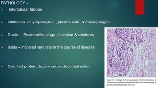
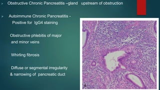
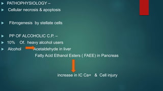
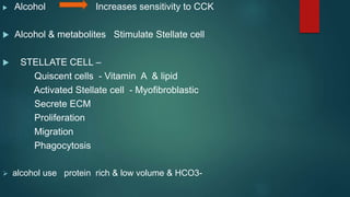
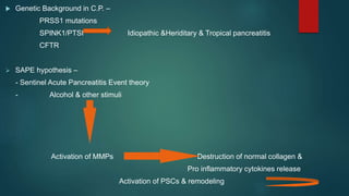
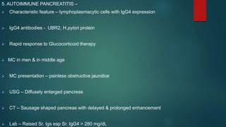
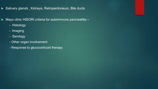
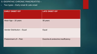
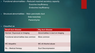
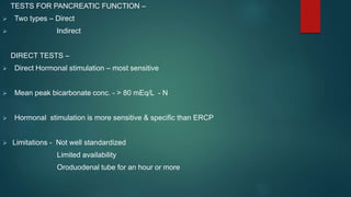
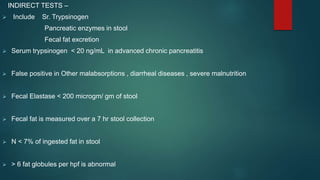
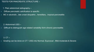
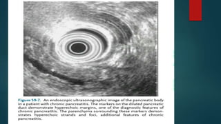
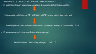
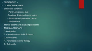
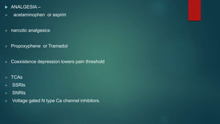
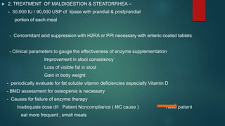
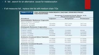
CHRONIC
PANCREATITIS
 DEFINITION
 EPIDEMIOLOGY
 PATHOLOGY
 PATHOPHYSIOLOGY
 ETIOLOGY
 CLINICAL FEATURES
 COMPLICATIONS
 PHYSICAL EXAMINATION
 DIAGNOSIS
 TREATMENT
 DEFINITION :
Permanent & irreversible damage to pancreas with inflammation ,
fibrosis & gland destruction
 Acute & Chronic pancreatitis
two ends of same spectrum
 Histological evidence
without c/fs
EPIDEMIOLOGY –
 Alcohol abuse – 2/3rd of cases
 Geographic variation
 Mortality Rate – 3.6
 Continued alcohol use – increased mortality risk by 60%
 Cause of death – conditions
associated with smoking , alcoholism , pancreatic cancer
PATHOLOGY –
 Interlobular fibrosis
 Infiltration of lymphocytes , plasma cells & macrophages
 Ducts – Eosinophilic plugs , dialation & strictures
 Islets – involved very late in the course of disease
 Calcified protein plugs – cause duct obstruction
 Obstructive Chronic Pancreatitis –gland upstream of obstruction
 Autoimmune Chronic Pancreatitis -
Positive for IgG4 staining
Obstructive phlebitis of major
and minor veins
Whirling fibrosis
Diffuse or segmental irregularity
& narrowing of pancreatic duct
 PATHOPHYSIOLOGY –
 Cellular necrosis & apoptosis
 Fibrogenesis by stellate cells
 PP OF ALCOHOLIC C.P. –
 10% Of. heavy alcohol users
 Alcohol Acetaldehyde in liver
Fatty Acid Ethanol Esters ( FAEE) in Pancreas
increase in IC Ca+ & Cell injury
 Alcohol Increases sensitivity to CCK
 Alcohol & metabolites Stimulate Stellate cell
 STELLATE CELL –
Quiscent cells - Vitamin A & lipid
Activated Stellate cell - Myofibroblastic
Secrete ECM
Proliferation
Migration
Phagocytosis
 alcohol use protein rich & low volume & HCO3-
 Genetic Background in C.P. –
PRSS1 mutations
SPINK1/PTSI Idiopathic &Heriditary & Tropical pancreatitis
CFTR
 SAPE hypothesis –
- Sentinel Acute Pancreatitis Event theory
- Alcohol & other stimuli
Activation of MMPs Destruction of normal collagen &
Pro inflammatory cytokines release
Activation of PSCs & remodeling
 TIGAR –O CLASSIFICATION SYSTEM FOR ETIOLOGY OF CHRONIC PANCREATITIS –
- Toxic – Metabolic
Alcohol
Tobacco Smoking
Hypercalcemia
Hyperlipidemia
- Idiopathic
Early onset
Late onset
Tropical
- Genetic
PRSS1 , CFTR , CASR, CTRC, SPINK1 gene mutations
- Autoimmune
Type 1
Type 2
- Recurrent , Severe Acute Pancreatitis
Post necrotic
Vascular diseases
Radiation
- Obstructive
Pancreas Divisum
Duct Obstruction
Duodenal wall cysts
duct scars
ETIOLOGY –
1. ALCOHOL –
 5 years of intake of >150gm/day
 Cofactors - Smoking ,
Genetic polymorphisms ,
Protein & fat rich diet
trace element deficiency
 Recurrent acute pancreatitis for 5-6years
 Prognosis - poor
2. TOBACCO –
 Independent risk factor & increases r/o pancreatic cancer
3. TROPICAL PANCREATITIS –
 Mean age – 24years
 Classical – pain abdomen , severe malnutrition , endocrine & exocrine insufficiency
 endocrine insufficiency - inevitable
 Steatorrhea – rare
 Mutations in SPINK1 & Chymotrypsinogen gene
 environmental triggers
Cyanogenic glycosides in cassava
Trace element deficiency
Oxidative stress
PEM
4. GENETIC –
 PRSS1/Cationic Trypsinogen Gene
 Rest - cofactors or modifier genes
5. AUTOIMMUNE PANCREATITIS –
 Characteristic feature – lymphoplasmacytic cells with IgG4 expression
 IgG4 antibodies - UBR2, H.pylori protein
 Rapid response to Glucocorticoid therapy
 MC in men & in middle age
 MC presentation – painless obstructive jaundice
 USG – Diffusely enlarged pancreas
 CT – Sausage shaped pancreas with delayed & prolonged enhancement
 Lab – Raised Sr. Igs esp Sr. IgG4 > 280 mg/dL
 Salivary glands , Kidneys, Retroperitoneum, Bile ducts
 Mayo clinic HISORt criteria for autoimmune pancreatitis –
- Histology
- Imaging
- Serology
- Other organ involvement
- Response to glucocorticoid therapy
6. OBSTRUCTIVE CHRONIC PANCREATTITS –
 Tumors , Scars , Stones, Strictures
 Pancreas divisum
7. RECURRENT OR SEVERE ACUTE PANCREATITIS –
 Pancreatic necrosis or Surgical debridement
 Hypertriglyceridemia >1000mg/dL
8. ASYMPTOMATIC PANCREATIC FIBROSIS –
 Elderly,
 Chronic renal failure ,
 hemodialysis
9. IDIOPATHIC CHRONIC PANCREATITIS –
 Two types – Early onset & Late onset
EARLY ONSET ICP LATE ONSET ICP
Mean Age – 20 years 56 years
Gender Distribution – Equal Equal
Predominant c/f – Pain Exocrine & endocrine insufficiency
CLINICAL FEATURES OF CHRONIC PANCREATITIS –
 Abdominal Pain
 Steatorrhea
 DM
ABDOMINAL PAIN –
 Epigastric,
 deep boring & penetrating
 worsened after a meal ,
 relieved by sitting or leaning forward ,assuming knee chest position on one side & clasping knees onto
chest
 Causes of pain –
1. tissue ischemia
2. Altered peripheral & central nociception
3. Complications
4. Hyperstimulation by CCK
STEATORRHEA –
 lipase secretion is reduced to <10% of maximal output
 Bulky foul smelling stools or passage of frank oil droplets
 Vs small bowel diarrhea – No Watery diarrhea & abdominal cramps
 Fat maldigestion earlier & severe compared to protein & carbohydrate maldigestion
R – 1. Lipase secretion decreases earlier
2. Lipase more sensitive to acid destruction & pancreatic proteases
3. Bile salt precipitation
 Median time for development of exocrine insufficiency – 13 – 26 years
 Weight loss – less common
 Weight loss can occur with
1. painful flares
2. Small bowel bacterial overgrowth
3. Pancreatic or extrapancreatic malignancy
 Fat soluble vitamin deficiencies – esp. Vit D def
 DIABETES MELLITUS –
 MC after pancreatic resection & tropical pancreatitis
 Vs Type 1 or Type 2 DM –
1. Loss of hepatocyte insulin receptor Decreased hepatic
glucose output d/t IAPP
2. Decrease in stimulated glucagon secretion due to coexistent alpha cell injury
prolonged & severe hypoglycemia with insulin Rx
 Mean time to development of DM - 19 – 26 years
 COMPLICATIONS OF CHRONIC PANCREATITIS –
- Chronic Abdominal Pain
- DM / IGT
- Gastroparesis
- Malabsorption
- GI bleeding
- Cholestatic Jaundice
- Biliary stricture / Biliary Cirrhosis
- Metabolic bone disease
- Pancreatic Fistulas
- Pancreatic Cancer
PHYSICAL EXAMINATION IN CHRONIC PANCREATITIS –
 Abdominal tenderness
 Weight loss
 Jaundice
 Palpable spleen
 Coexistent autoimmune features
DIAGNOSIS OF CHRONIC PANCREATITIS –
 Two categories
1. Tests for pancreatic function
2. Tests for pancreatic structure
 All diagnostic tests more accurate in far advanced disease
 Functional abnormalities – Reduced maximal secretory capacity
Exocrine insufficiency
Endocrine insufficiency
 Structural abnormalities - Main pancreatic duct
Side branches
Parenchyma
 Classified as
Small duct disease Big duct disease
Normal / Equivocal on Imaging Abnormalities in duct on imaging
Functional abnormalities less common More common
MC Idiopathic MC d/t Alcohol abuse
Rx – Medical therapy Duct Decompression
TESTS FOR PANCREATIC FUNCTION –
 Two types – Direct
 Indirect
DIRECT TESTS –
 Direct Hormonal stimulation – most sensitive
 Mean peak bicarbonate conc. - > 80 mEq/L - N
 Hormonal stimulation is more sensitive & specific than ERCP
 Limitations - Not well standardized
Limited availability
Oroduodenal tube for an hour or more
INDIRECT TESTS –
 Include Sr. Trypsinogen
Pancreatic enzymes in stool
Fecal fat excretion
 Serum trypsinogen < 20 ng/mL in advanced chronic pancreatitis
 False positive in Other malabsorptions , diarrheal diseases , severe malnutrition
 Fecal Elastase < 200 microgm/ gm of stool
 Fecal fat is measured over a 7 hr stool collection
 N < 7% of ingested fat in stool
 > 6 fat globules per hpf is abnormal
TESTS FOR PANCREATIC STRUCTURE –
1. Plain abdominal radiography –
 Diffuse pancreatic calcification is specific
 MC in alcoholic , late onset idiopathic , hereditary , tropical pancreatitis
2. Abdominal USG –
 Difficult to distinguish age related variability form chronic pancreatitis
3. CT –
 Grading can be done on CT / USG into Normal, Equivocal , Mild-moderate & Severe
 Findings in chronic pancreatitis – Duct dilatation
Duct obstruction
Parenchymal heterogenity
Irregular contour
Calcification
4. MRI –
 Duct Visualisation improved by secertin administration
5. ERCP –
 Diagnostic as well as therapeutic
 Risk - 5%
 Diagnosis based on abnormalities in main pancreatic duct & side branches
 CHAIN OF LAKES Appearance
 Changes similar can also be seen with normal ageing
 CAMBRIDGE GRADING OF CHRONIC PANCREATITIS ON ERCP
GRADE MAIN PANCREATIC DUCT SIDE BRANCHES
NORMAL Normal Normal
EQUIVOCAL Normal <3 Abnormal
MILD Normal >/= 3 Abnormal
MODERATE Abnormal “
SEVERE Abnormal with
Large cavity >10mm)
Obstruction
Filling defects
Severe dilatation /
irregularity
“
6. ENDOSCOPIC USG –
 Presence of >/= 3 features is positive
 Rosemont criteria -
 Score >5 highly specific
 Score of 3 or 4 is indeterminate
 EUS abnormalities in chronic pancreatitis -
PARENCHYMAL ABNORMALITIES DUCTAL ABNORMALITIES
Hyperechoic foci Main duct dilatation & irregularity
Hyperechoic strands Hyperechoic ductal walls
Lobularity of contour Visible side branches
Cysts Calcification
DIAGNOSTIC STRATEGY IN CHRONIC PANCREATITIS –
 In patients with pain as primary complaint & suspected chronic pancreatitis
High quality multidetector CT / MRI With MRCP is best initial diagnostic test
If nondiagnostic , Hormal stimulation direct pancreatic testing , If unavailable , EUS
 If exocrine or endocrine insufficiency is suspected
Fecal Elastase / Serum Trypsinogen / USG / CT
 TREATMENT -
 1. ABDOMINAL PAIN
 associated conditions
- Pancreatic pseudo cyst
- Duodenal & bile duct compression
- Superimposed pancreatic cancer
- Gastroparesis
 Identify patients with big duct pancreatitis
 MEDICAL THERAPY –
1. Analgesics
2. Cessation of Alcohol & Tobacco
3. Antioxidants
4. Pancreatic enzyme therapy
5. Octreotide
 ANALGESIA –
 acetaminophen or aspirin
 narcotic analgesics
 Propoxyphene or Tramadol
 Coexistence depression lowers pain threshold
 TCAs
 SSRIs
 SNRIs
 Voltage gated N type Ca channel inhibitors.
 CESSATION OF ALCOHOL & TOBACCO
 ANTIOXIDANTS –
- Selenium , beta carotene , Vitamin C, Vitamin E & methionine
 PANCREATIC ENZYME THERAPY –
- deliver proteases to duodenum or very proximal jejunum
- Enteric coated preparations may not release majority of proteases until they reach the
more distal small bowel
- Non Enteric coated preparations
- Non enteric coated enzymes cinactivated by gastric acid , concomitant use of
acid suppressant
- Enzyme therapy is not successful in pain a/w big duct chronic pancreatitis
 OCTREOTIDE –
- Reduces pancreatic secretion & circulating CCK levels
- It also has some direct anti nociceptive effect
 ENDOSCOPIC THERAPY –
- Most appropriate in patient with dominant stricture & ductal stone
- Options include
Pancreatic duct Sphincterotomy
Stent Placement
Pancreatic duct stone removal
I
 SURGICAL THERAPY –
- Considered for intractable pain abdomen with failed medical therapy
- Other indications
Other organ involvement
Failure of endoscopic therapy for pseudo cysts
Internal pancreatic fistulas
Exclusion of malignancy
- For pain , Pancreatic duct drainage
Resection of all or part of pancreas
 NERVE BLOCK & NEUROLYSIS –
- Celiac plexus block with a glucocorticoid & LA Under
- Celiac plexus neurolysis with absolute alcohol EUS or CT guidance
 2. TREATMENT OF MALDIGESTION & STEATORRHEA –
- 30,000 IU / 90,000 USP of lipase with prandial & postprandial
portion of each meal
- Concomitant acid suppression with H2RA or PPI necessary with enteric coated tablets
- Clinical parameters to gauge the effectiveness of enzyme supplementation
Improvement in stool consistency
Loss of visible fat in stool
Gain in body weight
- periodically evaluate for fat soluble vitamin deficiencies especially Vitamin D
- BMD assessment for osteopenia is necessary
- Causes for failure of enzyme therapy
Inadequate dose d/t Patient Noncompliance ( MC cause ) Have patient
eat more frequent , small meals
- If. fail , search for an alternative cause for malabsorption
- If all measures fail , replace diet fat with medium chain TGs
 3. TREATMENT OF DIABETES MELLITUS –
- Therapy is directed at control of urinary losses of glucose
- Insulin requirement is lower than that with T1 DM patients
- Overvigorous BG control are a/w hypoglycemia
- Tight BG control are indicated in patients with hyperlipidemic
pancreatitis
 TREATMENT OF AUTOIMMUNE PANCREATITIS –
- Glucocorticoids have clinical , histolopathological & morphological efficacy
- Start with Prednisone at 40 mg/ day for 4 weeks
- Parameters to be followed include
Symptomatic relief
Serial changes in pancreas & bile ducts on imaging
Reduced serum gamma globulin & IgG4 levels
Improvement in liver tests
- Poor response to GCs in 2 to 4 weeks should raise the suspicion of pancreatic cancer or
other causes of Chronic pancreatitis
- For relapse , agents useful are
6- Mercaptapurine , Azathioprine , Rituximab, Cyclosporine & Cyclophosphamide
 REFERENCES –
- Sleisenger & Fordtran GI and Liver Disease , 9th Ed
- Harrison Principles of Internal Medicine 19th Ed
THANK YOU

Chronic pancreatitis seminar

  • 1.
  • 2.
     DEFINITION  EPIDEMIOLOGY PATHOLOGY  PATHOPHYSIOLOGY  ETIOLOGY  CLINICAL FEATURES  COMPLICATIONS  PHYSICAL EXAMINATION  DIAGNOSIS  TREATMENT
  • 3.
     DEFINITION : Permanent& irreversible damage to pancreas with inflammation , fibrosis & gland destruction  Acute & Chronic pancreatitis two ends of same spectrum  Histological evidence without c/fs
  • 6.
    EPIDEMIOLOGY –  Alcoholabuse – 2/3rd of cases  Geographic variation  Mortality Rate – 3.6  Continued alcohol use – increased mortality risk by 60%  Cause of death – conditions associated with smoking , alcoholism , pancreatic cancer
  • 7.
    PATHOLOGY –  Interlobularfibrosis  Infiltration of lymphocytes , plasma cells & macrophages  Ducts – Eosinophilic plugs , dialation & strictures  Islets – involved very late in the course of disease  Calcified protein plugs – cause duct obstruction
  • 8.
     Obstructive ChronicPancreatitis –gland upstream of obstruction  Autoimmune Chronic Pancreatitis - Positive for IgG4 staining Obstructive phlebitis of major and minor veins Whirling fibrosis Diffuse or segmental irregularity & narrowing of pancreatic duct
  • 9.
     PATHOPHYSIOLOGY – Cellular necrosis & apoptosis  Fibrogenesis by stellate cells  PP OF ALCOHOLIC C.P. –  10% Of. heavy alcohol users  Alcohol Acetaldehyde in liver Fatty Acid Ethanol Esters ( FAEE) in Pancreas increase in IC Ca+ & Cell injury
  • 10.
     Alcohol Increasessensitivity to CCK  Alcohol & metabolites Stimulate Stellate cell  STELLATE CELL – Quiscent cells - Vitamin A & lipid Activated Stellate cell - Myofibroblastic Secrete ECM Proliferation Migration Phagocytosis  alcohol use protein rich & low volume & HCO3-
  • 11.
     Genetic Backgroundin C.P. – PRSS1 mutations SPINK1/PTSI Idiopathic &Heriditary & Tropical pancreatitis CFTR  SAPE hypothesis – - Sentinel Acute Pancreatitis Event theory - Alcohol & other stimuli Activation of MMPs Destruction of normal collagen & Pro inflammatory cytokines release Activation of PSCs & remodeling
  • 12.
     TIGAR –OCLASSIFICATION SYSTEM FOR ETIOLOGY OF CHRONIC PANCREATITIS – - Toxic – Metabolic Alcohol Tobacco Smoking Hypercalcemia Hyperlipidemia - Idiopathic Early onset Late onset Tropical - Genetic PRSS1 , CFTR , CASR, CTRC, SPINK1 gene mutations
  • 13.
    - Autoimmune Type 1 Type2 - Recurrent , Severe Acute Pancreatitis Post necrotic Vascular diseases Radiation - Obstructive Pancreas Divisum Duct Obstruction Duodenal wall cysts duct scars
  • 14.
    ETIOLOGY – 1. ALCOHOL–  5 years of intake of >150gm/day  Cofactors - Smoking , Genetic polymorphisms , Protein & fat rich diet trace element deficiency  Recurrent acute pancreatitis for 5-6years  Prognosis - poor 2. TOBACCO –  Independent risk factor & increases r/o pancreatic cancer
  • 15.
    3. TROPICAL PANCREATITIS–  Mean age – 24years  Classical – pain abdomen , severe malnutrition , endocrine & exocrine insufficiency  endocrine insufficiency - inevitable  Steatorrhea – rare  Mutations in SPINK1 & Chymotrypsinogen gene  environmental triggers Cyanogenic glycosides in cassava Trace element deficiency Oxidative stress PEM 4. GENETIC –  PRSS1/Cationic Trypsinogen Gene  Rest - cofactors or modifier genes
  • 16.
    5. AUTOIMMUNE PANCREATITIS–  Characteristic feature – lymphoplasmacytic cells with IgG4 expression  IgG4 antibodies - UBR2, H.pylori protein  Rapid response to Glucocorticoid therapy  MC in men & in middle age  MC presentation – painless obstructive jaundice  USG – Diffusely enlarged pancreas  CT – Sausage shaped pancreas with delayed & prolonged enhancement  Lab – Raised Sr. Igs esp Sr. IgG4 > 280 mg/dL
  • 17.
     Salivary glands, Kidneys, Retroperitoneum, Bile ducts  Mayo clinic HISORt criteria for autoimmune pancreatitis – - Histology - Imaging - Serology - Other organ involvement - Response to glucocorticoid therapy
  • 18.
    6. OBSTRUCTIVE CHRONICPANCREATTITS –  Tumors , Scars , Stones, Strictures  Pancreas divisum 7. RECURRENT OR SEVERE ACUTE PANCREATITIS –  Pancreatic necrosis or Surgical debridement  Hypertriglyceridemia >1000mg/dL 8. ASYMPTOMATIC PANCREATIC FIBROSIS –  Elderly,  Chronic renal failure ,  hemodialysis
  • 19.
    9. IDIOPATHIC CHRONICPANCREATITIS –  Two types – Early onset & Late onset EARLY ONSET ICP LATE ONSET ICP Mean Age – 20 years 56 years Gender Distribution – Equal Equal Predominant c/f – Pain Exocrine & endocrine insufficiency
  • 20.
    CLINICAL FEATURES OFCHRONIC PANCREATITIS –  Abdominal Pain  Steatorrhea  DM
  • 21.
    ABDOMINAL PAIN – Epigastric,  deep boring & penetrating  worsened after a meal ,  relieved by sitting or leaning forward ,assuming knee chest position on one side & clasping knees onto chest  Causes of pain – 1. tissue ischemia 2. Altered peripheral & central nociception 3. Complications 4. Hyperstimulation by CCK
  • 22.
    STEATORRHEA –  lipasesecretion is reduced to <10% of maximal output  Bulky foul smelling stools or passage of frank oil droplets  Vs small bowel diarrhea – No Watery diarrhea & abdominal cramps  Fat maldigestion earlier & severe compared to protein & carbohydrate maldigestion R – 1. Lipase secretion decreases earlier 2. Lipase more sensitive to acid destruction & pancreatic proteases 3. Bile salt precipitation  Median time for development of exocrine insufficiency – 13 – 26 years  Weight loss – less common
  • 23.
     Weight losscan occur with 1. painful flares 2. Small bowel bacterial overgrowth 3. Pancreatic or extrapancreatic malignancy  Fat soluble vitamin deficiencies – esp. Vit D def
  • 24.
     DIABETES MELLITUS–  MC after pancreatic resection & tropical pancreatitis  Vs Type 1 or Type 2 DM – 1. Loss of hepatocyte insulin receptor Decreased hepatic glucose output d/t IAPP 2. Decrease in stimulated glucagon secretion due to coexistent alpha cell injury prolonged & severe hypoglycemia with insulin Rx  Mean time to development of DM - 19 – 26 years
  • 25.
     COMPLICATIONS OFCHRONIC PANCREATITIS – - Chronic Abdominal Pain - DM / IGT - Gastroparesis - Malabsorption - GI bleeding - Cholestatic Jaundice - Biliary stricture / Biliary Cirrhosis - Metabolic bone disease - Pancreatic Fistulas - Pancreatic Cancer
  • 27.
    PHYSICAL EXAMINATION INCHRONIC PANCREATITIS –  Abdominal tenderness  Weight loss  Jaundice  Palpable spleen  Coexistent autoimmune features
  • 28.
    DIAGNOSIS OF CHRONICPANCREATITIS –  Two categories 1. Tests for pancreatic function 2. Tests for pancreatic structure  All diagnostic tests more accurate in far advanced disease
  • 29.
     Functional abnormalities– Reduced maximal secretory capacity Exocrine insufficiency Endocrine insufficiency  Structural abnormalities - Main pancreatic duct Side branches Parenchyma  Classified as Small duct disease Big duct disease Normal / Equivocal on Imaging Abnormalities in duct on imaging Functional abnormalities less common More common MC Idiopathic MC d/t Alcohol abuse Rx – Medical therapy Duct Decompression
  • 30.
    TESTS FOR PANCREATICFUNCTION –  Two types – Direct  Indirect DIRECT TESTS –  Direct Hormonal stimulation – most sensitive  Mean peak bicarbonate conc. - > 80 mEq/L - N  Hormonal stimulation is more sensitive & specific than ERCP  Limitations - Not well standardized Limited availability Oroduodenal tube for an hour or more
  • 31.
    INDIRECT TESTS – Include Sr. Trypsinogen Pancreatic enzymes in stool Fecal fat excretion  Serum trypsinogen < 20 ng/mL in advanced chronic pancreatitis  False positive in Other malabsorptions , diarrheal diseases , severe malnutrition  Fecal Elastase < 200 microgm/ gm of stool  Fecal fat is measured over a 7 hr stool collection  N < 7% of ingested fat in stool  > 6 fat globules per hpf is abnormal
  • 32.
    TESTS FOR PANCREATICSTRUCTURE – 1. Plain abdominal radiography –  Diffuse pancreatic calcification is specific  MC in alcoholic , late onset idiopathic , hereditary , tropical pancreatitis 2. Abdominal USG –  Difficult to distinguish age related variability form chronic pancreatitis 3. CT –  Grading can be done on CT / USG into Normal, Equivocal , Mild-moderate & Severe
  • 33.
     Findings inchronic pancreatitis – Duct dilatation Duct obstruction Parenchymal heterogenity Irregular contour Calcification 4. MRI –  Duct Visualisation improved by secertin administration 5. ERCP –  Diagnostic as well as therapeutic  Risk - 5%  Diagnosis based on abnormalities in main pancreatic duct & side branches
  • 35.
     CHAIN OFLAKES Appearance  Changes similar can also be seen with normal ageing  CAMBRIDGE GRADING OF CHRONIC PANCREATITIS ON ERCP GRADE MAIN PANCREATIC DUCT SIDE BRANCHES NORMAL Normal Normal EQUIVOCAL Normal <3 Abnormal MILD Normal >/= 3 Abnormal MODERATE Abnormal “ SEVERE Abnormal with Large cavity >10mm) Obstruction Filling defects Severe dilatation / irregularity “
  • 36.
    6. ENDOSCOPIC USG–  Presence of >/= 3 features is positive  Rosemont criteria -  Score >5 highly specific  Score of 3 or 4 is indeterminate  EUS abnormalities in chronic pancreatitis - PARENCHYMAL ABNORMALITIES DUCTAL ABNORMALITIES Hyperechoic foci Main duct dilatation & irregularity Hyperechoic strands Hyperechoic ductal walls Lobularity of contour Visible side branches Cysts Calcification
  • 38.
    DIAGNOSTIC STRATEGY INCHRONIC PANCREATITIS –  In patients with pain as primary complaint & suspected chronic pancreatitis High quality multidetector CT / MRI With MRCP is best initial diagnostic test If nondiagnostic , Hormal stimulation direct pancreatic testing , If unavailable , EUS  If exocrine or endocrine insufficiency is suspected Fecal Elastase / Serum Trypsinogen / USG / CT
  • 39.
     TREATMENT - 1. ABDOMINAL PAIN  associated conditions - Pancreatic pseudo cyst - Duodenal & bile duct compression - Superimposed pancreatic cancer - Gastroparesis  Identify patients with big duct pancreatitis  MEDICAL THERAPY – 1. Analgesics 2. Cessation of Alcohol & Tobacco 3. Antioxidants 4. Pancreatic enzyme therapy 5. Octreotide
  • 40.
     ANALGESIA – acetaminophen or aspirin  narcotic analgesics  Propoxyphene or Tramadol  Coexistence depression lowers pain threshold  TCAs  SSRIs  SNRIs  Voltage gated N type Ca channel inhibitors.
  • 41.
     CESSATION OFALCOHOL & TOBACCO  ANTIOXIDANTS – - Selenium , beta carotene , Vitamin C, Vitamin E & methionine
  • 42.
     PANCREATIC ENZYMETHERAPY – - deliver proteases to duodenum or very proximal jejunum - Enteric coated preparations may not release majority of proteases until they reach the more distal small bowel - Non Enteric coated preparations - Non enteric coated enzymes cinactivated by gastric acid , concomitant use of acid suppressant - Enzyme therapy is not successful in pain a/w big duct chronic pancreatitis
  • 43.
     OCTREOTIDE – -Reduces pancreatic secretion & circulating CCK levels - It also has some direct anti nociceptive effect  ENDOSCOPIC THERAPY – - Most appropriate in patient with dominant stricture & ductal stone - Options include Pancreatic duct Sphincterotomy Stent Placement Pancreatic duct stone removal I
  • 44.
     SURGICAL THERAPY– - Considered for intractable pain abdomen with failed medical therapy - Other indications Other organ involvement Failure of endoscopic therapy for pseudo cysts Internal pancreatic fistulas Exclusion of malignancy - For pain , Pancreatic duct drainage Resection of all or part of pancreas  NERVE BLOCK & NEUROLYSIS – - Celiac plexus block with a glucocorticoid & LA Under - Celiac plexus neurolysis with absolute alcohol EUS or CT guidance
  • 45.
     2. TREATMENTOF MALDIGESTION & STEATORRHEA – - 30,000 IU / 90,000 USP of lipase with prandial & postprandial portion of each meal - Concomitant acid suppression with H2RA or PPI necessary with enteric coated tablets - Clinical parameters to gauge the effectiveness of enzyme supplementation Improvement in stool consistency Loss of visible fat in stool Gain in body weight - periodically evaluate for fat soluble vitamin deficiencies especially Vitamin D - BMD assessment for osteopenia is necessary - Causes for failure of enzyme therapy Inadequate dose d/t Patient Noncompliance ( MC cause ) Have patient eat more frequent , small meals
  • 46.
    - If. fail, search for an alternative cause for malabsorption - If all measures fail , replace diet fat with medium chain TGs
  • 47.
     3. TREATMENTOF DIABETES MELLITUS – - Therapy is directed at control of urinary losses of glucose - Insulin requirement is lower than that with T1 DM patients - Overvigorous BG control are a/w hypoglycemia - Tight BG control are indicated in patients with hyperlipidemic pancreatitis
  • 48.
     TREATMENT OFAUTOIMMUNE PANCREATITIS – - Glucocorticoids have clinical , histolopathological & morphological efficacy - Start with Prednisone at 40 mg/ day for 4 weeks - Parameters to be followed include Symptomatic relief Serial changes in pancreas & bile ducts on imaging Reduced serum gamma globulin & IgG4 levels Improvement in liver tests - Poor response to GCs in 2 to 4 weeks should raise the suspicion of pancreatic cancer or other causes of Chronic pancreatitis - For relapse , agents useful are 6- Mercaptapurine , Azathioprine , Rituximab, Cyclosporine & Cyclophosphamide
  • 49.
     REFERENCES – -Sleisenger & Fordtran GI and Liver Disease , 9th Ed - Harrison Principles of Internal Medicine 19th Ed
  • 50.