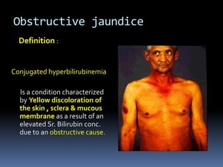
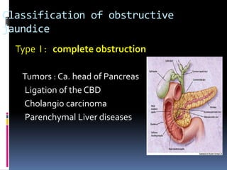
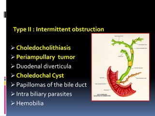
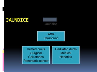
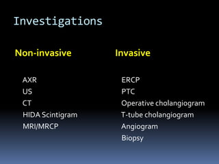
OBSTRUCTIVE JAUNDICE
Obstructive jaundice
Definition :
Conjugated hyperbilirubinemia
Is a condition characterized
by Yellow discoloration of
the skin , sclera & mucous
membrane as a result of an
elevated Sr. Bilirubin conc.
due to an obstructive cause.
Classification of obstructive
jaundice
Type I : complete obstruction
Tumors : Ca. head of Pancreas
Ligation of the CBD
Cholangio carcinoma
Parenchymal Liver diseases
Type II : Intermittent obstruction
 Choledocholithiasis
 Periampullary tumor
 Duodenal diverticula
 Choledochal Cyst
 Papillomas of the bile duct
 Intra biliary parasites
 Hemobilia
TYPE III : Chronic incomplete
obstruction
 Strictures of the CBD
Congenital
Traumatic
Sclerosing cholangitis
Post radiotherapy
 Stenosed biliary enteric
anastamosis
 Cystic fibrosis
 Chronic pancreatitis
 Stenosis of the Sphincter of
Oddi
ERCP showing distal common bile duct
stricture with proximal dilation
TYPE IV : Segmental Obstruction
 Traumatic
 Hepatodocholithiasis
 Sclerosing cholangitis
 Cholangio carcinoma
PATHOPHYSIOLOGY OF OBSTRUCTION
Alterations in
– Systemic and renal hemodynamics
– Hepatic function
– Hemostatic mechanism
– Gastrointestinal barrier
– Immune function
–Wound healing
• Protein synthesis,
• Reticulo-endothelial function
• Hepatic metabolism
Coagulation system
 Prolonged bile duct obstruction leads to significant
defects in clotting factors
 Before surgery these defects should be corrected by
Fresh frozen plasma andVitamin K
 Even if there is no measurable coagulation
dysfunctionVitamin K should be given to all patients
with obstructive jaundice
Abnormal LFTs
Obstruction Hepatitis Cirrhosis
Bilirubin
  
Alk phos
  /  /
ALT/AST
/   /
gGT
   /
PT (INR)
  
• Fever, persistent (90%)
• Abdominal pain (70%)
• Jaundice (60%)
•Tea-colored urine/pale stools
• Altered mental status (10-20%)
• Hypotension (30%)
• RUQ tenderness
reliable signs & symptoms 90% certainty)
a patient need urgent intervention ?
 Obstructive Jaundice
• Relief of Obstruction
• Prevent Complication
• Prevent Recurrence
Goal ofTreatment
Are the ducts dilated
What is the level of obstruction
What is the cause
What is the best therapeutic approach
The role of Radiology
JAUNDICE
Jaundice
Dilated ducts
Surgical
Gall stones
Pancreatic cancer
Undilated ducts
Medical
Hepatitis
AXR
Ultrasound
Investigations
Non-invasive
AXR
US
CT
HIDA Scintigram
MRI/MRCP
Invasive
ERCP
PTC
Operative cholangiogram
T-tube cholangiogram
Angiogram
Biopsy
Obstructive Jaundice
CBD stones (Choledocholithiasis) vs. tumor
 Clinical features favoring CBD stones:
 Age < 45
 Biliary colic
 Fever
 Transient spike in AST or amylase
 Clinical features favoring cancer:
 Painless jaundice
 Weight loss
 Palpable gallbladder
 Bilirubin > 10
Unconjugated vs. Conjugated
Unconjugated
  production exceeds
ability of liver to
conjugate
Ex. Hemolytic anemia's,
hemoglobinopathies,
in-born errors of
metab., transfusion
rxn.
Conjugated
 Can produce but not
excrete
 Intra- or extra hepatic
obstruction Metabolic
defect
Choledocholithiasis
Defined as stones in the CBD
Patho physiology : intermittent obstruction of CBD
 Often asymptomatic
 Symptoms are indistinguishable from other causes of
Biliary pain
 Predisposes to Cholangitis & Acute Pancreatitis
 Elevated sr. bilirubin & Alk. Phos.
Evaluation
ERCP
 Primary diagnostic and
therapeutic modality
 Sphincterotomy and stone
extraction
 Placement of stent if
stone extraction
unsuccessful
 Mortality rate 1.5%
Open CBD Exploration
Indications
 Presence of multiple stones (more than 5)
Stones > 1 cm
 Multiple intra hepatic stones
 Distal bile duct strictures
 Failure of ERCP
 Recurrence of CBD stones after sphincterotomy
CBD Exploration – Surgical Options
 Common bile duct exploration withT-tube
decompression
 Choledochoduodenostomy
 Transduodenal sphincterotomy and
sphincterplasty
 Roux-en-Y Choledocho jejunostomy
CHOLEDOCHAL CYSTS
 Congenital anomalies of the biliary tract that
manifest as cystic dilatation of the extra hepatic and
intra hepatic bile ducts
 Females are most commonly affected
ETIOLOGY :
 Congenital weakness of the bile duct wall
 Congenital obstruction of the bile ducts
 Reo virus association is seen in 78% of patients
 40% of anomalies are seen at the junction of
pancreatic and common bile ducts
CLASSIFICATION OF CHOLEDOCHAL CYST
 Proposed byTodani & colleagues
 TYPE I : accounts for 80 – 90 % of cases
exhibit segmental or diffuse fusiform dilatation of the CBD.
 TYPE II : consists of a true Choledochal diverticulum
 TYPE III : consists of dilatation of the intra duodenal portion of the CBD.
 TYPE IV : multiple intra hepatic & extra hepatic cysts
 TYPEV or CAROLIS disease : consists of single or multiple dilatation of
the intra hepatic ductal system
Clinical features :
 Disease often appears during first months of life
 80% of pts. have cholestatic jaundice & acholic stools
 Vomiting , irritability & failure to thrive may occur
 Spontaneous perforation of a Choledochal cysts may occur
 Progressive hepatic injury due to biliary obstruction
DIAGNOSIS :
 BEST established by USG Abdomen
 In Older children PTC or ERCP may help define the anatomy
of the cyst.
TREATMENT
 Surgical excision of the cyst with Reconstruction
of the extra hepatic biliary tree
 Biliary drainage is accomplished by Choledocho
– jejunostomy with a Roux – en –Y anastamosis
 Long term follow up is necessary because of
complications like cholangitis , lithiasis ,
anastomotic stricture
Cholangiocarcinoma
 90% are extra-hepatic
 60’s and 70’s
 Highest incidence in Japan, Israel, and Native
Americans
 Increased 3 fold in the last 30yrs in the USA
 M/F=3/2
Cholangiocarcinoma
Etiology
Ulcerative Colitis Thorotrast Exposure
Sclerosing Cholangitis Typhoid Carrier
Choledochal Cysts
Adult Polycystic Kidney
Disease
Hepatolithiasis
Liver Flukes
Papillomatosis of Bile
Ducts
Cholangiocarcinoma
Extra-hepatic: Distribution
 Right or left hepatic duct = 10%
 Bifurcation = 20%
 Proximal CBD = 30%
 Distal CBD = 30%
Cholangiocarcinoma
Diagnosis and Initial Workup
 Jaundice
 Wt loss, anorexia, abdominal pain, fever
 US then CT (CTA?) Followed by ERCP, PTC or
MRCP
 CEA and CA 19-9 can be elevated
Cholangiocarcinoma
Intra-hepatic Disease
 Suspicious mass on CT. Quadruple phase CT with 0.5 cm
cuts through the liver and portal hepatitis. Consider CTA
reconstruction.
Treatment
 If adenoncarcinoma: look for primary with a chest CT and
upper/lower endoscopy.
 Colon, pancreas, and stomach are common primary sites.
Cholangiocarcinoma
Intra-hepatic Disease-Surgery/Ablation
 Extent of surgical therapy is determined by the location,
hepatic function, and underlying cirrhosis.
 Anatomic resections have lowest recurrence rates.
However non anatomic resection increases potential
surgical candidates and improves survival
 Hepatic devascularization prior to resection is preferred
 Ablative therapy gives good local control.
MRCP of Extra-hepatic Cholangiocarcinoma at
the Bifurcation
Klatskin tumor
ERCP: Distal CBD Cancer
Ca of CBD Bifurcation
PERIAMPULLARY CARCINOMA AND
THE WHIPPLE
Endoscopic View
Pathology
 Adeno carcinoma accounts for 95%
Arises from 4 different tissues of origin
 Head of pancreas
 Distal Bile duct
 Ampullary ofVater
 Periampullary duodenum
Pathology
 Prognosis for each of these are different.
 Five year survival for pancreas: 18%
 Five year for ampulla: 36%
 Five year for distal bile duct: 34%
 Five year for duodenum: 33%
 Determination of tissue origin is important for
prognosis, extent of resection.
Pathology
 Determination of tissue origin from FNA,
endoscopic biopsy.
 Also from thin section CT scan, ERCP
 Determination of k-Ras also helps (95% of
pancreatic cancer).
Spread
 Loco regional spread results from lymphatic
invasion and direct tumor spread to adjacent
soft tissue.
 Ampullary lesions spread to LN 33%, typically
to a single LN in the posterior
pancreatcoduodenal group.
 Duodenal has intermediate spread.
 Pancreas metastasizes 88% to multiple sites.
Treatment
 StandardWhipple pancreaticoduodenectomy
thought to provide adequate tumor clearance in
the case of non-pancreatic ampullary tumor,
because tumor spread is localized.
 Biopsy proven paraduodenal LN is thought by
most to preclude curative resection
Surgery and Chemotherapy
 Low risk patients had 5 year local control and
survival of 100% and 80% respectively.
 High risk patients had 5 year local control and
survival of 50% and 38%, respectively.
 Based on these findings, some have proposed a
course of preoperative chemoradiation to
improve local disease control in these high risk
patients.
Whipple Procedure
Five basic techniques are used to resect pancreatic
cancers
 Standard pancreaticoduodenectomy
 Pylorus preserving pancreaticoduodenectomy
 Total pancreatectomy
 Regional pancreatectomy
 Extended resection (MD Anderson)
Kocherizing the Duodenum
SMA Involved?
SMV Identification
Dividing the Neck
The End Result
Adjuvant Therapy
 Autopsy series show that 85% of patients will
experience recurrence in operative field.
 70% have metastases to liver.
 So need to address local control (radiation) and
distant disease (chemotherapy).
 Most commonly used is 5 FU and this only has a
15-28% response on its own, but it’s a radio
sensitizer, so it improves response to chemo.
Obstructive Jaundice

Obstructive Jaundice

  • 1.
  • 2.
    Obstructive jaundice Definition : Conjugatedhyperbilirubinemia Is a condition characterized by Yellow discoloration of the skin , sclera & mucous membrane as a result of an elevated Sr. Bilirubin conc. due to an obstructive cause.
  • 3.
    Classification of obstructive jaundice TypeI : complete obstruction Tumors : Ca. head of Pancreas Ligation of the CBD Cholangio carcinoma Parenchymal Liver diseases
  • 4.
    Type II :Intermittent obstruction  Choledocholithiasis  Periampullary tumor  Duodenal diverticula  Choledochal Cyst  Papillomas of the bile duct  Intra biliary parasites  Hemobilia
  • 5.
    TYPE III :Chronic incomplete obstruction  Strictures of the CBD Congenital Traumatic Sclerosing cholangitis Post radiotherapy  Stenosed biliary enteric anastamosis  Cystic fibrosis  Chronic pancreatitis  Stenosis of the Sphincter of Oddi ERCP showing distal common bile duct stricture with proximal dilation
  • 6.
    TYPE IV :Segmental Obstruction  Traumatic  Hepatodocholithiasis  Sclerosing cholangitis  Cholangio carcinoma
  • 7.
    PATHOPHYSIOLOGY OF OBSTRUCTION Alterationsin – Systemic and renal hemodynamics – Hepatic function – Hemostatic mechanism – Gastrointestinal barrier – Immune function –Wound healing • Protein synthesis, • Reticulo-endothelial function • Hepatic metabolism
  • 8.
    Coagulation system  Prolongedbile duct obstruction leads to significant defects in clotting factors  Before surgery these defects should be corrected by Fresh frozen plasma andVitamin K  Even if there is no measurable coagulation dysfunctionVitamin K should be given to all patients with obstructive jaundice
  • 11.
    Abnormal LFTs Obstruction HepatitisCirrhosis Bilirubin    Alk phos   /  / ALT/AST /   / gGT    / PT (INR)   
  • 12.
    • Fever, persistent(90%) • Abdominal pain (70%) • Jaundice (60%) •Tea-colored urine/pale stools • Altered mental status (10-20%) • Hypotension (30%) • RUQ tenderness reliable signs & symptoms 90% certainty) a patient need urgent intervention ?
  • 13.
     Obstructive Jaundice •Relief of Obstruction • Prevent Complication • Prevent Recurrence Goal ofTreatment
  • 14.
    Are the ductsdilated What is the level of obstruction What is the cause What is the best therapeutic approach The role of Radiology
  • 15.
    JAUNDICE Jaundice Dilated ducts Surgical Gall stones Pancreaticcancer Undilated ducts Medical Hepatitis AXR Ultrasound
  • 16.
  • 17.
    Obstructive Jaundice CBD stones(Choledocholithiasis) vs. tumor  Clinical features favoring CBD stones:  Age < 45  Biliary colic  Fever  Transient spike in AST or amylase  Clinical features favoring cancer:  Painless jaundice  Weight loss  Palpable gallbladder  Bilirubin > 10
  • 18.
    Unconjugated vs. Conjugated Unconjugated  production exceeds ability of liver to conjugate Ex. Hemolytic anemia's, hemoglobinopathies, in-born errors of metab., transfusion rxn. Conjugated  Can produce but not excrete  Intra- or extra hepatic obstruction Metabolic defect
  • 19.
    Choledocholithiasis Defined as stonesin the CBD Patho physiology : intermittent obstruction of CBD  Often asymptomatic  Symptoms are indistinguishable from other causes of Biliary pain  Predisposes to Cholangitis & Acute Pancreatitis  Elevated sr. bilirubin & Alk. Phos.
  • 20.
    Evaluation ERCP  Primary diagnosticand therapeutic modality  Sphincterotomy and stone extraction  Placement of stent if stone extraction unsuccessful  Mortality rate 1.5%
  • 21.
    Open CBD Exploration Indications Presence of multiple stones (more than 5) Stones > 1 cm  Multiple intra hepatic stones  Distal bile duct strictures  Failure of ERCP  Recurrence of CBD stones after sphincterotomy
  • 23.
    CBD Exploration –Surgical Options  Common bile duct exploration withT-tube decompression  Choledochoduodenostomy  Transduodenal sphincterotomy and sphincterplasty  Roux-en-Y Choledocho jejunostomy
  • 24.
    CHOLEDOCHAL CYSTS  Congenitalanomalies of the biliary tract that manifest as cystic dilatation of the extra hepatic and intra hepatic bile ducts  Females are most commonly affected ETIOLOGY :  Congenital weakness of the bile duct wall  Congenital obstruction of the bile ducts  Reo virus association is seen in 78% of patients  40% of anomalies are seen at the junction of pancreatic and common bile ducts
  • 25.
    CLASSIFICATION OF CHOLEDOCHALCYST  Proposed byTodani & colleagues  TYPE I : accounts for 80 – 90 % of cases exhibit segmental or diffuse fusiform dilatation of the CBD.  TYPE II : consists of a true Choledochal diverticulum  TYPE III : consists of dilatation of the intra duodenal portion of the CBD.  TYPE IV : multiple intra hepatic & extra hepatic cysts  TYPEV or CAROLIS disease : consists of single or multiple dilatation of the intra hepatic ductal system
  • 26.
    Clinical features : Disease often appears during first months of life  80% of pts. have cholestatic jaundice & acholic stools  Vomiting , irritability & failure to thrive may occur  Spontaneous perforation of a Choledochal cysts may occur  Progressive hepatic injury due to biliary obstruction DIAGNOSIS :  BEST established by USG Abdomen  In Older children PTC or ERCP may help define the anatomy of the cyst.
  • 27.
    TREATMENT  Surgical excisionof the cyst with Reconstruction of the extra hepatic biliary tree  Biliary drainage is accomplished by Choledocho – jejunostomy with a Roux – en –Y anastamosis  Long term follow up is necessary because of complications like cholangitis , lithiasis , anastomotic stricture
  • 28.
    Cholangiocarcinoma  90% areextra-hepatic  60’s and 70’s  Highest incidence in Japan, Israel, and Native Americans  Increased 3 fold in the last 30yrs in the USA  M/F=3/2
  • 29.
    Cholangiocarcinoma Etiology Ulcerative Colitis ThorotrastExposure Sclerosing Cholangitis Typhoid Carrier Choledochal Cysts Adult Polycystic Kidney Disease Hepatolithiasis Liver Flukes Papillomatosis of Bile Ducts
  • 30.
    Cholangiocarcinoma Extra-hepatic: Distribution  Rightor left hepatic duct = 10%  Bifurcation = 20%  Proximal CBD = 30%  Distal CBD = 30%
  • 31.
    Cholangiocarcinoma Diagnosis and InitialWorkup  Jaundice  Wt loss, anorexia, abdominal pain, fever  US then CT (CTA?) Followed by ERCP, PTC or MRCP  CEA and CA 19-9 can be elevated
  • 32.
    Cholangiocarcinoma Intra-hepatic Disease  Suspiciousmass on CT. Quadruple phase CT with 0.5 cm cuts through the liver and portal hepatitis. Consider CTA reconstruction. Treatment  If adenoncarcinoma: look for primary with a chest CT and upper/lower endoscopy.  Colon, pancreas, and stomach are common primary sites.
  • 33.
    Cholangiocarcinoma Intra-hepatic Disease-Surgery/Ablation  Extentof surgical therapy is determined by the location, hepatic function, and underlying cirrhosis.  Anatomic resections have lowest recurrence rates. However non anatomic resection increases potential surgical candidates and improves survival  Hepatic devascularization prior to resection is preferred  Ablative therapy gives good local control.
  • 34.
    MRCP of Extra-hepaticCholangiocarcinoma at the Bifurcation Klatskin tumor
  • 35.
  • 36.
    Ca of CBDBifurcation
  • 37.
  • 38.
  • 39.
    Pathology  Adeno carcinomaaccounts for 95% Arises from 4 different tissues of origin  Head of pancreas  Distal Bile duct  Ampullary ofVater  Periampullary duodenum
  • 40.
    Pathology  Prognosis foreach of these are different.  Five year survival for pancreas: 18%  Five year for ampulla: 36%  Five year for distal bile duct: 34%  Five year for duodenum: 33%  Determination of tissue origin is important for prognosis, extent of resection.
  • 41.
    Pathology  Determination oftissue origin from FNA, endoscopic biopsy.  Also from thin section CT scan, ERCP  Determination of k-Ras also helps (95% of pancreatic cancer).
  • 42.
    Spread  Loco regionalspread results from lymphatic invasion and direct tumor spread to adjacent soft tissue.  Ampullary lesions spread to LN 33%, typically to a single LN in the posterior pancreatcoduodenal group.  Duodenal has intermediate spread.  Pancreas metastasizes 88% to multiple sites.
  • 44.
    Treatment  StandardWhipple pancreaticoduodenectomy thoughtto provide adequate tumor clearance in the case of non-pancreatic ampullary tumor, because tumor spread is localized.  Biopsy proven paraduodenal LN is thought by most to preclude curative resection
  • 45.
    Surgery and Chemotherapy Low risk patients had 5 year local control and survival of 100% and 80% respectively.  High risk patients had 5 year local control and survival of 50% and 38%, respectively.  Based on these findings, some have proposed a course of preoperative chemoradiation to improve local disease control in these high risk patients.
  • 46.
    Whipple Procedure Five basictechniques are used to resect pancreatic cancers  Standard pancreaticoduodenectomy  Pylorus preserving pancreaticoduodenectomy  Total pancreatectomy  Regional pancreatectomy  Extended resection (MD Anderson)
  • 47.
  • 48.
  • 49.
  • 50.
  • 51.
  • 52.
    Adjuvant Therapy  Autopsyseries show that 85% of patients will experience recurrence in operative field.  70% have metastases to liver.  So need to address local control (radiation) and distant disease (chemotherapy).  Most commonly used is 5 FU and this only has a 15-28% response on its own, but it’s a radio sensitizer, so it improves response to chemo.