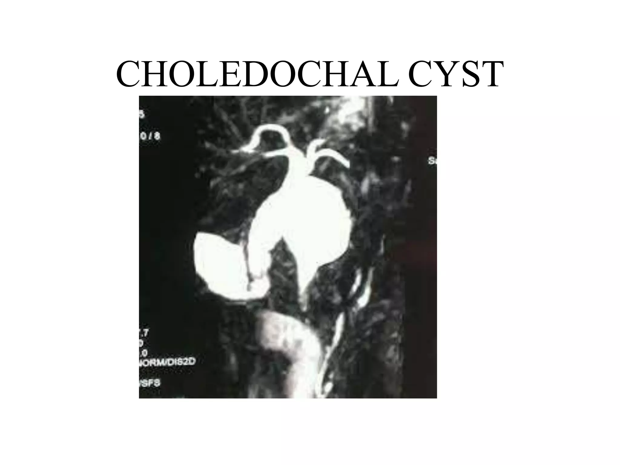
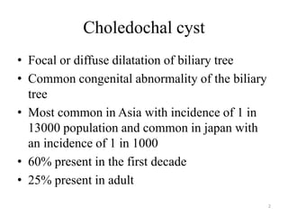
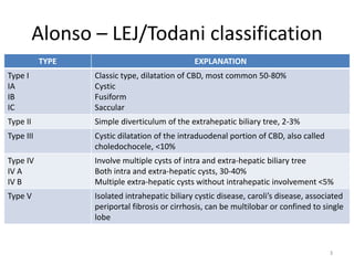
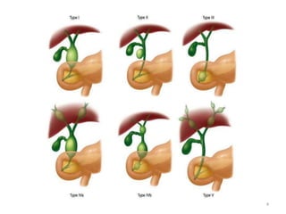
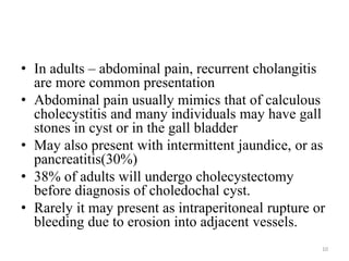
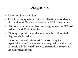
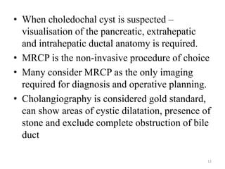
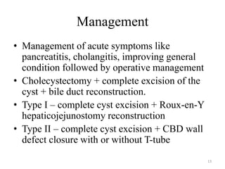
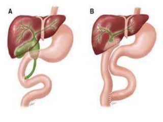
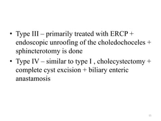
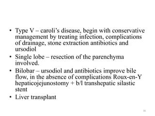
CHOLEDOCHAL CYST
Choledochal cyst
• Focal or diffuse dilatation of biliary tree
• Common congenital abnormality of the biliary
tree
• Most common in Asia with incidence of 1 in
13000 population and common in japan with
an incidence of 1 in 1000
• 60% present in the first decade
• 25% present in adult
2
Alonso – LEJ/Todani classification
TYPE EXPLANATION
Type I
IA
IB
IC
Classic type, dilatation of CBD, most common 50-80%
Cystic
Fusiform
Saccular
Type II Simple diverticulum of the extrahepatic biliary tree, 2-3%
Type III Cystic dilatation of the intraduodenal portion of CBD, also called
choledochocele, <10%
Type IV
IV A
IV B
Involve multiple cysts of intra and extra-hepatic biliary tree
Both intra and extra-hepatic cysts, 30-40%
Multiple extra-hepatic cysts without intrahepatic involvement <5%
Type V Isolated intrahepatic biliary cystic disease, caroli’s disease, associated
periportal fibrosis or cirrhosis, can be multilobar or confined to single
lobe
3
4
Pathogenesis
• The cause is unknown. There are theories
proposed.
• Theory I – defect in maturation with ductal plate
malformation.
– Describe development of intrahepatic liver progenitor
cells that are in contact with the mesenchyme of portal
vein and are remodeled intomature ducts.
– Defective bile duct plate remodeling in embryogenesis
results in inflammation and ulceration forming larger
ducts
5
• Theory II
– Bile duct obstruction or distention in the prenatal
or neonatal period leading to cyst formation
– Obstruction may be secondary to stricture, web ,or
sphincter of Oddi dysfunction
– Pancreatic juice reflux into biliary tree causing
chronic inflammation and increased bile duct
pressure leading to cyst formation
– Animal models
6
• Theory III
– Most common proposed model for choledochal
cyst
– Related to pancreaticobiliary maljunction
– Defined as an extramural junction of pancreatic
and biliary duct in the duodenum beyond the
intramural sphincter function
– Characterised by long common channel.
– Significant risk for developing
cholangiocarcinoma
7
Pancreaticobiliary
maljunction
Long common channel
Pancreatic reflux and mixing
with bile
Mixed juice potentially
stagnates in bile tree/gall
bladder
Cycles of inflammation,
activation of proteolytic
enzymes, alteration in bile
composition
Damage to biliary tree
epithelium
Duct distention and may
progress to malignancy
8
Presentation
• Classic triad of jaundice, abdominal pain and
Rt upper quadrant mass in children
• Infants present with elevated conjugated
bilirubin(80%), failure to thrive or abdominal
mass(30%)
• Abdominal mass becomes less comman with
increasing age
9
• In adults – abdominal pain, recurrent cholangitis
are more common presentation
• Abdominal pain usually mimics that of calculous
cholecystitis and many individuals may have gall
stones in cyst or in the gall bladder
• May also present with intermittent jaundice, or as
pancreatitis(30%)
• 38% of adults will undergo cholecystectomy
before diagnosis of choledochal cyst.
• Rarely it may present as intraperitoneal rupture or
bleeding due to erosion into adjacent vessels.
10
Diagnosis
• Requires high suspicion
• Type I cyst may mimics biliary dilatation secondary to
obstruction, difference is elevated ALP in obstruction
• USG is most common first line imaging used in 93% of
pediatric and 72% of adults
• CT is appropriate in adults in whom the differential
diagnosis is broader
• Important consideration in CT is assessing the
hepatobiliary and pancreatic anatomy, with evaluation
of possible biliary malignancy, metastatic disease and
vascular encasement.
11
• When choledochal cyst is suspected –
visualisation of the pancreatic, extrahepatic
and intrahepatic ductal anatomy is required.
• MRCP is the non-invasive procedure of choice
• Many consider MRCP as the only imaging
required for diagnosis and operative planning.
• Cholangiography is considered gold standard,
can show areas of cystic dilatation, presence of
stone and exclude complete obstruction of bile
duct
12
Management
• Management of acute symptoms like
pancreatitis, cholangitis, improving general
condition followed by operative management
• Cholecystectomy + complete excision of the
cyst + bile duct reconstruction.
• Type I – complete cyst excision + Roux-en-Y
hepaticojejunostomy reconstruction
• Type II – complete cyst excision + CBD wall
defect closure with or without T-tube
13
• Type III – primarily treated with ERCP +
endoscopic unroofing of the choledochoceles +
sphincterotomy is done
• Type IV – similar to type I , cholecystectomy +
complete cyst excision + biliary enteric
anastamosis
15
• Type V – caroli’s disease, begin with conservative
management by treating infection, complications
of drainage, stone extraction antibiotics and
ursodiol
• Single lobe – resection of the parenchyma
involved.
• Bilobar – ursodiol and antibiotics improve bile
flow, in the absence of complications Roux-en-Y
hepaticojejunostomy + b/l transhepatic silastic
stent
• Liver transplant
16
17

Choledochal cyst

  • 1.
  • 2.
    Choledochal cyst • Focalor diffuse dilatation of biliary tree • Common congenital abnormality of the biliary tree • Most common in Asia with incidence of 1 in 13000 population and common in japan with an incidence of 1 in 1000 • 60% present in the first decade • 25% present in adult 2
  • 3.
    Alonso – LEJ/Todaniclassification TYPE EXPLANATION Type I IA IB IC Classic type, dilatation of CBD, most common 50-80% Cystic Fusiform Saccular Type II Simple diverticulum of the extrahepatic biliary tree, 2-3% Type III Cystic dilatation of the intraduodenal portion of CBD, also called choledochocele, <10% Type IV IV A IV B Involve multiple cysts of intra and extra-hepatic biliary tree Both intra and extra-hepatic cysts, 30-40% Multiple extra-hepatic cysts without intrahepatic involvement <5% Type V Isolated intrahepatic biliary cystic disease, caroli’s disease, associated periportal fibrosis or cirrhosis, can be multilobar or confined to single lobe 3
  • 4.
  • 5.
    Pathogenesis • The causeis unknown. There are theories proposed. • Theory I – defect in maturation with ductal plate malformation. – Describe development of intrahepatic liver progenitor cells that are in contact with the mesenchyme of portal vein and are remodeled intomature ducts. – Defective bile duct plate remodeling in embryogenesis results in inflammation and ulceration forming larger ducts 5
  • 6.
    • Theory II –Bile duct obstruction or distention in the prenatal or neonatal period leading to cyst formation – Obstruction may be secondary to stricture, web ,or sphincter of Oddi dysfunction – Pancreatic juice reflux into biliary tree causing chronic inflammation and increased bile duct pressure leading to cyst formation – Animal models 6
  • 7.
    • Theory III –Most common proposed model for choledochal cyst – Related to pancreaticobiliary maljunction – Defined as an extramural junction of pancreatic and biliary duct in the duodenum beyond the intramural sphincter function – Characterised by long common channel. – Significant risk for developing cholangiocarcinoma 7
  • 8.
    Pancreaticobiliary maljunction Long common channel Pancreaticreflux and mixing with bile Mixed juice potentially stagnates in bile tree/gall bladder Cycles of inflammation, activation of proteolytic enzymes, alteration in bile composition Damage to biliary tree epithelium Duct distention and may progress to malignancy 8
  • 9.
    Presentation • Classic triadof jaundice, abdominal pain and Rt upper quadrant mass in children • Infants present with elevated conjugated bilirubin(80%), failure to thrive or abdominal mass(30%) • Abdominal mass becomes less comman with increasing age 9
  • 10.
    • In adults– abdominal pain, recurrent cholangitis are more common presentation • Abdominal pain usually mimics that of calculous cholecystitis and many individuals may have gall stones in cyst or in the gall bladder • May also present with intermittent jaundice, or as pancreatitis(30%) • 38% of adults will undergo cholecystectomy before diagnosis of choledochal cyst. • Rarely it may present as intraperitoneal rupture or bleeding due to erosion into adjacent vessels. 10
  • 11.
    Diagnosis • Requires highsuspicion • Type I cyst may mimics biliary dilatation secondary to obstruction, difference is elevated ALP in obstruction • USG is most common first line imaging used in 93% of pediatric and 72% of adults • CT is appropriate in adults in whom the differential diagnosis is broader • Important consideration in CT is assessing the hepatobiliary and pancreatic anatomy, with evaluation of possible biliary malignancy, metastatic disease and vascular encasement. 11
  • 12.
    • When choledochalcyst is suspected – visualisation of the pancreatic, extrahepatic and intrahepatic ductal anatomy is required. • MRCP is the non-invasive procedure of choice • Many consider MRCP as the only imaging required for diagnosis and operative planning. • Cholangiography is considered gold standard, can show areas of cystic dilatation, presence of stone and exclude complete obstruction of bile duct 12
  • 13.
    Management • Management ofacute symptoms like pancreatitis, cholangitis, improving general condition followed by operative management • Cholecystectomy + complete excision of the cyst + bile duct reconstruction. • Type I – complete cyst excision + Roux-en-Y hepaticojejunostomy reconstruction • Type II – complete cyst excision + CBD wall defect closure with or without T-tube 13
  • 15.
    • Type III– primarily treated with ERCP + endoscopic unroofing of the choledochoceles + sphincterotomy is done • Type IV – similar to type I , cholecystectomy + complete cyst excision + biliary enteric anastamosis 15
  • 16.
    • Type V– caroli’s disease, begin with conservative management by treating infection, complications of drainage, stone extraction antibiotics and ursodiol • Single lobe – resection of the parenchyma involved. • Bilobar – ursodiol and antibiotics improve bile flow, in the absence of complications Roux-en-Y hepaticojejunostomy + b/l transhepatic silastic stent • Liver transplant 16
  • 17.