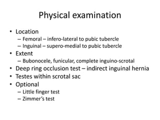
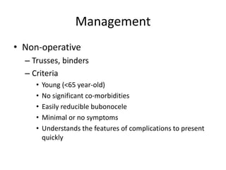
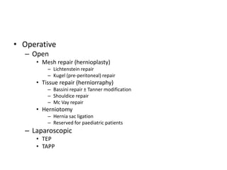
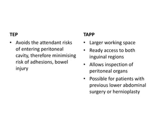
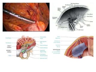
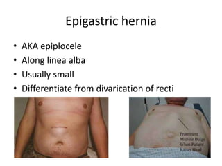
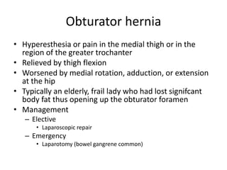
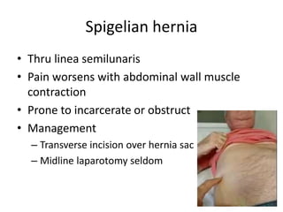
Hernia
Chea Chan Hooi
Surgeon
Sibu Hospital
Content
• Definition
• Epidemiology
• Risk factors
• Classification
• Clinical features
• Management
• Specific configurations
• Other types
• Q&A
Definition
• Protrusion, bulge or projection of organ
through the body wall which normally
contains it
Epidemiology
• Between 5 – 10% in the US
• Inguinal > femoral > other abdominal wall
hernias
Risk factors
Modifiable Non-modifiable
Obesity
Chronic straining on defecation or
urination
Chronic coughing
Chronic heavy lifting
Ascites
Previous surgery
Male
Prematurity
Patent procesus vaginalis
Maldescended testis
Associated urogenital disorders
(ambiguous genitalia, epispadias,
hypospadias)
Connective tissue disease
Aging
Family history of hernia
Classification
• Anatomical
– External
• Anterior abdominal wall
– Epigastric
– Spigelian
– Umbilical
– Inguinal
– Femoral
– Obturator
• Posterior abdominal wall
– Lumbar
– Sciatic
– Internal
• Hiatal hernia
• Cerebral herniation
• Clinical features
– Uncomplicated
– Complicated
Clinical features
• Impulse on coughing/straining
• Uncomplicated – easily reducible
• Complicated – usually irreducible
– Incarcerated
• Contents entrapped within hernia sac, usually large and for
years without much symptoms
– Obstructed
• Bowel luminal obstruction
– Strangulated
• Blood supply compromised
• Causative factors
Physical examination
• Location
– Femoral – infero-lateral to pubic tubercle
– Inguinal – supero-medial to pubic tubercle
• Extent
– Bubonocele, funicular, complete inguino-scrotal
• Deep ring occlusion test – indirect inguinal hernia
• Testes within scrotal sac
• Optional
– Little finger test
– Zimmer’s test
Management
• Non-operative
– Trusses, binders
– Criteria
• Young (<65 year-old)
• No significant co-morbidities
• Easily reducible bubonocele
• Minimal or no symptoms
• Understands the features of complications to present
quickly
• Operative
– Open
• Mesh repair (hernioplasty)
– Lichtenstein repair
– Kugel (pre-peritoneal) repair
• Tissue repair (herniorraphy)
– Bassini repair ± Tanner modification
– Shouldice repair
– Mc Vay repair
• Herniotomy
– Hernia sac ligation
– Reserved for paediatric patients
– Laparoscopic
• TEP
• TAPP
TEP
• Avoids the attendant risks
of entering peritoneal
cavity, therefore minimising
risk of adhesions, bowel
injury
TAPP
• Larger working space
• Ready access to both
inguinal regions
• Allows inspection of
peritoneal organs
• Possible for patients with
previous lower abdominal
surgery or hernioplasty
Specific configurations
• Richter
• Amyand
• Littre
• Maydl
• Reduction en-masse
• Intestinal stenosis of Garre
Other hernia types
n.b. incisional & parastomal hernia not included
Giant inguinal hernia
• Inguinal hernia that extends below the
midpoint of inner thigh when the patient is in
standing position
• Complications of reduction
– Abdominal compartment syndrome (loss of
domain >20%)
– Seroma
– Haematoma
Management
• Pre-operatively
– Increase intra-abdominal volume
• Progressive pneumoperitoneum
– Ensure adequate room in abdominal cavity by pneumoperitoneum before reduction of the
hernia contents
– Gradually insufflating gas into abdominal cavity via placed catheter in situ, usually in increments
of between 500 – 2000cc/day over 7 –14 days4
– Ambient air, oxygen, carbon dioxide and nitrous oxide
– Multiple sittings needed
• Intra-operatively
– Resection of contents
• Resected organs usually the colon, small bowel or omentum
• Single-stage operation
• Risk of anatomotic leak & mesh infection
– Rotation of viable tissue5, 6
• Scrotal skin flap, tensor fascia latae musculocutaneous flap & component separation
technique
• Single-stage procedure but specific surgical expertise required to prevent complications
Umbilical hernia
• Mid-abdominal location, centered around the
umbilicus
• Typically small
Umbilical (Direct) Paraumbilical (Indirect)
Patient Paediatric majority Adult majority
Clinical The whole umbilicus is symmetrically
effaced & it loses its characteristic
shape
Umbilicus has crescent appearance due to
an asymmetrical effacement
Defect Thru the umbilical ring Superior or inferior to the umbilical ring
Pathophysiology Failure of umbilical ring to close Congenital weak point between umbiical
ring and linea alba
Differential
diagnosis
Omphalocele Port site hernia post laparoscopic surgery
Adapted from Compendium of 100 Surgical Cases in Sibu Hospital
• Management
– Elective
• Open
– Mesh repair: Onlay vs. inlay vs. sublay repair
– Non-mesh repair: anatomical vs. Mayo repair
• Laparoscopic intraperitoneal onlay mesh repair (IPOM)
– Emergency
• Transverse incision feasible
Type Pros Cons
Onlay
Mesh placed anterior to fascia Relatively simple
Acceptable recurrence rate 5 – 15%
Skin flaps created to accommodate mesh
might be devascularised, predisposing to
seroma and infection
For large defects (>10cm), repair in
combination with component separation
Inlay
Mesh placed to bridge the fascial defect Relatively simple Abdominal pressure exerted directly on
mesh, detaching it away from fascial edges
Need a composite mesh (expensive) as
mesh in direct contact with peritoneal
content
Does not allow tissue-mesh integration
Obsolete due to high (3x) recurrence & SSI
rates
Sublay
Mesh placed posterior to recti muscles
where the force of abdominal pressure
holds the mesh against the posterior
surface of muscles
Lowest recurrence rate (3.5%)
Tissue integration superficial & deep to
mesh
Mesh protected from superficial SSI &
intra-abdominal adhesion/contamination
Technically challenging
Large dead space posterior to recti with
resultant seroma
Adapted from Compendium of 100 Surgical Cases in Sibu Hospital
Epigastric hernia
• AKA epiplocele
• Along linea alba
• Usually small
• Differentiate from divarication of recti
Obturator hernia
• Hyperesthesia or pain in the medial thigh or in the
region of the greater trochanter
• Relieved by thigh flexion
• Worsened by medial rotation, adduction, or extension
at the hip
• Typically an elderly, frail lady who had lost signifcant
body fat thus opening up the obturator foramen
• Management
– Elective
• Laparoscopic repair
– Emergency
• Laparotomy (bowel gangrene common)
Spigelian hernia
• Thru linea semilunaris
• Pain worsens with abdominal wall muscle
contraction
• Prone to incarcerate or obstruct
• Management
– Transverse incision over hernia sac
– Midline laparotomy seldom
Lumbar hernia
• Thru lumbar triangles
– Superior (Grynfeltt-Lesshaft)
– Inferior (Petit)
• Vague flank discomfort + mass
• Seldom incarcerate
• Management
– Non-operative
– Open posterior mesh repair via skin-line oblique
incision from 12th rib – iliac crest
Sciatic hernia
• Intestinal obstruction
• Ureteric obstruction
• Sciatic pain
• Tender mass in the gluteal area
• Differentials
– Lipoma
– Tuberculoma
– Soft tissue malignancy
• Management
– Elective
• Open transgluteal
– Emergency
• Open transperitoneal
TQ!
Q&A?

Hernia

  • 1.
  • 2.
    Content • Definition • Epidemiology •Risk factors • Classification • Clinical features • Management • Specific configurations • Other types • Q&A
  • 3.
    Definition • Protrusion, bulgeor projection of organ through the body wall which normally contains it
  • 4.
    Epidemiology • Between 5– 10% in the US • Inguinal > femoral > other abdominal wall hernias
  • 5.
    Risk factors Modifiable Non-modifiable Obesity Chronicstraining on defecation or urination Chronic coughing Chronic heavy lifting Ascites Previous surgery Male Prematurity Patent procesus vaginalis Maldescended testis Associated urogenital disorders (ambiguous genitalia, epispadias, hypospadias) Connective tissue disease Aging Family history of hernia
  • 6.
    Classification • Anatomical – External •Anterior abdominal wall – Epigastric – Spigelian – Umbilical – Inguinal – Femoral – Obturator • Posterior abdominal wall – Lumbar – Sciatic – Internal • Hiatal hernia • Cerebral herniation • Clinical features – Uncomplicated – Complicated
  • 7.
    Clinical features • Impulseon coughing/straining • Uncomplicated – easily reducible • Complicated – usually irreducible – Incarcerated • Contents entrapped within hernia sac, usually large and for years without much symptoms – Obstructed • Bowel luminal obstruction – Strangulated • Blood supply compromised • Causative factors
  • 8.
    Physical examination • Location –Femoral – infero-lateral to pubic tubercle – Inguinal – supero-medial to pubic tubercle • Extent – Bubonocele, funicular, complete inguino-scrotal • Deep ring occlusion test – indirect inguinal hernia • Testes within scrotal sac • Optional – Little finger test – Zimmer’s test
  • 9.
    Management • Non-operative – Trusses,binders – Criteria • Young (<65 year-old) • No significant co-morbidities • Easily reducible bubonocele • Minimal or no symptoms • Understands the features of complications to present quickly
  • 10.
    • Operative – Open •Mesh repair (hernioplasty) – Lichtenstein repair – Kugel (pre-peritoneal) repair • Tissue repair (herniorraphy) – Bassini repair ± Tanner modification – Shouldice repair – Mc Vay repair • Herniotomy – Hernia sac ligation – Reserved for paediatric patients – Laparoscopic • TEP • TAPP
  • 11.
    TEP • Avoids theattendant risks of entering peritoneal cavity, therefore minimising risk of adhesions, bowel injury TAPP • Larger working space • Ready access to both inguinal regions • Allows inspection of peritoneal organs • Possible for patients with previous lower abdominal surgery or hernioplasty
  • 14.
    Specific configurations • Richter •Amyand • Littre • Maydl • Reduction en-masse • Intestinal stenosis of Garre
  • 15.
    Other hernia types n.b.incisional & parastomal hernia not included
  • 16.
    Giant inguinal hernia •Inguinal hernia that extends below the midpoint of inner thigh when the patient is in standing position • Complications of reduction – Abdominal compartment syndrome (loss of domain >20%) – Seroma – Haematoma
  • 17.
    Management • Pre-operatively – Increaseintra-abdominal volume • Progressive pneumoperitoneum – Ensure adequate room in abdominal cavity by pneumoperitoneum before reduction of the hernia contents – Gradually insufflating gas into abdominal cavity via placed catheter in situ, usually in increments of between 500 – 2000cc/day over 7 –14 days4 – Ambient air, oxygen, carbon dioxide and nitrous oxide – Multiple sittings needed • Intra-operatively – Resection of contents • Resected organs usually the colon, small bowel or omentum • Single-stage operation • Risk of anatomotic leak & mesh infection – Rotation of viable tissue5, 6 • Scrotal skin flap, tensor fascia latae musculocutaneous flap & component separation technique • Single-stage procedure but specific surgical expertise required to prevent complications
  • 20.
    Umbilical hernia • Mid-abdominallocation, centered around the umbilicus • Typically small Umbilical (Direct) Paraumbilical (Indirect) Patient Paediatric majority Adult majority Clinical The whole umbilicus is symmetrically effaced & it loses its characteristic shape Umbilicus has crescent appearance due to an asymmetrical effacement Defect Thru the umbilical ring Superior or inferior to the umbilical ring Pathophysiology Failure of umbilical ring to close Congenital weak point between umbiical ring and linea alba Differential diagnosis Omphalocele Port site hernia post laparoscopic surgery Adapted from Compendium of 100 Surgical Cases in Sibu Hospital
  • 22.
    • Management – Elective •Open – Mesh repair: Onlay vs. inlay vs. sublay repair – Non-mesh repair: anatomical vs. Mayo repair • Laparoscopic intraperitoneal onlay mesh repair (IPOM) – Emergency • Transverse incision feasible
  • 23.
    Type Pros Cons Onlay Meshplaced anterior to fascia Relatively simple Acceptable recurrence rate 5 – 15% Skin flaps created to accommodate mesh might be devascularised, predisposing to seroma and infection For large defects (>10cm), repair in combination with component separation Inlay Mesh placed to bridge the fascial defect Relatively simple Abdominal pressure exerted directly on mesh, detaching it away from fascial edges Need a composite mesh (expensive) as mesh in direct contact with peritoneal content Does not allow tissue-mesh integration Obsolete due to high (3x) recurrence & SSI rates Sublay Mesh placed posterior to recti muscles where the force of abdominal pressure holds the mesh against the posterior surface of muscles Lowest recurrence rate (3.5%) Tissue integration superficial & deep to mesh Mesh protected from superficial SSI & intra-abdominal adhesion/contamination Technically challenging Large dead space posterior to recti with resultant seroma Adapted from Compendium of 100 Surgical Cases in Sibu Hospital
  • 25.
    Epigastric hernia • AKAepiplocele • Along linea alba • Usually small • Differentiate from divarication of recti
  • 26.
    Obturator hernia • Hyperesthesiaor pain in the medial thigh or in the region of the greater trochanter • Relieved by thigh flexion • Worsened by medial rotation, adduction, or extension at the hip • Typically an elderly, frail lady who had lost signifcant body fat thus opening up the obturator foramen • Management – Elective • Laparoscopic repair – Emergency • Laparotomy (bowel gangrene common)
  • 28.
    Spigelian hernia • Thrulinea semilunaris • Pain worsens with abdominal wall muscle contraction • Prone to incarcerate or obstruct • Management – Transverse incision over hernia sac – Midline laparotomy seldom
  • 29.
    Lumbar hernia • Thrulumbar triangles – Superior (Grynfeltt-Lesshaft) – Inferior (Petit) • Vague flank discomfort + mass • Seldom incarcerate • Management – Non-operative – Open posterior mesh repair via skin-line oblique incision from 12th rib – iliac crest
  • 31.
    Sciatic hernia • Intestinalobstruction • Ureteric obstruction • Sciatic pain • Tender mass in the gluteal area • Differentials – Lipoma – Tuberculoma – Soft tissue malignancy • Management – Elective • Open transgluteal – Emergency • Open transperitoneal
  • 32.