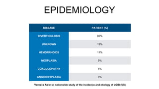
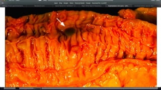
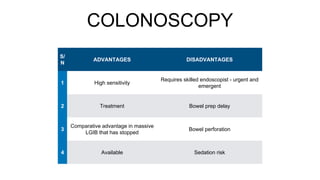
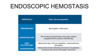
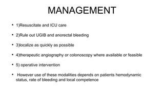
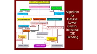
MANAGEMENT OF MASSIVE LOWER
GASTROINTESTINAL HEMORRHAGE
DR ADESIYAKAN A.A
OUTLINE
• INTRODUCTION
• EPIDEMIOLOGY
• RELEVANT ANATOMY
• ETIOLOGY
• PATHOGENESIS
• CLASSIFICATION
• CLINICAL FEATURES
• INVESTIGATIONS
• MANAGEMENT
• COMPLICATIONS
• PROGNOSIS
• CONCLUSION
INTRODUCTION
• Lower gastrointestinal hemorrhage refers to bleeding from the
gastrointestinal(GI) tract distal to the ligament of treitz
• Ranges from trivial to massive
• MASSIVE lower gastrointestinal bleeding is a life threatening condition
especially in the elderly in whom the disease is more common
INTRODUCTION (2)
• Criteria for diagnosing MASSIVE lower gastrointestinal bleeding(LGIB)
include;
• transfusion of at least 4 units of blood within 24 hours
• Hemodynamic instability and shock
• Decrease in hematocrit to 6g/dl or less
• Bleeding continues for 3 days
• Significant rebleeding within 1week
EPIDEMIOLOGY
• Worldwide, acute LGIB accounts for 1%-2% of hospital emergencies
• 15% of this fulfill the massive LGIB criteria with 5% of them requiring
emergent operative intervention
• Annual incidence rate of 20.5 patients per 100,000
• Male : female - 1.8 - 1
• AGE - 65
EPIDEMIOLOGY
DISEASE PATIENT (%)
DIVERTICULOSIS 60%
UNKNOWN 13%
HEMORRHOIDS 11%
NEOPLASIA 9%
COAGULOPATHY 4%
ANGIODYSPLASIA 3%
Vernava AM et al nationwide study of the incidence and etiology of LGIB (US)
38%
5%
17%
16%
6%
19%
hemorrhoids diverticulosis
carcinoma polyps
AVM UNIDENTIFIED
Akande et al-2015
Olookoba et al-2013
Dakubo et al -2008
Alatise et al- 2007
EPIDEMIOLOGY
RELEVANT ANATOMY
• Lower gastrointestinal tract extends from the ligament of trietz to the anus
• Conduit in which digestion absorption and secretion occurs
• Consists of the jejunum and ileum - small intestine
• Caecum, ascending colon, transverse , descending colon , sigmoid rectum
and anal canal
RELEVANT ANATOMY(2)
RELEVANT ANATOMY
(3)
• The superior rectal arteries and its branches
• The super and inferior hemorrhoidal
plexuses
• Hemorrhoids components- (a)mucosa
(b)stroma- blood vessels, smooth muscles
(c)anchoring connective tissue-secures the
hemorrhoids to the internal sphincter and
conjoined longitudinal coat.
CLASSIFICATIONS
CLASS EXAMPLES
ANATOMIC HEMORRHOIDS, DIVERTICULOSIS,
VASCULAR
ANGIODYSPLASIA,ISCHAEMIC, RADIATION
INDUCED,AORTOCOLONIC FISTULA
INFLAMMATORY INFECTIOUS AND NON-INFECTIOUS
NEOPLASTIC COLORECTAL CANCER,POLYPS
CLASSIFICATION
SITE PROPORTION %
COLON 80 - 85
SMALL INTESTINE 0.7 - 9
UPPER GIT 10
UPPER GIT(MASSIVE) 33
PATHOGENESIS
• DIVERTICULOSIS-
• Diverticulum herniates at the site of vasa recta
• The vessel becomes draped over the dome of the diverticulum, separated
by just mucosa
• Segmental weakening of the artery which ruptures and bleeds
• Bleeding is more common on the right ( Wong S.K et al found that right
sided diverticula are twice likely to bleed -similar to most studies
PATHOGENESIS
PATHOGENENESIS
• HEMORRHOIDS
• Deterioration of the connective tissue that anchors the hemorrhoids occur
with age
• begins at the 3rd decade and progresses depending on individual differences
and risk factors- Haaspa et al
• Morinaga K et al postulated that they imbalances between arterial supply and
drainage
• Thus patients who had ligation of the arterial supply had resolution of
symptoms
PATHOGENESIS
• ANGIODYSPLASIA
• Colonic angiodysplasias are acquired mucosal or submucosal telangiectasia of degenerative
origin.They are dilated and tortuous with the blood vessel walls lacking smooth muscles.
• Degenerative lesions from chronic intermittent low grade colonic contraction
• Leads to obstruction of mucosal venous drainage
• Over time, mucosal capillaries dilate become incompetent and form an AVM
• Bleeding more on the right
• Saperas et al found that the burden of the disease is in its tendency to re-bleed
• Associated with aortic stenosis, VWD, chronic renal failure
PATHOGENESIS
• NEOPLASIA
• Mucosal ulceration and erosion
• Vascular invasion
• Friable neovasculature
• OTHERS - IBD, ischemic colitis, infectious colitis, radiation induced colitis,
aorto- colonic, post procedure
CLINICAL FEATURES
• BIODATA
• Presenting complaints :Hematochezia, Melena ??, Hematemesis
• Anaemia - weakness, palpitation dizziness ,syncope
• Shock
• Abdominal complaints
• Past medical history and procedures- Recent polypectomy, colonoscopy, bleeding
dycrasias
• History of risk factors-previous bleeding, radiation, NSAIDS, anticoagulants , AAA graft,
Liver disease, HIV status, family history of colon cancer recent weight loss
CLINICAL FEATURES
PATHOLOGIC
CONDITION
CLASSIC PRESENTATION
EXTERNAL
HEMORRHOIDS
PAINFUL,SPORADIC LOW VOLUME BLEEDS WITH RED
BLOOD COATING STOOL
INTERNAL
HEMORHOIDS
PAINLESS, LOW- HIGH VOLUME
DIVERTICULAR
BLEEDING
PAINLESS/MILD CRAMPING,EPISODIC BURSTS OF
ARTERIAL BLEEDING,HIGH VOLUME,CLOTS,INCREASED
PREVALENCE WITH CERTAIN RISK FACTORS
ANGIODYSPLASIA
PAINLESS/MILD CRAMPING,EPISODIC VENOUS
BLEEDING,OVERT OR OCCULT,OFTEN AT LOW VOLUME(
BUT CAN BE MASSIVE), PREVALENT IN THE ELDERLY
COLON CANCER
CRAMPING OR PAINLESS,WEIGHT LOSS,MELENA,FAMILY
HISTORY
TRAUMA IATROGENIC, DOMESTIC
CLINICAL FEATURES
• Physical examination should be thorough and include the assessment of anorectum,
oropharynx, nasopharynx and the hemodynamic status of the patient ,tempo and extent of
blood loss, risk assessment
• Horizontal approach
• General physical examination - shock
• Abdominal examination
• Nasogastric tube aspiration and warm saline gastric lavage if necessary -
• Gentle DRE + proctoscopy
• Discovery of a benign pathology doesn’t rule out other etiology
RESUSCITATION
• ATLS
• RISK ASSESSMENT - HDU/ICU
• Vasopressors
• Urethral catheter
• Blood transfusion - target blood pressure
• Fresh frozen plasma - target INR
• Reassessment and monitoring
Ongoing Bleeding -
BLEED CRITERIA
Low SBP - < 100mmHG
Elevated PT ->1.2
Erratic mental status
Unstable Co-morbid Disease
INVESTIGATIONS
• FBC ( urgent PCV after resuscitation, normocytic vs microcytic)
• Blood group and cross match
• E/U/Cr - BUN/creatinine ->30:1 ,urea/creatinine ratio - 100:1
• Shock- base deficit, anion gap, serum lactate
• LFTs and clotting profile
• Nasogastric tube aspiration
• Proctoscopy
DIAGNOSTIC
INVESTIGATIONS
• Aim is to identify and localize
• Sequence depends on such factors as rate of bleeding, Hemodynamic
status of the patient, success at localization of the lesion
• Most of these overlap as diagnostic and therapeutic measures
• They include Computed tomography, Colonoscopy, angiography, CT-
angiography, radionuclide imaging each with its relative merit and demerit
COLONOSCOPY
• Fiberoptic flexible colonoscopy is the initial Diagnostic modality of choice
for stable patients
• Successfully identifies site in 80 -90 %
• Also offers opportunity of therapeutic interventions
• Relatively safe
COLONOSCOPY
• PATIENT CRITERIA - Hemodynamically stable, nil ongoing brisk bleeding (vs emergent )
• BOWEL PREPARATION-
- Blood is cathartic alone - poor cecal intubation rate (18%)
- Aggressive lavage - 4-6 liters of polyethylene glycol -(bleeding)
- Metoclopamide
- Hydro flush colonoscopy - mechanical suction devices and water jet pumps-
- TIMING-
- urgent colonoscopy(<12hrs) has comparative advantage in detection of the source of hemorrhage vs expectant colonoscopy but its advantage
in terms of length of hospital stay and re-bleed rates is unclear.
- Emergent has much Lower diagnostic yield but may be the only option
COLONOSCOPY
• Large national study by Jensen and machicado, urgent colonoscopy yielded a definitive
diagnosis in 90% of patients with also therapeutic opportunity as an added benefit
• Patients also had significantly shorter hospital stay
• Urgent colonoscopy can be performed in the operating suite
• Therapeutic colonoscopy - bipolar probe coagulation, Epinephrine injection, hemospray,
clip application , argon plasma coagulation, application of formalin and a combination of
these
• Tattooing
• Incidentally discovered lesions are left alone
COLONOSCOPY
S/
N
ADVANTAGES DISADVANTAGES
1 High sensitivity
Requires skilled endoscopist - urgent and
emergent
2 Treatment Bowel prep delay
3
Comparative advantage in massive
LGIB that has stopped
Bowel perforation
4 Available Sedation risk
ENDOSCOPIC HEMOSTASIS
DIVERTICULI Clips, thermocoagulation
HEMORRHOIDS Band ligation, sclerosants,
ANGIODYSPLASIA
Non-contact thermal therapy using argon plasma
coagulation(APC),injection sclerotherapy
POST
POLYPECTOMY
Mechanical clips, thermal coagulation, vasoconstrictors
hemospray
VASOPRESSORS
Initial control of active bleeding and improve visualization
Combined with second hemostatic mechanism
NO PLACE FOR COLONOSCOPY IN ACTIVELY BLEEDING OR
HEMODYNAMICALLY UNSTABLE PATIENTS
ANGIOGRAPHY
• In 1965 Baum al first described selective mesenteric angiography
• It is useful for patients in whom active bleeding precludes colonoscopy or where colonoscopy fails to
identify the bleeding site
• May be emergent or elective
• Elective angiography -patients with multiple episodes of LGIB without a known source or diagnosis.
• Emergency angiography is very useful in patients with massive ongoing bleeding
• Requires active bleeding at 0.5 - 1 ml/min ,creatinine <1.5mg/dl, eGFR>60ml/min/1.73m, INR- <1.5,
platelets >50,000
• Evaluation begins at the superior mesenteric artery
ANGIOGRAPHY
FINDING PROBABLE DIAGNOSIS
EXTRAVASATION BLEEDING SITE
FILLING OF SPACES OUTSIDE
THE BOWEL LUMEN
ANEURYSM,PSEUDOANEURYSM
EARLY ARTERIAL FILLING
,ACCUMULATION IN VASCULAR
TUFTS,SLOW DRAINING
VESSELS ANGIODYSPLASIA
NEOVASCULARITY NEOPLASM
HYPEREMIA COLITIS
ANGIOGRAPHY
• Embolization- goal is to mechanically occlude the arterial blood supply to the bleeding site while
maintaining collateral perfusion to prevent ischemia
• Hyper selective process
• Agents - 1) TEMPORARY- e.g absorbable gelatin sponge(gelfoam)-long acting, bio degradable
and can be cut into small blocks mixed with saline and iodinated contrast
• may come in powdery form(unpredictable)
• 2) PERMANENT- coils, particles, glue, polymers of ethylene alcohol
• Vasopressin, propanolol, epinephrine ( rebound bleeding)-
• Initial rate of 0.2I/min - repeat angiography-observe for 24-48 hours, if not resolved or
complications occur -> surgery
ANGIOGRAPHY
ADVANTAGES DISADVANTAGES
DOES NOT REQUIRE BOWEL
PREPARATION
LOW SENSITIVITY
CAN BE USED IN EMERGENCY
SETTING
REQUIRES ACTIVE BLEEDING
THERAPEUTIC USES
COMPLICATION RATE ( thrombosis,
embolization, renal failure)
COMPUTED TOMOGRAPHY
• Triple phase helical CT scanning using IV contrast is safe, fast convenient
and accurate diagnostic tool
• Criteria for identification include :
• vascular dilatations, vascular extravasation, spontaneous hyper density of
the peribowel fat
• Presence of diverticula - this alone is not enough for localization
• Comparable identification rate to mesenteric angiography and colonoscopy
COMPUTED TOMOGRAPHY
CT- ANGIOGRAM
• This is different from CT with intravenous contrast
• Cost effective accurate and rapid tool in the diagnosis of acute LGIB
• Studies report a pooled sensitivity of 85.2%- 89% and a specificity of 74%-
96.9% and positive predictive value of 98.5%
• When combined with visceral arteriography it increases diagnostic and
localization yield- jacovides et al
• Can track vessel and identify exact bleeding vessel
• Requires bleeding at 0.3 - 0.5 ml/min
CT ANGIOGRAM
ADVANTAGES DISADVANTAGES
NON-INVASIVE REQUIRES ACTIVE BLEEDING
GOOD LOCALIZATION RATE NOT THERAPEUTIC
PROVIDES ANATOMIC DETAILS RADIATION EXPOSURE
NO BOWEL PREPARATION IV CONTRAST
RADIONUCLIDE
• Requires bleeding at 0.1-0.5 ml/min - sensitive but low specificity
• Two types - technetium sulfur and 99m Tc pertechnetate labelled red blood cell
• Technetium sulfur is cleared rapidly from the intravascular space
• 99m Tc remains over 24 hours - intermittent
• Though these identify bleeding, the draw back of this is the poor localization as blood can move in
a peristaltic or antiperistaltic direction to a dependent area if still images are considered
• Thus it’s better if the dynamic images are assessed
• Studies have demonstrated a localization rate of < 26% with misdiagnosis of up to 25%- thus
segmental colectomy based on this is discouraged. Majorly suitable for selecting patients for
angiography
ADDITIONAL INVESTIGATIONS
• Esophagogastroduodenoscopy + PUSH ENDOSCOPY
• Capsule endoscopy
• Double balloon enteroscopy
• Enteroclysis
• Merkels scanning
• Barium enema
• Provocation -ryan JM et al,provocative bleeding studies with intraarterial heparin and tolazoline -
50%
• Alternate clamping
MANAGEMENT
• 1)Resuscitate and ICU care
• 2)Rule out UGIB and anorectal bleeding
• 3)localize as quickly as possible
• 4)therapeutic angiography or colonoscopy where available or feasible
• 5) operative intervention
• However use of these modalities depends on patients hemodynamic
status, rate of bleeding and local competence
OPERATIVE
• Reserved for cases in which other modalities fail or are unavailable or
poorly developed
• Selective segmental colectomy following localization is the preferred
surgical option
• Active bleeding from unidentified bleeding point may require subtotal
colectomy and ileostomy
• Blind segmental resection is discouraged - due to its high re bleeding rate,
morbidity and mortality
OPERATIVE
• Pre-operative measures -
• Intra-operative - laparotomy
• if bleeding vessel has been identified segmental colectomy
• If not ; Serial clamping
• Surgeon guided enteroscopy and colonoscopy based on local competence, may
require on table mechanical lavage
• Hemicolectomy
OPERATIVE
• subtotal colectomy and ileostomy +rectal stump
• secondary ileoproctostomy
• Hemorrhoidectomy, diverticulectomy
• Features of complications from other measures should be looked for and
treated
• Postoperative- elective ICU ,close monitoring
COMPLICATIONS
• Blood transfusion
• Procedure complications
• Surgery complications
PROGNOSIS
• Mortality rate of 10 - 20 % in developed countries where the full
armamentaria of diagnostic, therapeutic and emergency services are
available.
• Self-limiting in about 70 - 80 %
• Rebleeding is common especially is localization isn’t done
• Rebleed rates - early( <30 days)- 14.5 %
• Newer modalities and approach have reduced morbidity and mortality
CONCLUSION
• Massive lower gastrointestinal hemorrhage is a life threatening condition
• Multidisciplinary team of endoscopist, intervention radiologist and surgeons
are necessary for optimal care
• Accurate localization of the site and cause of bleeding are critical for
focused care
• Surgery is increasingly reserved as the last resort.
REFERENCES
1. Savides TJ, Jensen DM. Colonoscopic hemostasis for recurrent diverticular hemorrhage associated with a visible vessel: a report
of three cases. Gastrointest Endosc 1994; 40:70.
2. Foutch PG, Zimmerman K. Diverticular bleeding and the pigmented protuberance (sentinel clot): clinical implications,
histopathological correlation, and results of endoscopic intervention. Am J Gastroenterol 1996; 91:2589.
3. Foutch PG. Angiodysplasia of the gastrointestinal tract. Am J Gastroenterol 1993; 88:807.
4. Jensen DM, Jutabha R, Machicado GA, et al. Prospective randomized comparative study of bipolar electrocoagulation versus
heater probe for treatment of chronically bleeding internal hemorrhoids. Gastrointest Endosc 1997; 46:435.
5. Trowers EA, Ganga U, Rizk R, et al. Endoscopic hemorrhoidal ligation: preliminary clinical experience. Gastrointest Endosc
1998; 48:49.
6. Barbatzas C, Spencer GM, Thorpe SM, et al. Nd:YAG laser treatment for bleeding from radiation proctitis. Endoscopy 1996;
28:497.
7. Strate et al, lower GI bleeding; epidemiology and diagnosis. Gastroenterol clin north am 2005; 34;643
8. F.charles brunicardi, Schwartz principle of surgery, lower GI bleeding mc GRAW hill, 10th edition
9. Osinowo et al, audit of colonoscopy practice in LUTH, jcsjournal, 2016, vol. 13
10.Ahidjo et al ,angiography in UMTH, research gate. 2015
THANK YOU

lower gastrointestinal bleeding ppt

  • 1.
    MANAGEMENT OF MASSIVELOWER GASTROINTESTINAL HEMORRHAGE DR ADESIYAKAN A.A
  • 2.
    OUTLINE • INTRODUCTION • EPIDEMIOLOGY •RELEVANT ANATOMY • ETIOLOGY • PATHOGENESIS • CLASSIFICATION • CLINICAL FEATURES • INVESTIGATIONS • MANAGEMENT • COMPLICATIONS • PROGNOSIS • CONCLUSION
  • 3.
    INTRODUCTION • Lower gastrointestinalhemorrhage refers to bleeding from the gastrointestinal(GI) tract distal to the ligament of treitz • Ranges from trivial to massive • MASSIVE lower gastrointestinal bleeding is a life threatening condition especially in the elderly in whom the disease is more common
  • 4.
    INTRODUCTION (2) • Criteriafor diagnosing MASSIVE lower gastrointestinal bleeding(LGIB) include; • transfusion of at least 4 units of blood within 24 hours • Hemodynamic instability and shock • Decrease in hematocrit to 6g/dl or less • Bleeding continues for 3 days • Significant rebleeding within 1week
  • 5.
    EPIDEMIOLOGY • Worldwide, acuteLGIB accounts for 1%-2% of hospital emergencies • 15% of this fulfill the massive LGIB criteria with 5% of them requiring emergent operative intervention • Annual incidence rate of 20.5 patients per 100,000 • Male : female - 1.8 - 1 • AGE - 65
  • 6.
    EPIDEMIOLOGY DISEASE PATIENT (%) DIVERTICULOSIS60% UNKNOWN 13% HEMORRHOIDS 11% NEOPLASIA 9% COAGULOPATHY 4% ANGIODYSPLASIA 3% Vernava AM et al nationwide study of the incidence and etiology of LGIB (US)
  • 7.
    38% 5% 17% 16% 6% 19% hemorrhoids diverticulosis carcinoma polyps AVMUNIDENTIFIED Akande et al-2015 Olookoba et al-2013 Dakubo et al -2008 Alatise et al- 2007
  • 8.
  • 9.
    RELEVANT ANATOMY • Lowergastrointestinal tract extends from the ligament of trietz to the anus • Conduit in which digestion absorption and secretion occurs • Consists of the jejunum and ileum - small intestine • Caecum, ascending colon, transverse , descending colon , sigmoid rectum and anal canal
  • 10.
  • 11.
    RELEVANT ANATOMY (3) • Thesuperior rectal arteries and its branches • The super and inferior hemorrhoidal plexuses • Hemorrhoids components- (a)mucosa (b)stroma- blood vessels, smooth muscles (c)anchoring connective tissue-secures the hemorrhoids to the internal sphincter and conjoined longitudinal coat.
  • 12.
    CLASSIFICATIONS CLASS EXAMPLES ANATOMIC HEMORRHOIDS,DIVERTICULOSIS, VASCULAR ANGIODYSPLASIA,ISCHAEMIC, RADIATION INDUCED,AORTOCOLONIC FISTULA INFLAMMATORY INFECTIOUS AND NON-INFECTIOUS NEOPLASTIC COLORECTAL CANCER,POLYPS
  • 13.
    CLASSIFICATION SITE PROPORTION % COLON80 - 85 SMALL INTESTINE 0.7 - 9 UPPER GIT 10 UPPER GIT(MASSIVE) 33
  • 14.
    PATHOGENESIS • DIVERTICULOSIS- • Diverticulumherniates at the site of vasa recta • The vessel becomes draped over the dome of the diverticulum, separated by just mucosa • Segmental weakening of the artery which ruptures and bleeds • Bleeding is more common on the right ( Wong S.K et al found that right sided diverticula are twice likely to bleed -similar to most studies
  • 16.
  • 17.
    PATHOGENENESIS • HEMORRHOIDS • Deteriorationof the connective tissue that anchors the hemorrhoids occur with age • begins at the 3rd decade and progresses depending on individual differences and risk factors- Haaspa et al • Morinaga K et al postulated that they imbalances between arterial supply and drainage • Thus patients who had ligation of the arterial supply had resolution of symptoms
  • 18.
    PATHOGENESIS • ANGIODYSPLASIA • Colonicangiodysplasias are acquired mucosal or submucosal telangiectasia of degenerative origin.They are dilated and tortuous with the blood vessel walls lacking smooth muscles. • Degenerative lesions from chronic intermittent low grade colonic contraction • Leads to obstruction of mucosal venous drainage • Over time, mucosal capillaries dilate become incompetent and form an AVM • Bleeding more on the right • Saperas et al found that the burden of the disease is in its tendency to re-bleed • Associated with aortic stenosis, VWD, chronic renal failure
  • 21.
    PATHOGENESIS • NEOPLASIA • Mucosalulceration and erosion • Vascular invasion • Friable neovasculature • OTHERS - IBD, ischemic colitis, infectious colitis, radiation induced colitis, aorto- colonic, post procedure
  • 22.
    CLINICAL FEATURES • BIODATA •Presenting complaints :Hematochezia, Melena ??, Hematemesis • Anaemia - weakness, palpitation dizziness ,syncope • Shock • Abdominal complaints • Past medical history and procedures- Recent polypectomy, colonoscopy, bleeding dycrasias • History of risk factors-previous bleeding, radiation, NSAIDS, anticoagulants , AAA graft, Liver disease, HIV status, family history of colon cancer recent weight loss
  • 23.
    CLINICAL FEATURES PATHOLOGIC CONDITION CLASSIC PRESENTATION EXTERNAL HEMORRHOIDS PAINFUL,SPORADICLOW VOLUME BLEEDS WITH RED BLOOD COATING STOOL INTERNAL HEMORHOIDS PAINLESS, LOW- HIGH VOLUME DIVERTICULAR BLEEDING PAINLESS/MILD CRAMPING,EPISODIC BURSTS OF ARTERIAL BLEEDING,HIGH VOLUME,CLOTS,INCREASED PREVALENCE WITH CERTAIN RISK FACTORS ANGIODYSPLASIA PAINLESS/MILD CRAMPING,EPISODIC VENOUS BLEEDING,OVERT OR OCCULT,OFTEN AT LOW VOLUME( BUT CAN BE MASSIVE), PREVALENT IN THE ELDERLY COLON CANCER CRAMPING OR PAINLESS,WEIGHT LOSS,MELENA,FAMILY HISTORY TRAUMA IATROGENIC, DOMESTIC
  • 24.
    CLINICAL FEATURES • Physicalexamination should be thorough and include the assessment of anorectum, oropharynx, nasopharynx and the hemodynamic status of the patient ,tempo and extent of blood loss, risk assessment • Horizontal approach • General physical examination - shock • Abdominal examination • Nasogastric tube aspiration and warm saline gastric lavage if necessary - • Gentle DRE + proctoscopy • Discovery of a benign pathology doesn’t rule out other etiology
  • 26.
    RESUSCITATION • ATLS • RISKASSESSMENT - HDU/ICU • Vasopressors • Urethral catheter • Blood transfusion - target blood pressure • Fresh frozen plasma - target INR • Reassessment and monitoring Ongoing Bleeding - BLEED CRITERIA Low SBP - < 100mmHG Elevated PT ->1.2 Erratic mental status Unstable Co-morbid Disease
  • 27.
    INVESTIGATIONS • FBC (urgent PCV after resuscitation, normocytic vs microcytic) • Blood group and cross match • E/U/Cr - BUN/creatinine ->30:1 ,urea/creatinine ratio - 100:1 • Shock- base deficit, anion gap, serum lactate • LFTs and clotting profile • Nasogastric tube aspiration • Proctoscopy
  • 28.
    DIAGNOSTIC INVESTIGATIONS • Aim isto identify and localize • Sequence depends on such factors as rate of bleeding, Hemodynamic status of the patient, success at localization of the lesion • Most of these overlap as diagnostic and therapeutic measures • They include Computed tomography, Colonoscopy, angiography, CT- angiography, radionuclide imaging each with its relative merit and demerit
  • 29.
    COLONOSCOPY • Fiberoptic flexiblecolonoscopy is the initial Diagnostic modality of choice for stable patients • Successfully identifies site in 80 -90 % • Also offers opportunity of therapeutic interventions • Relatively safe
  • 30.
    COLONOSCOPY • PATIENT CRITERIA- Hemodynamically stable, nil ongoing brisk bleeding (vs emergent ) • BOWEL PREPARATION- - Blood is cathartic alone - poor cecal intubation rate (18%) - Aggressive lavage - 4-6 liters of polyethylene glycol -(bleeding) - Metoclopamide - Hydro flush colonoscopy - mechanical suction devices and water jet pumps- - TIMING- - urgent colonoscopy(<12hrs) has comparative advantage in detection of the source of hemorrhage vs expectant colonoscopy but its advantage in terms of length of hospital stay and re-bleed rates is unclear. - Emergent has much Lower diagnostic yield but may be the only option
  • 31.
    COLONOSCOPY • Large nationalstudy by Jensen and machicado, urgent colonoscopy yielded a definitive diagnosis in 90% of patients with also therapeutic opportunity as an added benefit • Patients also had significantly shorter hospital stay • Urgent colonoscopy can be performed in the operating suite • Therapeutic colonoscopy - bipolar probe coagulation, Epinephrine injection, hemospray, clip application , argon plasma coagulation, application of formalin and a combination of these • Tattooing • Incidentally discovered lesions are left alone
  • 32.
    COLONOSCOPY S/ N ADVANTAGES DISADVANTAGES 1 Highsensitivity Requires skilled endoscopist - urgent and emergent 2 Treatment Bowel prep delay 3 Comparative advantage in massive LGIB that has stopped Bowel perforation 4 Available Sedation risk
  • 34.
    ENDOSCOPIC HEMOSTASIS DIVERTICULI Clips,thermocoagulation HEMORRHOIDS Band ligation, sclerosants, ANGIODYSPLASIA Non-contact thermal therapy using argon plasma coagulation(APC),injection sclerotherapy POST POLYPECTOMY Mechanical clips, thermal coagulation, vasoconstrictors hemospray VASOPRESSORS Initial control of active bleeding and improve visualization Combined with second hemostatic mechanism
  • 35.
    NO PLACE FORCOLONOSCOPY IN ACTIVELY BLEEDING OR HEMODYNAMICALLY UNSTABLE PATIENTS
  • 36.
    ANGIOGRAPHY • In 1965Baum al first described selective mesenteric angiography • It is useful for patients in whom active bleeding precludes colonoscopy or where colonoscopy fails to identify the bleeding site • May be emergent or elective • Elective angiography -patients with multiple episodes of LGIB without a known source or diagnosis. • Emergency angiography is very useful in patients with massive ongoing bleeding • Requires active bleeding at 0.5 - 1 ml/min ,creatinine <1.5mg/dl, eGFR>60ml/min/1.73m, INR- <1.5, platelets >50,000 • Evaluation begins at the superior mesenteric artery
  • 37.
    ANGIOGRAPHY FINDING PROBABLE DIAGNOSIS EXTRAVASATIONBLEEDING SITE FILLING OF SPACES OUTSIDE THE BOWEL LUMEN ANEURYSM,PSEUDOANEURYSM EARLY ARTERIAL FILLING ,ACCUMULATION IN VASCULAR TUFTS,SLOW DRAINING VESSELS ANGIODYSPLASIA NEOVASCULARITY NEOPLASM HYPEREMIA COLITIS
  • 38.
    ANGIOGRAPHY • Embolization- goalis to mechanically occlude the arterial blood supply to the bleeding site while maintaining collateral perfusion to prevent ischemia • Hyper selective process • Agents - 1) TEMPORARY- e.g absorbable gelatin sponge(gelfoam)-long acting, bio degradable and can be cut into small blocks mixed with saline and iodinated contrast • may come in powdery form(unpredictable) • 2) PERMANENT- coils, particles, glue, polymers of ethylene alcohol • Vasopressin, propanolol, epinephrine ( rebound bleeding)- • Initial rate of 0.2I/min - repeat angiography-observe for 24-48 hours, if not resolved or complications occur -> surgery
  • 39.
    ANGIOGRAPHY ADVANTAGES DISADVANTAGES DOES NOTREQUIRE BOWEL PREPARATION LOW SENSITIVITY CAN BE USED IN EMERGENCY SETTING REQUIRES ACTIVE BLEEDING THERAPEUTIC USES COMPLICATION RATE ( thrombosis, embolization, renal failure)
  • 41.
    COMPUTED TOMOGRAPHY • Triplephase helical CT scanning using IV contrast is safe, fast convenient and accurate diagnostic tool • Criteria for identification include : • vascular dilatations, vascular extravasation, spontaneous hyper density of the peribowel fat • Presence of diverticula - this alone is not enough for localization • Comparable identification rate to mesenteric angiography and colonoscopy
  • 42.
  • 43.
    CT- ANGIOGRAM • Thisis different from CT with intravenous contrast • Cost effective accurate and rapid tool in the diagnosis of acute LGIB • Studies report a pooled sensitivity of 85.2%- 89% and a specificity of 74%- 96.9% and positive predictive value of 98.5% • When combined with visceral arteriography it increases diagnostic and localization yield- jacovides et al • Can track vessel and identify exact bleeding vessel • Requires bleeding at 0.3 - 0.5 ml/min
  • 44.
    CT ANGIOGRAM ADVANTAGES DISADVANTAGES NON-INVASIVEREQUIRES ACTIVE BLEEDING GOOD LOCALIZATION RATE NOT THERAPEUTIC PROVIDES ANATOMIC DETAILS RADIATION EXPOSURE NO BOWEL PREPARATION IV CONTRAST
  • 46.
    RADIONUCLIDE • Requires bleedingat 0.1-0.5 ml/min - sensitive but low specificity • Two types - technetium sulfur and 99m Tc pertechnetate labelled red blood cell • Technetium sulfur is cleared rapidly from the intravascular space • 99m Tc remains over 24 hours - intermittent • Though these identify bleeding, the draw back of this is the poor localization as blood can move in a peristaltic or antiperistaltic direction to a dependent area if still images are considered • Thus it’s better if the dynamic images are assessed • Studies have demonstrated a localization rate of < 26% with misdiagnosis of up to 25%- thus segmental colectomy based on this is discouraged. Majorly suitable for selecting patients for angiography
  • 48.
    ADDITIONAL INVESTIGATIONS • Esophagogastroduodenoscopy+ PUSH ENDOSCOPY • Capsule endoscopy • Double balloon enteroscopy • Enteroclysis • Merkels scanning • Barium enema • Provocation -ryan JM et al,provocative bleeding studies with intraarterial heparin and tolazoline - 50% • Alternate clamping
  • 49.
    MANAGEMENT • 1)Resuscitate andICU care • 2)Rule out UGIB and anorectal bleeding • 3)localize as quickly as possible • 4)therapeutic angiography or colonoscopy where available or feasible • 5) operative intervention • However use of these modalities depends on patients hemodynamic status, rate of bleeding and local competence
  • 51.
    OPERATIVE • Reserved forcases in which other modalities fail or are unavailable or poorly developed • Selective segmental colectomy following localization is the preferred surgical option • Active bleeding from unidentified bleeding point may require subtotal colectomy and ileostomy • Blind segmental resection is discouraged - due to its high re bleeding rate, morbidity and mortality
  • 52.
    OPERATIVE • Pre-operative measures- • Intra-operative - laparotomy • if bleeding vessel has been identified segmental colectomy • If not ; Serial clamping • Surgeon guided enteroscopy and colonoscopy based on local competence, may require on table mechanical lavage • Hemicolectomy
  • 54.
    OPERATIVE • subtotal colectomyand ileostomy +rectal stump • secondary ileoproctostomy • Hemorrhoidectomy, diverticulectomy • Features of complications from other measures should be looked for and treated • Postoperative- elective ICU ,close monitoring
  • 55.
    COMPLICATIONS • Blood transfusion •Procedure complications • Surgery complications
  • 56.
    PROGNOSIS • Mortality rateof 10 - 20 % in developed countries where the full armamentaria of diagnostic, therapeutic and emergency services are available. • Self-limiting in about 70 - 80 % • Rebleeding is common especially is localization isn’t done • Rebleed rates - early( <30 days)- 14.5 % • Newer modalities and approach have reduced morbidity and mortality
  • 57.
    CONCLUSION • Massive lowergastrointestinal hemorrhage is a life threatening condition • Multidisciplinary team of endoscopist, intervention radiologist and surgeons are necessary for optimal care • Accurate localization of the site and cause of bleeding are critical for focused care • Surgery is increasingly reserved as the last resort.
  • 58.
    REFERENCES 1. Savides TJ,Jensen DM. Colonoscopic hemostasis for recurrent diverticular hemorrhage associated with a visible vessel: a report of three cases. Gastrointest Endosc 1994; 40:70. 2. Foutch PG, Zimmerman K. Diverticular bleeding and the pigmented protuberance (sentinel clot): clinical implications, histopathological correlation, and results of endoscopic intervention. Am J Gastroenterol 1996; 91:2589. 3. Foutch PG. Angiodysplasia of the gastrointestinal tract. Am J Gastroenterol 1993; 88:807. 4. Jensen DM, Jutabha R, Machicado GA, et al. Prospective randomized comparative study of bipolar electrocoagulation versus heater probe for treatment of chronically bleeding internal hemorrhoids. Gastrointest Endosc 1997; 46:435. 5. Trowers EA, Ganga U, Rizk R, et al. Endoscopic hemorrhoidal ligation: preliminary clinical experience. Gastrointest Endosc 1998; 48:49. 6. Barbatzas C, Spencer GM, Thorpe SM, et al. Nd:YAG laser treatment for bleeding from radiation proctitis. Endoscopy 1996; 28:497. 7. Strate et al, lower GI bleeding; epidemiology and diagnosis. Gastroenterol clin north am 2005; 34;643 8. F.charles brunicardi, Schwartz principle of surgery, lower GI bleeding mc GRAW hill, 10th edition 9. Osinowo et al, audit of colonoscopy practice in LUTH, jcsjournal, 2016, vol. 13 10.Ahidjo et al ,angiography in UMTH, research gate. 2015
  • 59.