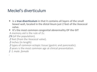
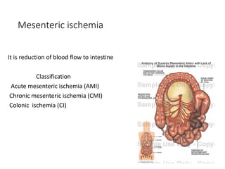
GAMBELLA UNIVERSITY
DEPARTMENT OF PUBLIC HEALTH (4rth year)
SEMINAR FOR SURGICAL WARD STUDENTS C-2
ATTACHMENT
TOPIC: GASTROINTESTINAL BLEEDING
PRESENTERS BY:1: (HO) Shaban
2: (HO) Gatwech chuol
PRESENTED TO: DR BHAN(ass...professor surgeon)
Introduction
•GI bleeding is a common clinical problem accounting
for 1-2% of all acute admissions with diverse
manifestations.
•May range from trivial to massive
•Can originate from any region of the GIT including
the esophagus, stomach ,Intestines, pancreas, liver
and biliary tree.
History
•On set , volume and frequency ,color- estimating
blood loss
•Hematemesis
•epistaxis
•Melena( foul smelling tarry stool, UGIB)
•Hematochezia is bright red bleeding(LGIB)
➢Antecedent vomiting, - Mallory-Weiss tear
➢ Weight ,appetite loss - malignancy
➢ Age - elderly patients bleed from lesions such as
angiodysplasias, diverticula, ischemic colitis, and
cancer,
➢Younger patients bleed from peptic ulcers, varices,
and Meckel's diverticulum
Antecedent Epigastric pain ►peptic ulcer
previous aortic surgery ►aorto-enteric
fistula
A history of liver disease, Jaundice
►variceal bleeding,
Hx of ingestion of salicylates, NSAIDs, SSRIs
►GI mucosal erosions
P/E
Postural hypotension
Pallor
PR –elevation by 20 beats/minute reflects at least
a 20% blood loss
Obtundation, agitation, and hypotension + cool,
clammy extremities, consistent with hemorrhagic
shock and suggest a loss of > 40% of the patient's
BV
❑Palpable left supra clavicular LN and
abdominal mass-----GI cancer.
❑Epigastric tenderness-----PUD.
❑A rectal exam and anoscope are performed
to exclude a low-lying rectal cancer or
bleeding from hemorrhoids
Risk Factors for Morbidity and Mortality in Acute Gastrointestinal Hemorrhage
Age >60 yr
Comorbid disease
Renal failure
Liver disease
Respiratory insufficiency
Cardiac disease
Magnitude of the hemorrhage
Systolic blood pressure <100 mm Hg on presentation
Transfusion requirement
Persistent or recurrent hemorrhage
Onset of hemorrhage during hospitalization
Need for surgery
UPPER GI BLEEDING
Definition
•Upper GI bleeding is bleeding that arises from the GI
tract proximal to the ligament of Treitz.
•Accounts for ~80% of significant GI bleeding.
Causes of Upper GI bleeding
•Can be classified based on their relation to portal
HTN into:-
Non variceal Bleeding(80%) Variceal Bleeding(20%)
PUD (30-50%) Gastroesophageal Varices (>90%)
Mallory Weiss syndrome (15-20%) Hypertensive Portal gastropathy(<5%)
Gastritis or duodenitis (10-15%) Isolated gastric varices (rare)
Esophagitis (5-10%)
Arteriovenous malformation(5%)
Tumours (2%)
Others (5%)
Non variceal bleeding
• ~10-15% of patients with PUD develop bleeding.
• It is the most frequent indication for operation and the principal cause
of death.
• Duodenal ulcers are more common than gastric ulcers.
• massive bleeding
PUD
• If the patient is not at risk of rebleeding
-Assess for H. pylori
-Treat with PPI.
Endoscopic therapy –
- epinephrine injection,
- Thermal coagulation&
- Clipping at ulcer site to constrict or
compress
surgical treatment –for patients at high risk of failed
endoscopic therapy.
➢Indications for Surgery in GI
hemorrhage
=› Hemodynamic instability despite vigorous resuscitation
=› Failure of endoscopic techniques to arrest hemorrhage
=› Recurrent hemorrhage after initial stabilization
=› Shock associated with recurrent hemorrhage
=› Continued slow bleeding .
CTD….
◆ Measures to prevent re-bleeding;
✓ H. pylori; most common cause of PUD should be treated.
✓ NSAIDs,SSRI should be discontinued.
Mallory Weiss syndrome
• common in alcoholic patients
• Most tears occur along the lesser curvature
• Dx by history(may be missed wz endoscopy)
• 90% of bleeding are self limited
• Recurrent bleeding is uncommon
management
Mallory Weiss syndrome
❖ resuscitation
❖Local Endoscopic therapy wz injection
❖ angiographic embolization
❖If this mauver failed high gastrotomy and
sutring of z mucosal tear
❖Avoid alcohol
Gastritis
•Results from acid and pepsin injury due to ischemia
from hypoperfusion states.
•Also caused by NSAIDs, ,stress, alcohol
•Cxzed by the appearance of multiple superficial
erosion of the entire stomach.
•Clinical presentation-painless upper GI bleeding.
management
❖Treatment of underlying sepsis
❖Discontinuation of NSAIDs
❖saline lavage & remove any pooled blood
❖Ng tube decompression
❖H2 antagonists
❖PPIs or Sucralfate
❖If failed, Vasopressin Or Surgical Tx- Vagotomy and
pyloroplasty
❖Prophylactic antiacid
Gastritis
Esophagitis
• Due to repeated exposure of esophageal mucosa to the acid
gastric secretion in GERD.
• Results in inflammatory response which can involve chronic
blood loss.
• Also caused by a variety of infectious agents particularly in
immunocompromised patients
• Medications, Crohn's disease, radiation are additional causes.
Management
❖Acid suppressive therapy.
❖Give antibiotic for infections.
❖Endoscopic control of the hemorrhage.
❖ Operation is seldomly necessary.
Esophagitis
Tumors
❖usually associated with chronic anemia
❖ can also present ulcerative lesions that bleed
persistently
❖ endoscopic therapy is often successful in
controlling hemorrhage
❖surgical resection.
Variceal bleeding
-Are almost always emergencies
causing hemodynamic shock &
multi organ failure
❖High risk of rebleeding,need for
transfusion & increased mortality
Complications of variceal bleeding
• The complications of variceal bleeding are due to either the bleeding
itself or the procedures used to control bleeding.
• Bleeding-related complications include vascular collapse and
hypotension, encephalopathy, aspiration, and subacute bacterial
peritonitis.
Management of Variceal Bleeding
❖General Resuscitation
❖Air way protection
❖Correct coagulopathy
❖Drug Therapy
❖Endoscopic Tx
✓ sclerotherapy using ethanolamine oleate
or butyl cyanoacrylate
✓ Banding
Esophageal ballon
tamponade(Sengstaken-Balkemore
tube)
✓reserved for patients with
massive hemorrhage to permit
more definitive therapies.
Prevention of variceal bleeding
•Treating the underlying cause of
bleeding varices
•Treating liver diseases earlier
• prophylactic b-blockers
LOWER GASTROINTESTINAL
BLEEDING
Lower GI Bleeding
❖LGI bleeding is defined as hemorrhage into the lumen of
the bowel from a source distal to the ligament of Treitz.
Causes of lower GI bleeding
❖Colorectal Cancer
❖IBD (UC, Crohn’s Disease)
❖Diverticular disease
❖ Anorectal disease
❖Mesenteric ischemia
❖Angiodysplasia
Diverticular Disease
Diverticulum: herniation of mucosa
through the circular muscle at the
site of penetration of blood vessels.
60% of people over 70 yo,very rare
before 35 yo
Most common segment affected is
sigmoid colon.
Its related to high intraluminal
pressure
Types of diverticuli
True diverticulum
• - congenital, young
• - proximal colon
• - pouch contain all layer
False diverticulum
• - acquired, elderly
• - distal colon
• pouch contain only mucosa
Meckel's diverticulum
 is a true diverticulum in that it contains all layers of the small
bowel wall, located in the distal ileum just 2 feet of the ileocecal
valve.
 It’s the most common congenital abnormality OF the GIT
A memory aid is the rule of 2s:
2% (of the population).
2 feet (from the ileocecal valve).
2 inches (in length)
2 types of common ectopic tissue (gastric and pancreatic).
2 years is the most common age at clinical presentation.
2 :1 male: female
.
5% symptomatic
o Abdominal pain, painless rectal bleeding, vomiting.
 Bleeding from Meckel's diverticulum is usually from an ulcerative
lesion on the ileal wall opposite the diverticulum, caused by acid
production by ectopic gastric mucosa.
Management
• T he best method for diagnosis and treatment is colonoscopy
✓If bleeding diverticula is identified
➢Epinephrine injection
➢Electrocautery
➢Endoscopic clips
✓If bleeding persists
➢Angiography with embolization
❖Indications for resection of the adjacent bowel:
➢ Bleeding
➢ strangulation
➢ bowel obstruction
Ulcerative Colitis
• A relapsing and remitting inflammatory disease
originating in the colonic mucosa and submucosa.
• UC is limited to the colon. Begins in the rectum and can
extend proximally, but rarely beyond ileocaecal junction.
• M = F but 2 peaks:
• i) 14-40 years old
• ii) 15% of new pts >60 years old at diagnosis
Complications of UC
❖Perforation,
➢Haemorrhage,
➢Toxic Megacolon (hypotonic, grossly distended bowel > 5cm)
➢Colorectal Ca
N.B death if complication is inadequately treated
Extraintestinal Manifestations in IBD
Crohn’s disease
❖Chronic transmural inflammatory GI Disease which is
non-caseating & granulomatous in nature. Associated
with recurrence in severe disease and extraintestinal
manifestations.
❖Can affect any portion of GI tract →Terminal Ileum
(50%) & Proximal Colon common. Inflammation is
discontinuous and separated by normal gut mucosa
(‘skip lesions’).
❖M = F affects any age but majority of cases between
11-35 years old
Complications in CD
➢Full-thickness mucosal inflammation→ Stricture→ Small bowel
obstruction
➢Abscess formation between loops of intestine
➢Perforation
➢Fistula formation: Enteroenteric, Enterocutaneous and Vesicocolic
Colorectal Cancer
❖3rd commonest malignancy in UK
❖M:F = 3:1 peak age 45-70yo
❖Risk Factor’s:
➢FH of Colorectal Ca, FAP, HNPCC,
➢Prev Hx of Colon, Breast, Ovarian or Uterine Ca
➢Prev Hx of Adenomatous Polyps
➢Chronic UC or Colonic Crohn’s disease
➢Western diet, Obesity, Smoking
❖Presentation depends on site:
➢Left-sided: Altered bowel habit (constipation & diarrhoea), PR ( bright
red bleeding coating the stool), Tenesmus, Painful defecation ,Small
diameter of Left Colon → Tendency towards obstruction
➢Right-sided: Present later. Weight loss, Right abdo pain/mass,
Tendency to bleed,PR (Blood mixed in with stools), high incidence of
IDA
❖Emergency (40%): Obstruction, Perforation with
Peritonitis, Acute Haemorrhage
Mesenteric ischemia
It is reduction of blood flow to intestine
Classification
Acute mesenteric ischemia (AMI)
Chronic mesenteric ischemia (CMI)
Colonic ischemia (CI)
PATHOPHYSILOGY
Impaired microcirculation leads to activation of inflammatory cells ,
and bacterial translocation
RISK FACTORS
age > 65 year
atherosclerotic disease
preexisting cardiovascular diseases (e.g. CHF)
known hypercoagulable state
Medication (e.g. vasopressor)
Inraabdomina malignancy
Cont’d…………..
Commonest site of ischemic colitis
Watershed areas –Splenic flexure
-Rectosegmoid junction
Clinical features
ON HX
• Sever abdominal pain of abrupt onset
• Bloody diarrhea
• Nausea
• Vomiting
• FEVER
ON P/E
–tachycardia
-abdominal tenderness
-Rebound tenderness
-blood on DRE
Clinical stages of ischemic colitis
Hyperactive phases
sever pain ,bloody diarrhea
Paralytic phase
pain become diffuse and continuous ,
Distended abdomen ,tender with absent
Bowel sound
Shock phase (10-15%)
rapid HR and metabolic acidosis ,fever , electrolyte
disturbance
bowel become gangrenes
* Rapid surgical intervention is required
Angiodysplasia
It is acquired arteriovenous malformation in the colonic submucosa
is responsible for 3-20% LGI Bleeding
It is degenerative disease secondary to progressive dilatation of
normal blood vessels in submucosa
Cont’d….....
• More common >50 years of age patients
• Mostly associated with renal failure and aortic stenosis
• presents with recurrent fresh blood bleeding
• Often asymptomatic
diagnosis
COLONSCOPY
Red , flat lesions about 2-10 cm of diameter
ANGIOGRAPHY
Dilated, slowly emptying veins
management
• Bleeding stops spontaneously in most cases with high recurrence rate
within 5 years
• Arterial embolization
• Endoscopic coagulation
if bleeding fails to stop surgical resection is effective
Gi bleeding seminar topic .pdfinjury.pdf

Gi bleeding seminar topic .pdfinjury.pdf

  • 1.
    GAMBELLA UNIVERSITY DEPARTMENT OFPUBLIC HEALTH (4rth year) SEMINAR FOR SURGICAL WARD STUDENTS C-2 ATTACHMENT TOPIC: GASTROINTESTINAL BLEEDING PRESENTERS BY:1: (HO) Shaban 2: (HO) Gatwech chuol PRESENTED TO: DR BHAN(ass...professor surgeon)
  • 2.
    Introduction •GI bleeding isa common clinical problem accounting for 1-2% of all acute admissions with diverse manifestations. •May range from trivial to massive •Can originate from any region of the GIT including the esophagus, stomach ,Intestines, pancreas, liver and biliary tree.
  • 3.
    History •On set ,volume and frequency ,color- estimating blood loss •Hematemesis •epistaxis •Melena( foul smelling tarry stool, UGIB) •Hematochezia is bright red bleeding(LGIB)
  • 4.
    ➢Antecedent vomiting, -Mallory-Weiss tear ➢ Weight ,appetite loss - malignancy ➢ Age - elderly patients bleed from lesions such as angiodysplasias, diverticula, ischemic colitis, and cancer, ➢Younger patients bleed from peptic ulcers, varices, and Meckel's diverticulum
  • 5.
    Antecedent Epigastric pain►peptic ulcer previous aortic surgery ►aorto-enteric fistula A history of liver disease, Jaundice ►variceal bleeding, Hx of ingestion of salicylates, NSAIDs, SSRIs ►GI mucosal erosions
  • 6.
    P/E Postural hypotension Pallor PR –elevationby 20 beats/minute reflects at least a 20% blood loss Obtundation, agitation, and hypotension + cool, clammy extremities, consistent with hemorrhagic shock and suggest a loss of > 40% of the patient's BV
  • 7.
    ❑Palpable left supraclavicular LN and abdominal mass-----GI cancer. ❑Epigastric tenderness-----PUD. ❑A rectal exam and anoscope are performed to exclude a low-lying rectal cancer or bleeding from hemorrhoids
  • 8.
    Risk Factors forMorbidity and Mortality in Acute Gastrointestinal Hemorrhage Age >60 yr Comorbid disease Renal failure Liver disease Respiratory insufficiency Cardiac disease Magnitude of the hemorrhage Systolic blood pressure <100 mm Hg on presentation Transfusion requirement Persistent or recurrent hemorrhage Onset of hemorrhage during hospitalization Need for surgery
  • 9.
  • 10.
    Definition •Upper GI bleedingis bleeding that arises from the GI tract proximal to the ligament of Treitz. •Accounts for ~80% of significant GI bleeding.
  • 11.
    Causes of UpperGI bleeding •Can be classified based on their relation to portal HTN into:- Non variceal Bleeding(80%) Variceal Bleeding(20%) PUD (30-50%) Gastroesophageal Varices (>90%) Mallory Weiss syndrome (15-20%) Hypertensive Portal gastropathy(<5%) Gastritis or duodenitis (10-15%) Isolated gastric varices (rare) Esophagitis (5-10%) Arteriovenous malformation(5%) Tumours (2%) Others (5%)
  • 12.
    Non variceal bleeding •~10-15% of patients with PUD develop bleeding. • It is the most frequent indication for operation and the principal cause of death. • Duodenal ulcers are more common than gastric ulcers. • massive bleeding PUD
  • 13.
    • If thepatient is not at risk of rebleeding -Assess for H. pylori -Treat with PPI. Endoscopic therapy – - epinephrine injection, - Thermal coagulation& - Clipping at ulcer site to constrict or compress surgical treatment –for patients at high risk of failed endoscopic therapy.
  • 14.
    ➢Indications for Surgeryin GI hemorrhage =› Hemodynamic instability despite vigorous resuscitation =› Failure of endoscopic techniques to arrest hemorrhage =› Recurrent hemorrhage after initial stabilization =› Shock associated with recurrent hemorrhage =› Continued slow bleeding .
  • 15.
    CTD…. ◆ Measures toprevent re-bleeding; ✓ H. pylori; most common cause of PUD should be treated. ✓ NSAIDs,SSRI should be discontinued.
  • 16.
    Mallory Weiss syndrome •common in alcoholic patients • Most tears occur along the lesser curvature • Dx by history(may be missed wz endoscopy) • 90% of bleeding are self limited • Recurrent bleeding is uncommon
  • 17.
    management Mallory Weiss syndrome ❖resuscitation ❖Local Endoscopic therapy wz injection ❖ angiographic embolization ❖If this mauver failed high gastrotomy and sutring of z mucosal tear ❖Avoid alcohol
  • 18.
    Gastritis •Results from acidand pepsin injury due to ischemia from hypoperfusion states. •Also caused by NSAIDs, ,stress, alcohol •Cxzed by the appearance of multiple superficial erosion of the entire stomach. •Clinical presentation-painless upper GI bleeding.
  • 19.
    management ❖Treatment of underlyingsepsis ❖Discontinuation of NSAIDs ❖saline lavage & remove any pooled blood ❖Ng tube decompression ❖H2 antagonists ❖PPIs or Sucralfate ❖If failed, Vasopressin Or Surgical Tx- Vagotomy and pyloroplasty ❖Prophylactic antiacid Gastritis
  • 20.
    Esophagitis • Due torepeated exposure of esophageal mucosa to the acid gastric secretion in GERD. • Results in inflammatory response which can involve chronic blood loss. • Also caused by a variety of infectious agents particularly in immunocompromised patients • Medications, Crohn's disease, radiation are additional causes.
  • 21.
    Management ❖Acid suppressive therapy. ❖Giveantibiotic for infections. ❖Endoscopic control of the hemorrhage. ❖ Operation is seldomly necessary. Esophagitis
  • 22.
    Tumors ❖usually associated withchronic anemia ❖ can also present ulcerative lesions that bleed persistently ❖ endoscopic therapy is often successful in controlling hemorrhage ❖surgical resection.
  • 23.
    Variceal bleeding -Are almostalways emergencies causing hemodynamic shock & multi organ failure ❖High risk of rebleeding,need for transfusion & increased mortality
  • 24.
    Complications of varicealbleeding • The complications of variceal bleeding are due to either the bleeding itself or the procedures used to control bleeding. • Bleeding-related complications include vascular collapse and hypotension, encephalopathy, aspiration, and subacute bacterial peritonitis.
  • 25.
    Management of VaricealBleeding ❖General Resuscitation ❖Air way protection ❖Correct coagulopathy ❖Drug Therapy ❖Endoscopic Tx ✓ sclerotherapy using ethanolamine oleate or butyl cyanoacrylate ✓ Banding
  • 26.
    Esophageal ballon tamponade(Sengstaken-Balkemore tube) ✓reserved forpatients with massive hemorrhage to permit more definitive therapies.
  • 27.
    Prevention of varicealbleeding •Treating the underlying cause of bleeding varices •Treating liver diseases earlier • prophylactic b-blockers
  • 29.
  • 31.
    Lower GI Bleeding ❖LGIbleeding is defined as hemorrhage into the lumen of the bowel from a source distal to the ligament of Treitz.
  • 32.
    Causes of lowerGI bleeding ❖Colorectal Cancer ❖IBD (UC, Crohn’s Disease) ❖Diverticular disease ❖ Anorectal disease ❖Mesenteric ischemia ❖Angiodysplasia
  • 33.
    Diverticular Disease Diverticulum: herniationof mucosa through the circular muscle at the site of penetration of blood vessels. 60% of people over 70 yo,very rare before 35 yo Most common segment affected is sigmoid colon. Its related to high intraluminal pressure
  • 34.
    Types of diverticuli Truediverticulum • - congenital, young • - proximal colon • - pouch contain all layer False diverticulum • - acquired, elderly • - distal colon • pouch contain only mucosa
  • 36.
    Meckel's diverticulum  isa true diverticulum in that it contains all layers of the small bowel wall, located in the distal ileum just 2 feet of the ileocecal valve.  It’s the most common congenital abnormality OF the GIT A memory aid is the rule of 2s: 2% (of the population). 2 feet (from the ileocecal valve). 2 inches (in length) 2 types of common ectopic tissue (gastric and pancreatic). 2 years is the most common age at clinical presentation. 2 :1 male: female .
  • 37.
    5% symptomatic o Abdominalpain, painless rectal bleeding, vomiting.  Bleeding from Meckel's diverticulum is usually from an ulcerative lesion on the ileal wall opposite the diverticulum, caused by acid production by ectopic gastric mucosa.
  • 38.
    Management • T hebest method for diagnosis and treatment is colonoscopy ✓If bleeding diverticula is identified ➢Epinephrine injection ➢Electrocautery ➢Endoscopic clips ✓If bleeding persists ➢Angiography with embolization
  • 39.
    ❖Indications for resectionof the adjacent bowel: ➢ Bleeding ➢ strangulation ➢ bowel obstruction
  • 41.
    Ulcerative Colitis • Arelapsing and remitting inflammatory disease originating in the colonic mucosa and submucosa. • UC is limited to the colon. Begins in the rectum and can extend proximally, but rarely beyond ileocaecal junction. • M = F but 2 peaks: • i) 14-40 years old • ii) 15% of new pts >60 years old at diagnosis
  • 42.
    Complications of UC ❖Perforation, ➢Haemorrhage, ➢ToxicMegacolon (hypotonic, grossly distended bowel > 5cm) ➢Colorectal Ca N.B death if complication is inadequately treated
  • 43.
  • 44.
    Crohn’s disease ❖Chronic transmuralinflammatory GI Disease which is non-caseating & granulomatous in nature. Associated with recurrence in severe disease and extraintestinal manifestations. ❖Can affect any portion of GI tract →Terminal Ileum (50%) & Proximal Colon common. Inflammation is discontinuous and separated by normal gut mucosa (‘skip lesions’). ❖M = F affects any age but majority of cases between 11-35 years old
  • 45.
    Complications in CD ➢Full-thicknessmucosal inflammation→ Stricture→ Small bowel obstruction ➢Abscess formation between loops of intestine ➢Perforation ➢Fistula formation: Enteroenteric, Enterocutaneous and Vesicocolic
  • 46.
    Colorectal Cancer ❖3rd commonestmalignancy in UK ❖M:F = 3:1 peak age 45-70yo ❖Risk Factor’s: ➢FH of Colorectal Ca, FAP, HNPCC, ➢Prev Hx of Colon, Breast, Ovarian or Uterine Ca ➢Prev Hx of Adenomatous Polyps ➢Chronic UC or Colonic Crohn’s disease ➢Western diet, Obesity, Smoking
  • 47.
    ❖Presentation depends onsite: ➢Left-sided: Altered bowel habit (constipation & diarrhoea), PR ( bright red bleeding coating the stool), Tenesmus, Painful defecation ,Small diameter of Left Colon → Tendency towards obstruction ➢Right-sided: Present later. Weight loss, Right abdo pain/mass, Tendency to bleed,PR (Blood mixed in with stools), high incidence of IDA ❖Emergency (40%): Obstruction, Perforation with Peritonitis, Acute Haemorrhage
  • 49.
    Mesenteric ischemia It isreduction of blood flow to intestine Classification Acute mesenteric ischemia (AMI) Chronic mesenteric ischemia (CMI) Colonic ischemia (CI)
  • 50.
    PATHOPHYSILOGY Impaired microcirculation leadsto activation of inflammatory cells , and bacterial translocation RISK FACTORS age > 65 year atherosclerotic disease preexisting cardiovascular diseases (e.g. CHF) known hypercoagulable state Medication (e.g. vasopressor) Inraabdomina malignancy
  • 51.
    Cont’d………….. Commonest site ofischemic colitis Watershed areas –Splenic flexure -Rectosegmoid junction
  • 52.
    Clinical features ON HX •Sever abdominal pain of abrupt onset • Bloody diarrhea • Nausea • Vomiting • FEVER ON P/E –tachycardia -abdominal tenderness -Rebound tenderness -blood on DRE
  • 53.
    Clinical stages ofischemic colitis Hyperactive phases sever pain ,bloody diarrhea Paralytic phase pain become diffuse and continuous , Distended abdomen ,tender with absent Bowel sound Shock phase (10-15%) rapid HR and metabolic acidosis ,fever , electrolyte disturbance bowel become gangrenes * Rapid surgical intervention is required
  • 54.
    Angiodysplasia It is acquiredarteriovenous malformation in the colonic submucosa is responsible for 3-20% LGI Bleeding It is degenerative disease secondary to progressive dilatation of normal blood vessels in submucosa
  • 55.
    Cont’d…..... • More common>50 years of age patients • Mostly associated with renal failure and aortic stenosis • presents with recurrent fresh blood bleeding • Often asymptomatic
  • 56.
    diagnosis COLONSCOPY Red , flatlesions about 2-10 cm of diameter ANGIOGRAPHY Dilated, slowly emptying veins
  • 57.
    management • Bleeding stopsspontaneously in most cases with high recurrence rate within 5 years • Arterial embolization • Endoscopic coagulation if bleeding fails to stop surgical resection is effective