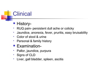
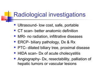
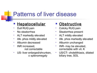
LFT
Liver function tests
Liver disease patterns
Hepatocellular
or
Cholestatic
Clinical
 History-
 RUQ pain- persistent dull ache or colicky
 Jaundice, anorexia, fever, pruritis, easy bruisability
 Color of stool & urine
 Personal & family history
 Examination-
 Pallor, jaundice, purpura
 Signs of CLD
 Liver, gall bladder, spleen, ascitis
Investigations-Ix-1
Bilirubin- a product of Hb metabolism
Indirect- lipid soluble
Direct- water soluble
Ix-2
 Aminotransferases- hepatocellular damage
 ALT/SGPT- found mainly in liver, more specific
 AST/SGOT- found in liver/muscle/heart/kidney; less specific
 Usually parallel rise/fall, exception being alcoholic
liver disease where AST >> ALT due to deficiency of
cofactor pyridoxine-5- phosphate
 Serial measurements more important
 Persistent elevation needs evaluation
Ix-3
 Alkaline phosphatase-
 Present in liver, bone, intestine, placenta
 In liver, mainly in biliary tract epithelium
 Marker of cholestasis/obstruction of biliary tract
 5’ nucleotidase-
 To confirm liver origin of alkaline phosphatase
 γ-glutamyl transpeptidase-
 Most sensitive indicator of biliary tract disease
 A marker of alcoholism
Ix-4
 Albumin-
 Half-life ~2-3 weeks
 Good marker of CLD
 Levels influenced by nutritional status, GI/renal loss,
redistribution in acute illness
 Clotting factors-
 Liver synthesizes factors I, II, V, VII, IX, X
 Need vitamin K- II, VII, IX, X
 Clotting impaired in acute or CLD
 Measured by elevated PT/INR
 Elevation a poor prognostic marker in liver disease
Ix-5
Ammonia- a product of urea cycle
Levels suggest hepatocellular function
Marker for hepatic encephalopathy
Ix-6
 Other-
 Markers for viral hepatitis
 Immunological-
Autoimmune hepatitis- ANA, LKM-1 Ab
PBC- anti-mitochondrial Ab
 AFP- Hepatocellular carcinoma
 Ferritin- hemochromatosis
 Ceruloplasmin- Wilson’s disease
 Liver biopsy
Radiological investigations
 Ultrasound- low cost, safe, portable
 CT scan- better anatomic definition
 MRI- no radiation, infiltrative diseases
 ERCP- biliary pathology, Dx & Rx
 PTC- dilated biliary tree, proximal disease
 HIDA scan- Dx of acute cholecystitis
 Angiography- Dx, resectability, palliation of
hepatic tumors or vascular lesions
Patterns of liver disease
 Hepatocellular
 Dull RUQ pain
 No steatorrhea
 ALT markedly elevated
 Alk. phos mildly elevated
 Albumin decreased
 INR increased,
not correctable
 US- liver enlarged/shrunken,
± splenomegaly
 Obstructive
 Colicky RUQ pain
 Steatorrhea present
 ALT mildly elevated
 Alk. phos markedly elevated
 Albumin unchanged
 INR- may be elevated,
correctable with vit. K
 US/CT- cholelithiasis, dilated
biliary tree, SOL

Liver function test

  • 1.
  • 2.
  • 3.
    Clinical  History-  RUQpain- persistent dull ache or colicky  Jaundice, anorexia, fever, pruritis, easy bruisability  Color of stool & urine  Personal & family history  Examination-  Pallor, jaundice, purpura  Signs of CLD  Liver, gall bladder, spleen, ascitis
  • 4.
    Investigations-Ix-1 Bilirubin- a productof Hb metabolism Indirect- lipid soluble Direct- water soluble
  • 5.
    Ix-2  Aminotransferases- hepatocellulardamage  ALT/SGPT- found mainly in liver, more specific  AST/SGOT- found in liver/muscle/heart/kidney; less specific  Usually parallel rise/fall, exception being alcoholic liver disease where AST >> ALT due to deficiency of cofactor pyridoxine-5- phosphate  Serial measurements more important  Persistent elevation needs evaluation
  • 6.
    Ix-3  Alkaline phosphatase- Present in liver, bone, intestine, placenta  In liver, mainly in biliary tract epithelium  Marker of cholestasis/obstruction of biliary tract  5’ nucleotidase-  To confirm liver origin of alkaline phosphatase  γ-glutamyl transpeptidase-  Most sensitive indicator of biliary tract disease  A marker of alcoholism
  • 7.
    Ix-4  Albumin-  Half-life~2-3 weeks  Good marker of CLD  Levels influenced by nutritional status, GI/renal loss, redistribution in acute illness  Clotting factors-  Liver synthesizes factors I, II, V, VII, IX, X  Need vitamin K- II, VII, IX, X  Clotting impaired in acute or CLD  Measured by elevated PT/INR  Elevation a poor prognostic marker in liver disease
  • 8.
    Ix-5 Ammonia- a productof urea cycle Levels suggest hepatocellular function Marker for hepatic encephalopathy
  • 9.
    Ix-6  Other-  Markersfor viral hepatitis  Immunological- Autoimmune hepatitis- ANA, LKM-1 Ab PBC- anti-mitochondrial Ab  AFP- Hepatocellular carcinoma  Ferritin- hemochromatosis  Ceruloplasmin- Wilson’s disease  Liver biopsy
  • 10.
    Radiological investigations  Ultrasound-low cost, safe, portable  CT scan- better anatomic definition  MRI- no radiation, infiltrative diseases  ERCP- biliary pathology, Dx & Rx  PTC- dilated biliary tree, proximal disease  HIDA scan- Dx of acute cholecystitis  Angiography- Dx, resectability, palliation of hepatic tumors or vascular lesions
  • 11.
    Patterns of liverdisease  Hepatocellular  Dull RUQ pain  No steatorrhea  ALT markedly elevated  Alk. phos mildly elevated  Albumin decreased  INR increased, not correctable  US- liver enlarged/shrunken, ± splenomegaly  Obstructive  Colicky RUQ pain  Steatorrhea present  ALT mildly elevated  Alk. phos markedly elevated  Albumin unchanged  INR- may be elevated, correctable with vit. K  US/CT- cholelithiasis, dilated biliary tree, SOL