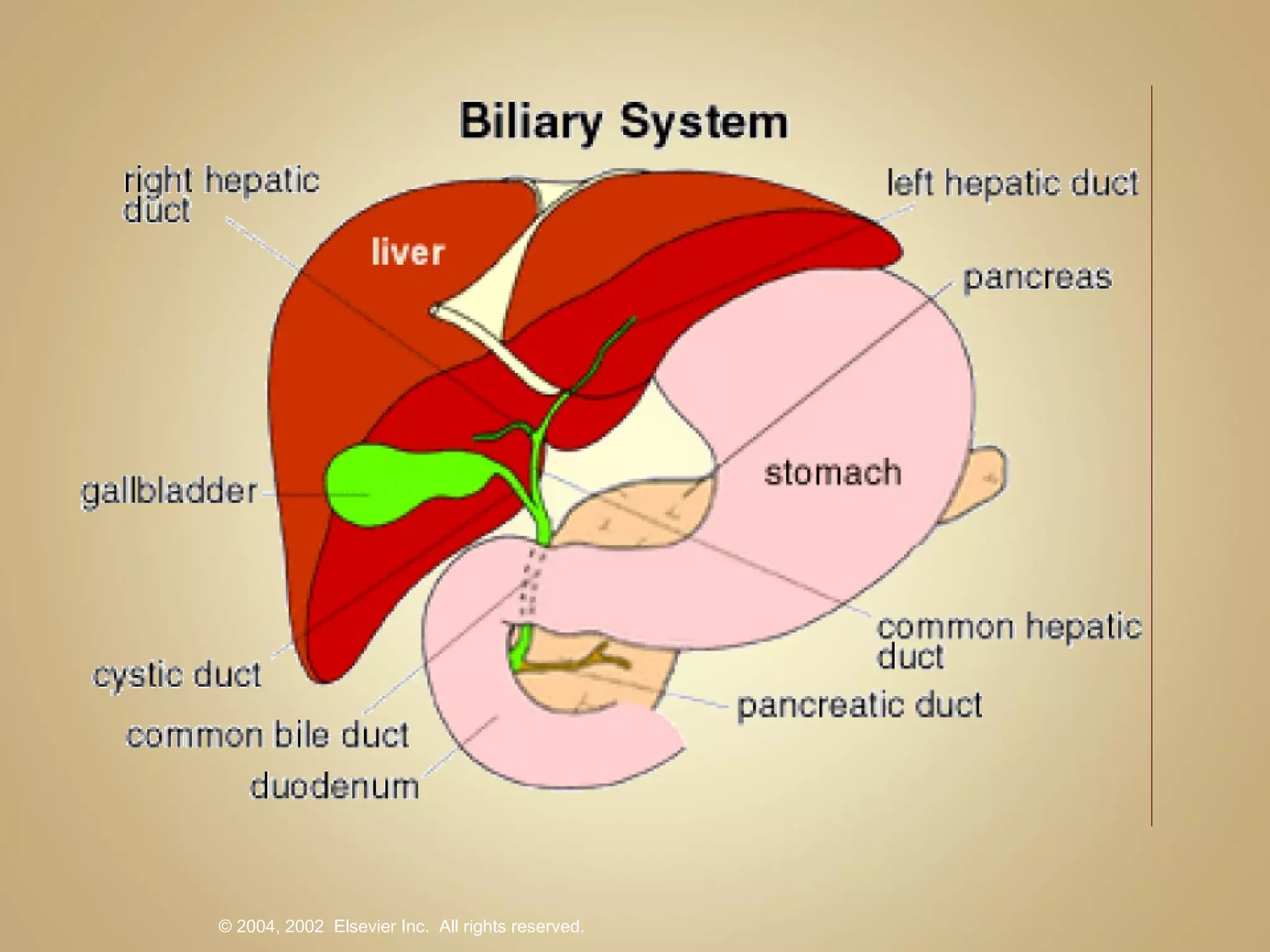
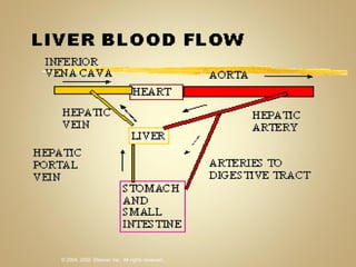
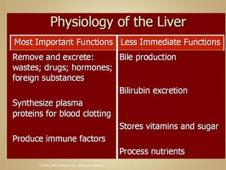
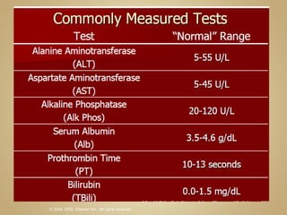
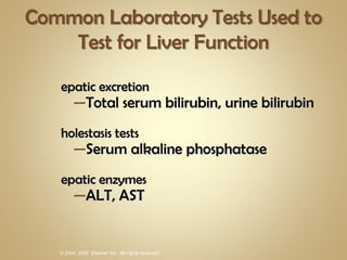
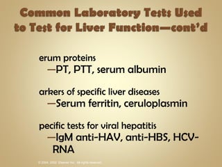
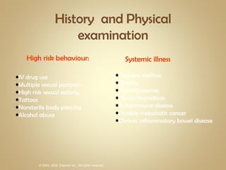
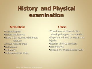
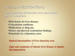
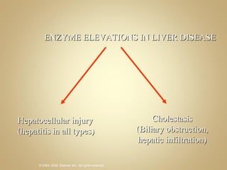
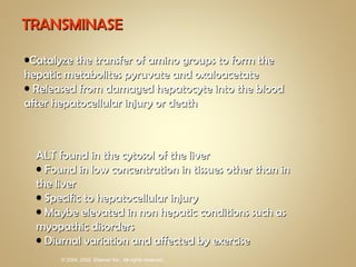
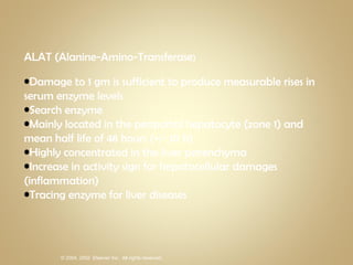
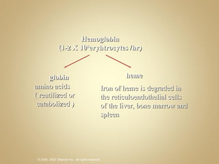
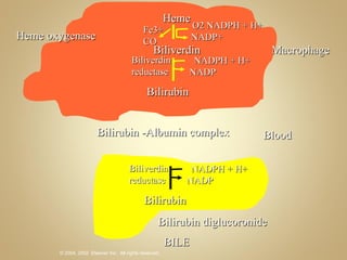
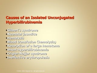
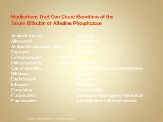
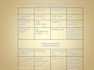
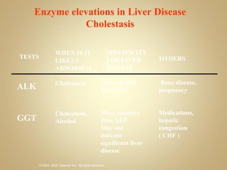
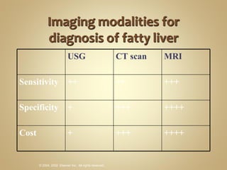
Hepatic excretion Total serum bilirubin, urine bilirubin Cholestasis tests Serum alkaline phosphatase Hepatic enzymes ALT, AST
Serum proteins PT, PTT, serum albumin Markers of specific liver diseases Serum ferritin, ceruloplasmin Specific tests for viral hepatitis IgM anti-HAV, anti-HBS, HCV-RNA
High risk behaviour:  IV drug use Multiple sexual partners High risk sexual activity Tattoos Nonsterile body piercing  Alcohol abuse Systemic illness Diabetis mellitus Obesity Hyperlipidemia Hemochromatosis Autoimmune disease Possible metastatic cancer Chronic inflammatory bowel disease
Medications Acetaminophen Herbal medications HMG-COA reductase inhibitors  (statins) Anticonvulsants drugs Isotretinoin Antibiotics Antituberculosis drugs Others Travel to or residence in less  developed regions or countries Exposure to blood or needle stick injuries Receipt of blood products Hemodialysis Ingesting of contaminated foods
INTIAL INVESTIGATIONS Initial evaluation of laboratory tests must involve patient’s symptoms: Risk factors for liver disease,  Concomitant conditions  Medications or drug use,  history and physical examination findings,  Potentially be a laboratory error. Marked abnormalities of liver chemistry tests   +  Signs and symptoms of chronic liver disease or hepatic decompensation
Test for hepatocellular damage   AST  ALT Test for cholestasis Alkaline Phosphatase Test for Biliary Excretion Total Bilirubin, Direct Bilirubin Test for liver synthetic function Albumin  Protime
ENZYME ELEVATIONS IN LIVER DISEASE Hepatocellular injury (hepatitis in all types) Cholestasis (Biliary obstruction, hepatic infiltration)
TRANSMINASE Catalyze the transfer of amino groups to form the hepatic metabolites pyruvate and oxaloacetate Released from damaged hepatocyte into the blood after hepatocellular injury or death ALT found in the cytosol of the liver Found in low concentration in tissues other than in the liver Specific to hepatocellular injury Maybe elevated in non hepatic conditions such as myopathic disorders Diurnal variation and affected by exercise
ALAT (Alanine-Amino-Transferase ) Damage to 1 gm is sufficient to produce measurable rises in serum enzyme levels Search enzyme Mainly located in the periportal hepatocyte (zone 1) and mean half life of 48 hours (+/- 10 h) Highly concentrated in the liver parenchyma Increase in activity sign for hepatocellular damages (inflammation) Tracing enzyme for liver diseases
AST  Found in the cytosol and mitochondria of the liver Abundantly expressed in several non-hepatic tissues including heart, skeletal muschle and blood
ASAT (Aspartate-Amino-Transferase) Mitochondrial enzymes (GOT, GLDH) are released for cell necrosis accompanied by destruction of mitochondria SGOT > SGPT Necrosis type (SGPT>SGOT Inflammation Type) SGOT: SGPT DeRitis Quotient: more severe   Prognosis all the more serious the more the activity of SGOT exceeds SGPT Mean half life SGOT is 17+/- 5 hours High activity in hepatocytes, myocardium, and in the skeletal muscles Increase in activity sign for liver disorders, heart attacks and damages in skeletal muscles
Additional test to identify the cause of elevated aminotransferase in asymptomatic patient
Bilirubin  Normal heme degradation product that is excreted from the body predominately via secretion into bile.  Insoluble in water and requires conjugation (glucuronidation) into the water-soluble bilirubin mono- and di-glucuronide forms before biliary secretion Bilirubin-UGT, the enzyme that conjugates bilirubin, is expressed shortly after birth.
JAUNDICE Most visible manifestation of liver and biliary tract Condition characterized by yellowish discoloration of skin,sclerae as a result of elevated bilirubin concentration
Hemoglobin (1-2 X 10 8 eryhtrocytes /hr) globin  heme Iron of heme is degraded in the reticuloendothelial cells of the liver, bone marrow and spleen  amino acids ( reutilized or catabolized )
Heme O2 NADPH + H+ NADP+ Fe3+ CO Biliverdin NADPH + H+ NADP Biliverdin reductase Bilirubin Macrophage Bilirubin -Albumin complex Blood Heme oxygenase NADPH + H+ NADP Biliverdin reductase Bilirubin Bilirubin diglucoronide BILE
Causes of an Isolated Unconjugated Hyperbilirubinemia Gilbert’s syndrome Neonatal jaundice Hemolysis Blood transfusion (hemolysis) Resorption of a large hematoma Shunt hyperbilirubinemia Crigler-Najjar syndrome Ineffective erythropoiesis
Causes of Conjugated Hyperbilirubinemia Bile duct obstruction Hepatitis Cirrhosis Medications/toxins Primary biliary cirrhosis Primary sclerosing cholangitis Sepsis Total parenteral nutrition Intrahepatic cholestasis of pregnancy Benign recurrent cholestasis Vanishing bile duct syndromes Dubin-Johnson syndrome Rotor syndrome
Medications That Can Cause Elevations of the Serum Bilirubin or Alkaline Phosphatase Anabolic steroids  Gold salts Allopurinol  Imipramine Amoxicillin-clavulanic acid  Indinivir Captopril  Iprindole Carbamazepine  Nevirapine Chlorpropamide  Methyltestosterone Cyproheptadine.  Methylenedioxymethamphetamine Diltiazem  Oxaprozin Erythromycin  Pizotyline Estrogens  Quinidine Floxuridine  Tolbutamide Flucloxacillin  Total parenteral hyperalimentation Fluphenazine  Trimethoprim-sulfamethoxazole
CHOLESTASIS Syndrome due to the impairment of the formation of bile secondary to drugs,  infectious autoimmune, metabolic or genetic disorder Secretion of bile depends on: Function of a number of membrane transport systems in hepatocytes and bile duct epithelial cells (Cholangiocytes) Structural function integrity of bile-secretory apparatus
Bile plugs
ALKALINE PHOSPHATASE Family of enzymes are zinc metalloenzymes  In the liver, the enzyme has been immuno-localized to the microvilli of the bile canaliculus.  Under normal conditions, it is predominantly is caused by liver and bone isoenzymes, (intestinal enzymes about 20% of total activity) Dependent  on a host of factors including the method of determination, patient age and gender, and the postprandial state. Elevated by cholestatic or infiltrative diseases of the liver and by diseases causing obstruction to the biliary system, bone diseases, medications, and tumors of hepatic and non-hepatic origin.  Important clinical issue is the determination of whether the alkaline phosphatase abnormality is of hepatobiliary or non-hepatic origin.
ALKALINE PHOSPHATASE Formed in the osteoblasts, small intestine, liver, bile duct system, duodenum kidneys, prostate, placenta Half live 3-7 days Physiologic increase occurs at growing age
Sources of elevated alkaline phosphatase: liver and bone Women in the third trimester of pregnancy  In persons with blood type O or B, serum alkaline phosphatase levels may increase after the ingestion of a fatty meal Vary with age. Rapidly growing adolescents can have serum alkaline phosphatase levels that are twice those of healthy adults (bone ) Normally increase  between the ages of 40 and 65 years, particularly in women.
Causes of Elevated Serum Alkaline Phosphatase Hepatobiliary Bile duct obstruction Primary biliary cirrhosis Primary sclerosing cholangitis Medications Infiltrating diseases of the liver Hepatic metastasis Hepatitis Cirrhosis Vanishing bile duct syndromes Benign recurrent cholestasis Nonhepatic Bone disease Pregnancy Chronic renal failure Lymphoma and other malignancies Congestive heart failure Childhood growth Infection/inflammation
ALK + AMA is positive: primary biliary cirrhosis. If:  the AMA  (-) ultrasonography (-),   alkaline phosphatase level remains >50% :liver biopsy  ERCP, MRCP. If the increase in the alkaline phosphatase level is <50%: observation
Infiltrating Diseases of the Liver That Can Cause Elevations of the Serum Alkaline Phosphatase Sarcoidosis Tuberculosis Fungal infection Other granulomatous diseases Amyloidosis Lymphoma Metastatic malignancy Hepatocellular carcinoma
 ‑ GT activity Does not correlate with the results for GPT and GOT Rise in   ‑GT is found in biliary tract diseases, liver-cell membrane damage and liver-cell regeneration, but also as a result of the induction of microsomal enzyme systems by  drugs. Not liver-cell specific Not cholestasis-specific (about 3% non-response). Not proof of liver-cell damage Not proof of alcohol abuse. It is instead:  Initially only a sign of increased activity of the detoxification function induced by alcohol, chemicals, medications or toxins, or evidence of cholestasis or liver-cell regeneration. Clinical experience shows that an increase in   ‑GT activity is found, for example, in some  intrahepatic and extrahepatic diseases
Graduated chemical laboratory program for the diagnosis or differential diagnosis of hepatobiliary diseases Minimum Necessary Maximum 1.Enzymatic activity: GPT,   -GT   GOT, GLDH   LDH 2.Synthesis output ChE, Quick value   Albumins   Ammonia, galactose 3.Excretory output: AP, bile pigments in urine   Bilirubin, LAP  Bile acids, iron, copper, cholesterol, indocyanine green, LP-X  Enzyme tracing grid Enzyme quotients  4.Mesenchymal activity:  -Globulins   Immunoglobulins,   A, G, M  Copper, procollagen III peptide 5.Immunological activity: HBs-Ag, HBc-Ab, AMA,   HA-Ab HBs-Ab, ANA   HBe-Ag, Hbe-Ab, HBV-DNA, HDV, HCV-Ab, SMA, LMA,   1 -fetoprotein
TESTS WHEN IS IT LIKELY ABNORMAL SPECIFICITY FOR LIVER DISEASE OTHERS Hepatitis (viral, autoimmune, drug induced, NAFLD, iron overload) Hepatitis (particularly alcoholic), hepatitic fibrosis, cirrhosis Sensitive and specific Less sensitive And specific than ALT Acute obstructive jaundice (within first 24 hours) Cardiac or skeletal muscle injury ALT AST Enzyme elevations in liver disease Hepatocellular injury
TESTS WHEN IS IT LIKELY ABNORMAL SPECIFICITY FOR LIVER DISEASE OTHERS More specific than GGT More sensitive than ALP May not indicate significant liver disease Bone disease, pregnancy Medications, hepatic congestion  ( CHF ) ALK GGT Enzyme elevations in Liver Disease Cholestasis Cholestasis Cholestasis, Alcohol
USG CT scan MRI Sensitivity ++ ++ +++ Specificity + +++ ++++ Cost + +++ ++++
Screening of High-risk Populations Tumor markers *Alpha-fetoprotein (AFP) Ultrasonography (US) *Easy *Simple *Low stress
Screening and Early Diagnosis of Primary Liver Cancer  Yang B et al. National Medical Journal of China (Zhonghua Yi Xue Za Zhi) 1999 AFP US AFP w/ or w/o US SENSITIVITY 69% 84% 92% SPECIFICITY 95% 97.1% 92.5% PPV 3.3% 6.6% 3.0% NPV 99.9% 99.9% 100%

Chapter31.liver

  • 1.
  • 2.
  • 3.
  • 4.
  • 5.
    Hepatic excretion Totalserum bilirubin, urine bilirubin Cholestasis tests Serum alkaline phosphatase Hepatic enzymes ALT, AST
  • 6.
    Serum proteins PT,PTT, serum albumin Markers of specific liver diseases Serum ferritin, ceruloplasmin Specific tests for viral hepatitis IgM anti-HAV, anti-HBS, HCV-RNA
  • 7.
    High risk behaviour: IV drug use Multiple sexual partners High risk sexual activity Tattoos Nonsterile body piercing Alcohol abuse Systemic illness Diabetis mellitus Obesity Hyperlipidemia Hemochromatosis Autoimmune disease Possible metastatic cancer Chronic inflammatory bowel disease
  • 8.
    Medications Acetaminophen Herbalmedications HMG-COA reductase inhibitors (statins) Anticonvulsants drugs Isotretinoin Antibiotics Antituberculosis drugs Others Travel to or residence in less developed regions or countries Exposure to blood or needle stick injuries Receipt of blood products Hemodialysis Ingesting of contaminated foods
  • 9.
    INTIAL INVESTIGATIONS Initialevaluation of laboratory tests must involve patient’s symptoms: Risk factors for liver disease, Concomitant conditions Medications or drug use, history and physical examination findings, Potentially be a laboratory error. Marked abnormalities of liver chemistry tests + Signs and symptoms of chronic liver disease or hepatic decompensation
  • 10.
    Test for hepatocellulardamage AST ALT Test for cholestasis Alkaline Phosphatase Test for Biliary Excretion Total Bilirubin, Direct Bilirubin Test for liver synthetic function Albumin Protime
  • 11.
    ENZYME ELEVATIONS INLIVER DISEASE Hepatocellular injury (hepatitis in all types) Cholestasis (Biliary obstruction, hepatic infiltration)
  • 12.
    TRANSMINASE Catalyze thetransfer of amino groups to form the hepatic metabolites pyruvate and oxaloacetate Released from damaged hepatocyte into the blood after hepatocellular injury or death ALT found in the cytosol of the liver Found in low concentration in tissues other than in the liver Specific to hepatocellular injury Maybe elevated in non hepatic conditions such as myopathic disorders Diurnal variation and affected by exercise
  • 13.
    ALAT (Alanine-Amino-Transferase )Damage to 1 gm is sufficient to produce measurable rises in serum enzyme levels Search enzyme Mainly located in the periportal hepatocyte (zone 1) and mean half life of 48 hours (+/- 10 h) Highly concentrated in the liver parenchyma Increase in activity sign for hepatocellular damages (inflammation) Tracing enzyme for liver diseases
  • 14.
    AST Foundin the cytosol and mitochondria of the liver Abundantly expressed in several non-hepatic tissues including heart, skeletal muschle and blood
  • 15.
    ASAT (Aspartate-Amino-Transferase) Mitochondrialenzymes (GOT, GLDH) are released for cell necrosis accompanied by destruction of mitochondria SGOT > SGPT Necrosis type (SGPT>SGOT Inflammation Type) SGOT: SGPT DeRitis Quotient: more severe Prognosis all the more serious the more the activity of SGOT exceeds SGPT Mean half life SGOT is 17+/- 5 hours High activity in hepatocytes, myocardium, and in the skeletal muscles Increase in activity sign for liver disorders, heart attacks and damages in skeletal muscles
  • 16.
    Additional test toidentify the cause of elevated aminotransferase in asymptomatic patient
  • 17.
    Bilirubin Normalheme degradation product that is excreted from the body predominately via secretion into bile. Insoluble in water and requires conjugation (glucuronidation) into the water-soluble bilirubin mono- and di-glucuronide forms before biliary secretion Bilirubin-UGT, the enzyme that conjugates bilirubin, is expressed shortly after birth.
  • 18.
    JAUNDICE Most visiblemanifestation of liver and biliary tract Condition characterized by yellowish discoloration of skin,sclerae as a result of elevated bilirubin concentration
  • 19.
    Hemoglobin (1-2 X10 8 eryhtrocytes /hr) globin heme Iron of heme is degraded in the reticuloendothelial cells of the liver, bone marrow and spleen amino acids ( reutilized or catabolized )
  • 20.
    Heme O2 NADPH+ H+ NADP+ Fe3+ CO Biliverdin NADPH + H+ NADP Biliverdin reductase Bilirubin Macrophage Bilirubin -Albumin complex Blood Heme oxygenase NADPH + H+ NADP Biliverdin reductase Bilirubin Bilirubin diglucoronide BILE
  • 21.
    Causes of anIsolated Unconjugated Hyperbilirubinemia Gilbert’s syndrome Neonatal jaundice Hemolysis Blood transfusion (hemolysis) Resorption of a large hematoma Shunt hyperbilirubinemia Crigler-Najjar syndrome Ineffective erythropoiesis
  • 22.
    Causes of ConjugatedHyperbilirubinemia Bile duct obstruction Hepatitis Cirrhosis Medications/toxins Primary biliary cirrhosis Primary sclerosing cholangitis Sepsis Total parenteral nutrition Intrahepatic cholestasis of pregnancy Benign recurrent cholestasis Vanishing bile duct syndromes Dubin-Johnson syndrome Rotor syndrome
  • 23.
    Medications That CanCause Elevations of the Serum Bilirubin or Alkaline Phosphatase Anabolic steroids Gold salts Allopurinol Imipramine Amoxicillin-clavulanic acid Indinivir Captopril Iprindole Carbamazepine Nevirapine Chlorpropamide Methyltestosterone Cyproheptadine. Methylenedioxymethamphetamine Diltiazem Oxaprozin Erythromycin Pizotyline Estrogens Quinidine Floxuridine Tolbutamide Flucloxacillin Total parenteral hyperalimentation Fluphenazine Trimethoprim-sulfamethoxazole
  • 24.
    CHOLESTASIS Syndrome dueto the impairment of the formation of bile secondary to drugs, infectious autoimmune, metabolic or genetic disorder Secretion of bile depends on: Function of a number of membrane transport systems in hepatocytes and bile duct epithelial cells (Cholangiocytes) Structural function integrity of bile-secretory apparatus
  • 25.
  • 26.
    ALKALINE PHOSPHATASE Familyof enzymes are zinc metalloenzymes In the liver, the enzyme has been immuno-localized to the microvilli of the bile canaliculus. Under normal conditions, it is predominantly is caused by liver and bone isoenzymes, (intestinal enzymes about 20% of total activity) Dependent on a host of factors including the method of determination, patient age and gender, and the postprandial state. Elevated by cholestatic or infiltrative diseases of the liver and by diseases causing obstruction to the biliary system, bone diseases, medications, and tumors of hepatic and non-hepatic origin. Important clinical issue is the determination of whether the alkaline phosphatase abnormality is of hepatobiliary or non-hepatic origin.
  • 27.
    ALKALINE PHOSPHATASE Formedin the osteoblasts, small intestine, liver, bile duct system, duodenum kidneys, prostate, placenta Half live 3-7 days Physiologic increase occurs at growing age
  • 28.
    Sources of elevatedalkaline phosphatase: liver and bone Women in the third trimester of pregnancy In persons with blood type O or B, serum alkaline phosphatase levels may increase after the ingestion of a fatty meal Vary with age. Rapidly growing adolescents can have serum alkaline phosphatase levels that are twice those of healthy adults (bone ) Normally increase between the ages of 40 and 65 years, particularly in women.
  • 29.
    Causes of ElevatedSerum Alkaline Phosphatase Hepatobiliary Bile duct obstruction Primary biliary cirrhosis Primary sclerosing cholangitis Medications Infiltrating diseases of the liver Hepatic metastasis Hepatitis Cirrhosis Vanishing bile duct syndromes Benign recurrent cholestasis Nonhepatic Bone disease Pregnancy Chronic renal failure Lymphoma and other malignancies Congestive heart failure Childhood growth Infection/inflammation
  • 30.
    ALK + AMAis positive: primary biliary cirrhosis. If: the AMA (-) ultrasonography (-), alkaline phosphatase level remains >50% :liver biopsy ERCP, MRCP. If the increase in the alkaline phosphatase level is <50%: observation
  • 31.
    Infiltrating Diseases ofthe Liver That Can Cause Elevations of the Serum Alkaline Phosphatase Sarcoidosis Tuberculosis Fungal infection Other granulomatous diseases Amyloidosis Lymphoma Metastatic malignancy Hepatocellular carcinoma
  • 32.
     ‑ GTactivity Does not correlate with the results for GPT and GOT Rise in  ‑GT is found in biliary tract diseases, liver-cell membrane damage and liver-cell regeneration, but also as a result of the induction of microsomal enzyme systems by drugs. Not liver-cell specific Not cholestasis-specific (about 3% non-response). Not proof of liver-cell damage Not proof of alcohol abuse. It is instead: Initially only a sign of increased activity of the detoxification function induced by alcohol, chemicals, medications or toxins, or evidence of cholestasis or liver-cell regeneration. Clinical experience shows that an increase in  ‑GT activity is found, for example, in some intrahepatic and extrahepatic diseases
  • 33.
    Graduated chemical laboratoryprogram for the diagnosis or differential diagnosis of hepatobiliary diseases Minimum Necessary Maximum 1.Enzymatic activity: GPT,  -GT  GOT, GLDH  LDH 2.Synthesis output ChE, Quick value  Albumins  Ammonia, galactose 3.Excretory output: AP, bile pigments in urine  Bilirubin, LAP  Bile acids, iron, copper, cholesterol, indocyanine green, LP-X  Enzyme tracing grid Enzyme quotients  4.Mesenchymal activity:  -Globulins  Immunoglobulins,  A, G, M Copper, procollagen III peptide 5.Immunological activity: HBs-Ag, HBc-Ab, AMA,  HA-Ab HBs-Ab, ANA  HBe-Ag, Hbe-Ab, HBV-DNA, HDV, HCV-Ab, SMA, LMA,  1 -fetoprotein
  • 34.
    TESTS WHEN ISIT LIKELY ABNORMAL SPECIFICITY FOR LIVER DISEASE OTHERS Hepatitis (viral, autoimmune, drug induced, NAFLD, iron overload) Hepatitis (particularly alcoholic), hepatitic fibrosis, cirrhosis Sensitive and specific Less sensitive And specific than ALT Acute obstructive jaundice (within first 24 hours) Cardiac or skeletal muscle injury ALT AST Enzyme elevations in liver disease Hepatocellular injury
  • 35.
    TESTS WHEN ISIT LIKELY ABNORMAL SPECIFICITY FOR LIVER DISEASE OTHERS More specific than GGT More sensitive than ALP May not indicate significant liver disease Bone disease, pregnancy Medications, hepatic congestion ( CHF ) ALK GGT Enzyme elevations in Liver Disease Cholestasis Cholestasis Cholestasis, Alcohol
  • 36.
    USG CT scanMRI Sensitivity ++ ++ +++ Specificity + +++ ++++ Cost + +++ ++++
  • 37.
    Screening of High-riskPopulations Tumor markers *Alpha-fetoprotein (AFP) Ultrasonography (US) *Easy *Simple *Low stress
  • 38.
    Screening and EarlyDiagnosis of Primary Liver Cancer Yang B et al. National Medical Journal of China (Zhonghua Yi Xue Za Zhi) 1999 AFP US AFP w/ or w/o US SENSITIVITY 69% 84% 92% SPECIFICITY 95% 97.1% 92.5% PPV 3.3% 6.6% 3.0% NPV 99.9% 99.9% 100%

Editor's Notes

  • #18 Bilirubin is a normal heme degradation product that is excreted from the body predominately via secretion into bile. Bilirubin is insoluble in water and requires conjugation (glucuronidation) into the water-soluble bilirubin mono- and di-glucuronide forms before biliary secretion. In the second decade of the twentieth century, van den Bergh and Muller16 used Ehrlich’s diazo reagent to determine that two types of bilirubin were present in the serum of jaundiced patients: one that reacted directly with the reagent (direct bilirubin) and a second form that required the addition of alcohol for color development (indirect bilirubin). Four decades later, independent work by Billing and Schmid demonstrated that unconjugated bilirubin was the indirect form, whereas the direct form was a combination of the bilirubin monoand di-glucuronides (conjugated bilirubin).17 Although the methodologies in serum bilirubin determination have advanced since this time, the terminology of direct and indirect bilirubin have remained virtually synonymous with conjugated and unconjugated bilirubin, respectively. To secrete bilirubin into bile, unconjugated bilirubin must be taken up into the hepatocyte and conjugated into the glucuronide form by the endoplasmic reticulum enzyme bilirubin UDP-glucuronyltransferase (bilirubin- UGT), and the water-soluble bilirubin glucuronides must be secreted across the canalicular membrane into bile. The molecular mechanisms of these processes recently have been delineated and reviewed18–20 but are beyond the scope of this document. Bilirubin-UGT, the enzyme that conjugates bilirubin, is expressed shortly after birth. However, once enzyme expression occurs, it continues to be highly expressed and active even in severe liver disease and cirrhosis.21,22 Diminished expression of this enzyme is one of the defects causing Gilbert’s syndrome, a benign, unconjugated hyperbilirubinemia occurring in up to 5% of the normal population.23–27 Unconjugated hyperbilirubinemia also may result from hemolysis (increased heme breakdown) or in rare genetic diseases such as the Crigler-Najjar syndrome.27,28 After the neonatal period, most hepatic conditions that result in a conjugated hyperbilirubinemia are caused by either extrahepatic obstruction of bile flow, intrahepatic cholestasis, hepatitis, or cirrhosis, with a resultant impairment of hepatocellular bilirubin secretion into bile. Because bilirubin-UGT expression and bilirubin conjugation typically are well preserved, these pathophysiological states usually result in a conjugated hyperbilirubinemia. When conjugated hyperbilirubinemia occurs, significant amounts of bilirubin may also be excreted via the urine.
  • #27 alkaline phosphatase family of enzymes are zinc metalloenzymes that are present in nearly all tissues. In liver, the enzyme has been immunolocalized to the microvilli of the bile canaliculus. Under normal conditions, serum alkaline phosphatase predominantly is caused by liver and bone isoenzymes, with intestinal enzymes contributing up to 20% of total activity. The normal reference range is dependent on a host of factors including the method of determination, patient age and gender, and the postprandial state.29 During normal pregnancy, alkaline phosphatase activity begins to rise by the late first trimester (because of placental isoenzymes), may reach levels of twice normal by term, and can remain elevated for several weeks after delivery.4,5 Serum alkaline phosphatase levels can be elevated by cholestatic or infiltrative diseases of the liver and by diseases causing obstruction to the biliary system, as well as by bone diseases, numerous medications, and tumors of hepatic and nonhepatic origin. When evaluating serum liver chemistries, the important clinical issue is the determination of whether the alkaline phosphatase abnormality is of hepatobiliary or nonhepatic origin. Liver alkaline phosphatase is more heat stable than bone, and isoenzyme determination can be made based on heat sensitivity; however, this assay may be subject to considerable inaccuracy and therefore its clinical use may be “laboratory-specific.” Other isoenzyme determination methodologies may include assays using monoclonal antibodies or wheat germ lectin precipitation.
  • #36 Gamma glutamyl transpeptidase (GGT) is a protein found in especially high levels in special cells (epithelium) lining the bile ducts of the liver. The GGT can be an especially sensitive test of biliary disease in the liver but an isolated elevated GGT in and of itself may have no meaning at all.