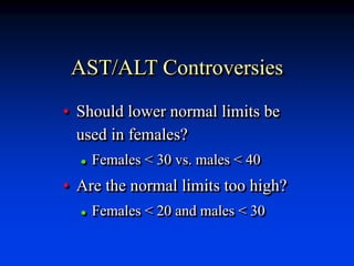
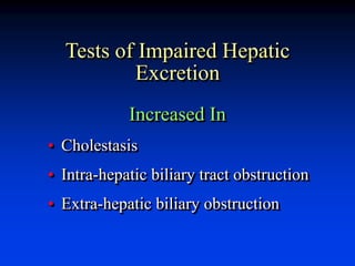
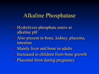
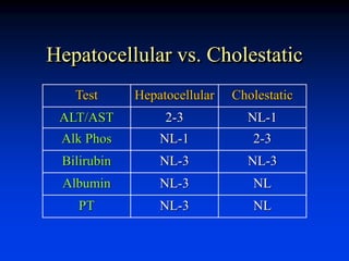
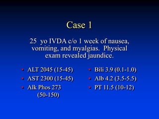
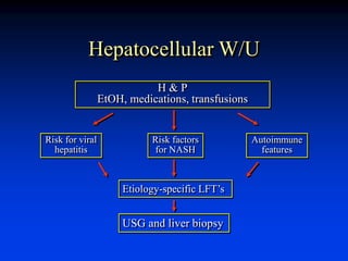
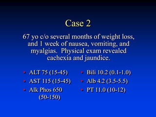
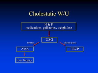
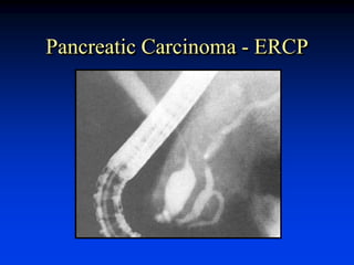
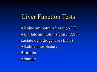
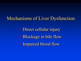
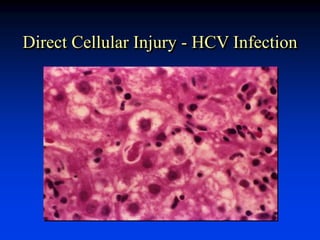
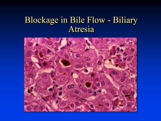
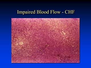
Evaluation of Liver Injury
Mark J. Czaja
Liver Research Center
Albert Einstein College of Medicine
Bronx, N.Y.
Liver Function Tests
• Alanine aminotransferase (ALT)
• Aspartate aminotransferase (AST)
• Lactate dehydrogenase (LDH)
• Alkaline phosphatase
• Bilirubin
• Albumin
Mechanisms of Liver Dysfunction
• Direct cellular injury
• Blockage in bile flow
• Impaired blood flow
Direct Cellular Injury - HCV Infection
Blockage in Bile Flow - Biliary
Atresia
Impaired Blood Flow - CHF
Consequences of Liver Injury
liver cell injury
liver cell death
proliferation matrix deposition
sufficient inadequate altered architecture
recovery liver failure cirrhosis
Types of Liver Tests
• True tests of liver function
• Biochemical markers of liver injury
• Biochemical markers of specific
liver diseases
Testable Biochemical Liver
Function
• Ability to transport organic anions
• Capacity to metabolize certain
substances
• Capability to synthesize various
proteins
Steps in Organic Anion Transport
• Delivery and uptake
• Metabolic alteration
• Secretion and excretion
Bilirubin
• Tetrapyrole
• Toxic in neonates - kernicterus
• Derived from:
 Senescent RBC (70-80%)
 Hemoproteins (20-30%)
 Ineffective erythropoiesis
Bilirubin Formation
heme biliverdin bilirubin
heme
oxygenase
biliverdin
reductase
Transport: hydrophobic due to internal H-bonding
circulates bound to albumin
Bilirubin Metabolism
UCB
ligandin
glucuronyl
transferase
BMG
BDG
Alb
UCB
BMG
BDG
Plasma Hepatocyte Bile
BMG
BDG
Bilirubin Elimination
Intestine
• BMG (20%) +
BDG (80%)
+UCB (trace)
• Deconjugated to
urobilinogen
• Excreted or reab-
sorbed (20%)
Urine
• BMG and BDG
• No UCB
Measurement of Serum
Bilirubin
• Normal concentration < 1 mg/dl
• Conjugated < 5%
• Jaundice if > 3 mg/dl
• Detected by diazo reaction - cleaved
to colored azo-dipyrole
 Conjugated reacts rapidly (direct)
 Unconjugated reacts slowly (indirect)
Differential Diagnosis I
• Prehepatic
• Intrahepatic
 Congenital
 Acquired
• Posthepatic
Differential Diagnosis II
• Unconjugated hyperbilirubinemia
 Increased bilirubin production
(hematological)
 Decreased uptake (drug)
 Decreased conjugation (congenital)
• Conjugated hyperbilirubinemia
 Congenital
 Drug
 Liver disease
 Biliary obstruction
Inherited Disorders Causing
Unconjugated Hyperbilirubinemia
• Crigler-Najjar syndrome
 Type 1 – absent GT
 Type 2 – reduced GT activity
• Gilbert’s syndrome – reduced GT
activity due to genetic defect in
TATAA element of GT promoter
Inherited Disorders Causing
Conjugated Hyperbilirubinemia
• Dubin-Johnson syndrome –
mutations in multidrug resistance
associated protein 2 (MRP2)
• Rotor’s syndrome – genetic
defect
Hepatic Metabolic Capacity
• Clearance must depend on total
functional mass or metabolic activity
• Hepatic drug metabolism -
[14C]amino-pyrine breath test
• Galactose elimination
• Not used clinically
Hepatic Synthetic Capacity
• Most major plasma proteins are made
in the liver
• Decreased hepatocytes = decreased
protein synthesis and release
• Albumin and coagulation factors are
clinically important
Albumin
• 50% of all synthesized hepatic
protein
• Determinant of plasma oncotic
pressure
• Important transport protein
Serum Albumin Levels
• Long half-life of 20 days
• Large hepatic synthetic reserve
• Decreased with persistent, large injury
• Decreased in chronic liver disease
• Poor prognostic sign
Non-hepatic Causes of
Hypoalbuminemia
• Severe malnutrition
• Renal or GI loss
 Glomerulopathy, HIV enteropathy
• High catabolism
 Infections, burns
Coagulation Factors
• Half-lives of hours to days
• Liver synthesizes I, II, V, VII,
IX, and X
• Large synthetic reserve
Prothrombin Time (PT)
• PT detects abnormalities in I, II,
V, VII and X (extrinsic pathway)
• PT is increased in liver disease
• Best prognostic indicator
 Acute liver disease
 Chronic liver disease
Non-hepatic Causes of
Elevated PT
• Congenital coagulation factor
deficiencies
• Consumptive coagulopathies
• Vitamin K deficiency (II, VII, IX, X)
To Rule Out Vitamin K Deficiency
• Any patient with an elevated PT
• Parental vitamin K for 3 days
• Normalization of PT - vitamin K
deficiency
• Failure to normalize - hepatocellular
disease
Serum Immunoglobulins
• Not produced by hepatocytes
• Frequently elevated in liver disease
• Secondary to inflammatory process
• ? produced by antigen shunting
Biochemical
Markers of Liver
Injury
Liver Enzymes
• Low levels always present in serum
• Leak out from cell after injury
• Very sensitive
• Magnitude of abnormality does not
correlate well with degree of injury
Aspartate Aminotransferase
(AST)
• Serum glutamic-oxaloacetic
transaminase (SGOT)
• Transfers an a-amino group of aspartate
to a-keto group of ketoglutaric acid
• Present in skeletal muscle, kidney, brain
Alanine Aminotransferase
(ALT)
• Serum glutamic-pyruvic transaminase
(SGPT)
• Transfers an a-amino group of alanine
to a-keto group of ketoglutaric acid
• Present principally in liver
AST and ALT
• Elevated in most liver diseases
• Highest levels are in acute liver
diseases
• Only slight elevations in chronic
liver diseases
• Usually increase in parallel
AST/ALT in Alcoholic
Hepatitis
• Transaminases rarely exceed 300
• AST:ALT >2
Factors Affecting AST/ALT
• Depressed by pyridoxine (vit. B6)
deficiency
• Decreased by uremia and renal dialysis
AST/ALT Controversies
• Should lower normal limits be
used in females?
 Females < 30 vs. males < 40
• Are the normal limits too high?
 Females < 20 and males < 30
Lactate Dehydrogenase
(LDH)
• Component of classic LFT’s
• Highly non-specific
Tests of Impaired Hepatic
Excretion
Increased In
• Cholestasis
• Intra-hepatic biliary tract obstruction
• Extra-hepatic biliary obstruction
Alkaline Phosphatase
• Hydrolyzes phosphate esters at
alkaline pH
• Also present in bone, kidney, placenta,
intestine
• Mainly liver and bone in adults
• Increased in children from bone growth
• Placental form during pregnancy
Elevated Alkaline Phosphatase
• Can occur in any liver disease
• Highest with cholestasis or biliary tract
obstruction
• Elevated in infiltrative diseases
• Due to increase synthesis and secretion
Alkaline Phosphatase Isoenzymes
Source
Heat
Inactivation 5' NT GGTP
Liver Moderate + +
Bone Rapid - -
Placenta Slow - -
Intestine Slow - -
5'-Nucleotidase
• Hydrolyzes 5'- adenosine monophosphate
• Mainly present in liver
• Increases along with alkaline phosphatase
g-Glutamyl Transpeptidase
(GGTP)
• Transfers g-glutamyl groups
• Widely distributed
• Sensitive correlate to alkaline phosphatase
• Non-specific (alcoholism, MI, DM,
pancreatic disease, renal failure)
Biochemical Markers
of Specific Liver
Diseases
Etiology-specific Liver Tests
• Viral hepatitis serologies
• Serum ferritin level
• Ceruloplasmin level
• Alpha1-antitrypsin level
• Antimitochondrial antibody titer
Viral Hepatitis Serology
• HAV – anti-HAV IgM and IgG
• HBV – HBsAg, anti-HBsAg,
and anti-HBcAg
• HCV – anti-HCV, HCV RNA
Serum Ferritin
• Widely distributed storage protein
• Levels reflect body iron stores
• Elevated in primary hemochromatosis
• Elevated in acute inflammation and
cirrhosis
Serum Ceruloplasmin
• Copper-binding protein
• Decreased in 95% of patients
with Wilson’s disease
• 20% of heterozygotes have
decreased levels
a1-Antitrypsin
• Inhibits serum trypsin
• Major component of a1-globulin
• Deficiency cause of neonatal
hepatitis
Antimitochondrial Antibody
(AMA)
• Directed against mitochondrial
enzyme pyruvate
dehydrogenase complex
• Positive in 90% of patients
with primary biliary cirrhosis
Interpretation of Abnormal
LFT’s
• Examine multiple tests
• Consider non-hepatic causes
• Determine the most abnormal tests
Hepatocellular vs. Cholestatic
Test Hepatocellular Cholestatic
ALT/AST 2-3 NL-1
Alk Phos NL-1 2-3
Bilirubin NL-3 NL-3
Albumin NL-3 NL
PT NL-3 NL
Case 1
• ALT 2045 (15-45)
• AST 2300 (15-45)
• Alk Phos 273
(50-150)
• Bili 3.9 (0.1-1.0)
• Alb 4.2 (3.5-5.5)
• PT 11.5 (10-12)
25 yo IVDA c/o 1 week of nausea,
vomiting, and myalgias. Physical
exam revealed jaundice.
Hepatocellular W/U
H & P
EtOH, medications, transfusions
Risk for viral
hepatitis
Risk factors
for NASH
Autoimmune
features
Etiology-specific LFT’s
USG and liver biopsy
HBV Infection - HBcAg Staining
Case 2
• ALT 75 (15-45)
• AST 115 (15-45)
• Alk Phos 650
(50-150)
• Bili 10.2 (0.1-1.0)
• Alb 4.2 (3.5-5.5)
• PT 11.0 (10-12)
67 yo c/o several months of weight loss,
and 1 week of nausea, vomiting, and
myalgias. Physical exam revealed
cachexia and jaundice.
Cholestatic W/U
H & P
medications, gallstones, weight loss
AMA
USG
ERCP
liver biopsy
dilated ducts
normal
Pancreatic Carcinoma - ERCP

20110722 - Czaja - LFTs.ppt

  • 1.
    Evaluation of LiverInjury Mark J. Czaja Liver Research Center Albert Einstein College of Medicine Bronx, N.Y.
  • 2.
    Liver Function Tests •Alanine aminotransferase (ALT) • Aspartate aminotransferase (AST) • Lactate dehydrogenase (LDH) • Alkaline phosphatase • Bilirubin • Albumin
  • 3.
    Mechanisms of LiverDysfunction • Direct cellular injury • Blockage in bile flow • Impaired blood flow
  • 4.
    Direct Cellular Injury- HCV Infection
  • 5.
    Blockage in BileFlow - Biliary Atresia
  • 6.
  • 7.
    Consequences of LiverInjury liver cell injury liver cell death proliferation matrix deposition sufficient inadequate altered architecture recovery liver failure cirrhosis
  • 8.
    Types of LiverTests • True tests of liver function • Biochemical markers of liver injury • Biochemical markers of specific liver diseases
  • 9.
    Testable Biochemical Liver Function •Ability to transport organic anions • Capacity to metabolize certain substances • Capability to synthesize various proteins
  • 10.
    Steps in OrganicAnion Transport • Delivery and uptake • Metabolic alteration • Secretion and excretion
  • 11.
    Bilirubin • Tetrapyrole • Toxicin neonates - kernicterus • Derived from:  Senescent RBC (70-80%)  Hemoproteins (20-30%)  Ineffective erythropoiesis
  • 12.
    Bilirubin Formation heme biliverdinbilirubin heme oxygenase biliverdin reductase Transport: hydrophobic due to internal H-bonding circulates bound to albumin
  • 13.
  • 14.
    Bilirubin Elimination Intestine • BMG(20%) + BDG (80%) +UCB (trace) • Deconjugated to urobilinogen • Excreted or reab- sorbed (20%) Urine • BMG and BDG • No UCB
  • 15.
    Measurement of Serum Bilirubin •Normal concentration < 1 mg/dl • Conjugated < 5% • Jaundice if > 3 mg/dl • Detected by diazo reaction - cleaved to colored azo-dipyrole  Conjugated reacts rapidly (direct)  Unconjugated reacts slowly (indirect)
  • 16.
    Differential Diagnosis I •Prehepatic • Intrahepatic  Congenital  Acquired • Posthepatic
  • 17.
    Differential Diagnosis II •Unconjugated hyperbilirubinemia  Increased bilirubin production (hematological)  Decreased uptake (drug)  Decreased conjugation (congenital) • Conjugated hyperbilirubinemia  Congenital  Drug  Liver disease  Biliary obstruction
  • 18.
    Inherited Disorders Causing UnconjugatedHyperbilirubinemia • Crigler-Najjar syndrome  Type 1 – absent GT  Type 2 – reduced GT activity • Gilbert’s syndrome – reduced GT activity due to genetic defect in TATAA element of GT promoter
  • 19.
    Inherited Disorders Causing ConjugatedHyperbilirubinemia • Dubin-Johnson syndrome – mutations in multidrug resistance associated protein 2 (MRP2) • Rotor’s syndrome – genetic defect
  • 20.
    Hepatic Metabolic Capacity •Clearance must depend on total functional mass or metabolic activity • Hepatic drug metabolism - [14C]amino-pyrine breath test • Galactose elimination • Not used clinically
  • 21.
    Hepatic Synthetic Capacity •Most major plasma proteins are made in the liver • Decreased hepatocytes = decreased protein synthesis and release • Albumin and coagulation factors are clinically important
  • 22.
    Albumin • 50% ofall synthesized hepatic protein • Determinant of plasma oncotic pressure • Important transport protein
  • 23.
    Serum Albumin Levels •Long half-life of 20 days • Large hepatic synthetic reserve • Decreased with persistent, large injury • Decreased in chronic liver disease • Poor prognostic sign
  • 24.
    Non-hepatic Causes of Hypoalbuminemia •Severe malnutrition • Renal or GI loss  Glomerulopathy, HIV enteropathy • High catabolism  Infections, burns
  • 25.
    Coagulation Factors • Half-livesof hours to days • Liver synthesizes I, II, V, VII, IX, and X • Large synthetic reserve
  • 26.
    Prothrombin Time (PT) •PT detects abnormalities in I, II, V, VII and X (extrinsic pathway) • PT is increased in liver disease • Best prognostic indicator  Acute liver disease  Chronic liver disease
  • 27.
    Non-hepatic Causes of ElevatedPT • Congenital coagulation factor deficiencies • Consumptive coagulopathies • Vitamin K deficiency (II, VII, IX, X)
  • 28.
    To Rule OutVitamin K Deficiency • Any patient with an elevated PT • Parental vitamin K for 3 days • Normalization of PT - vitamin K deficiency • Failure to normalize - hepatocellular disease
  • 29.
    Serum Immunoglobulins • Notproduced by hepatocytes • Frequently elevated in liver disease • Secondary to inflammatory process • ? produced by antigen shunting
  • 30.
  • 31.
    Liver Enzymes • Lowlevels always present in serum • Leak out from cell after injury • Very sensitive • Magnitude of abnormality does not correlate well with degree of injury
  • 32.
    Aspartate Aminotransferase (AST) • Serumglutamic-oxaloacetic transaminase (SGOT) • Transfers an a-amino group of aspartate to a-keto group of ketoglutaric acid • Present in skeletal muscle, kidney, brain
  • 33.
    Alanine Aminotransferase (ALT) • Serumglutamic-pyruvic transaminase (SGPT) • Transfers an a-amino group of alanine to a-keto group of ketoglutaric acid • Present principally in liver
  • 34.
    AST and ALT •Elevated in most liver diseases • Highest levels are in acute liver diseases • Only slight elevations in chronic liver diseases • Usually increase in parallel
  • 35.
    AST/ALT in Alcoholic Hepatitis •Transaminases rarely exceed 300 • AST:ALT >2
  • 36.
    Factors Affecting AST/ALT •Depressed by pyridoxine (vit. B6) deficiency • Decreased by uremia and renal dialysis
  • 37.
    AST/ALT Controversies • Shouldlower normal limits be used in females?  Females < 30 vs. males < 40 • Are the normal limits too high?  Females < 20 and males < 30
  • 38.
    Lactate Dehydrogenase (LDH) • Componentof classic LFT’s • Highly non-specific
  • 39.
    Tests of ImpairedHepatic Excretion Increased In • Cholestasis • Intra-hepatic biliary tract obstruction • Extra-hepatic biliary obstruction
  • 40.
    Alkaline Phosphatase • Hydrolyzesphosphate esters at alkaline pH • Also present in bone, kidney, placenta, intestine • Mainly liver and bone in adults • Increased in children from bone growth • Placental form during pregnancy
  • 41.
    Elevated Alkaline Phosphatase •Can occur in any liver disease • Highest with cholestasis or biliary tract obstruction • Elevated in infiltrative diseases • Due to increase synthesis and secretion
  • 42.
    Alkaline Phosphatase Isoenzymes Source Heat Inactivation5' NT GGTP Liver Moderate + + Bone Rapid - - Placenta Slow - - Intestine Slow - -
  • 43.
    5'-Nucleotidase • Hydrolyzes 5'-adenosine monophosphate • Mainly present in liver • Increases along with alkaline phosphatase
  • 44.
    g-Glutamyl Transpeptidase (GGTP) • Transfersg-glutamyl groups • Widely distributed • Sensitive correlate to alkaline phosphatase • Non-specific (alcoholism, MI, DM, pancreatic disease, renal failure)
  • 45.
  • 46.
    Etiology-specific Liver Tests •Viral hepatitis serologies • Serum ferritin level • Ceruloplasmin level • Alpha1-antitrypsin level • Antimitochondrial antibody titer
  • 47.
    Viral Hepatitis Serology •HAV – anti-HAV IgM and IgG • HBV – HBsAg, anti-HBsAg, and anti-HBcAg • HCV – anti-HCV, HCV RNA
  • 48.
    Serum Ferritin • Widelydistributed storage protein • Levels reflect body iron stores • Elevated in primary hemochromatosis • Elevated in acute inflammation and cirrhosis
  • 49.
    Serum Ceruloplasmin • Copper-bindingprotein • Decreased in 95% of patients with Wilson’s disease • 20% of heterozygotes have decreased levels
  • 50.
    a1-Antitrypsin • Inhibits serumtrypsin • Major component of a1-globulin • Deficiency cause of neonatal hepatitis
  • 51.
    Antimitochondrial Antibody (AMA) • Directedagainst mitochondrial enzyme pyruvate dehydrogenase complex • Positive in 90% of patients with primary biliary cirrhosis
  • 52.
    Interpretation of Abnormal LFT’s •Examine multiple tests • Consider non-hepatic causes • Determine the most abnormal tests
  • 53.
    Hepatocellular vs. Cholestatic TestHepatocellular Cholestatic ALT/AST 2-3 NL-1 Alk Phos NL-1 2-3 Bilirubin NL-3 NL-3 Albumin NL-3 NL PT NL-3 NL
  • 54.
    Case 1 • ALT2045 (15-45) • AST 2300 (15-45) • Alk Phos 273 (50-150) • Bili 3.9 (0.1-1.0) • Alb 4.2 (3.5-5.5) • PT 11.5 (10-12) 25 yo IVDA c/o 1 week of nausea, vomiting, and myalgias. Physical exam revealed jaundice.
  • 55.
    Hepatocellular W/U H &P EtOH, medications, transfusions Risk for viral hepatitis Risk factors for NASH Autoimmune features Etiology-specific LFT’s USG and liver biopsy
  • 56.
    HBV Infection -HBcAg Staining
  • 57.
    Case 2 • ALT75 (15-45) • AST 115 (15-45) • Alk Phos 650 (50-150) • Bili 10.2 (0.1-1.0) • Alb 4.2 (3.5-5.5) • PT 11.0 (10-12) 67 yo c/o several months of weight loss, and 1 week of nausea, vomiting, and myalgias. Physical exam revealed cachexia and jaundice.
  • 58.
    Cholestatic W/U H &P medications, gallstones, weight loss AMA USG ERCP liver biopsy dilated ducts normal
  • 59.

Editor's Notes

  • #3 Really tests indicating injury and not necessarily function. Ideal test would be highly specific and sensitive. No test fulfills that criteria.