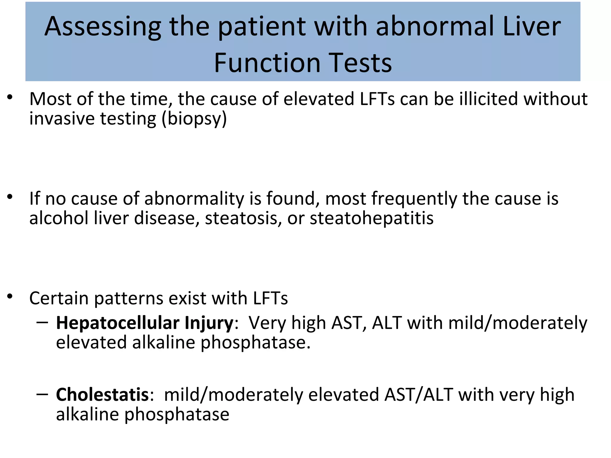
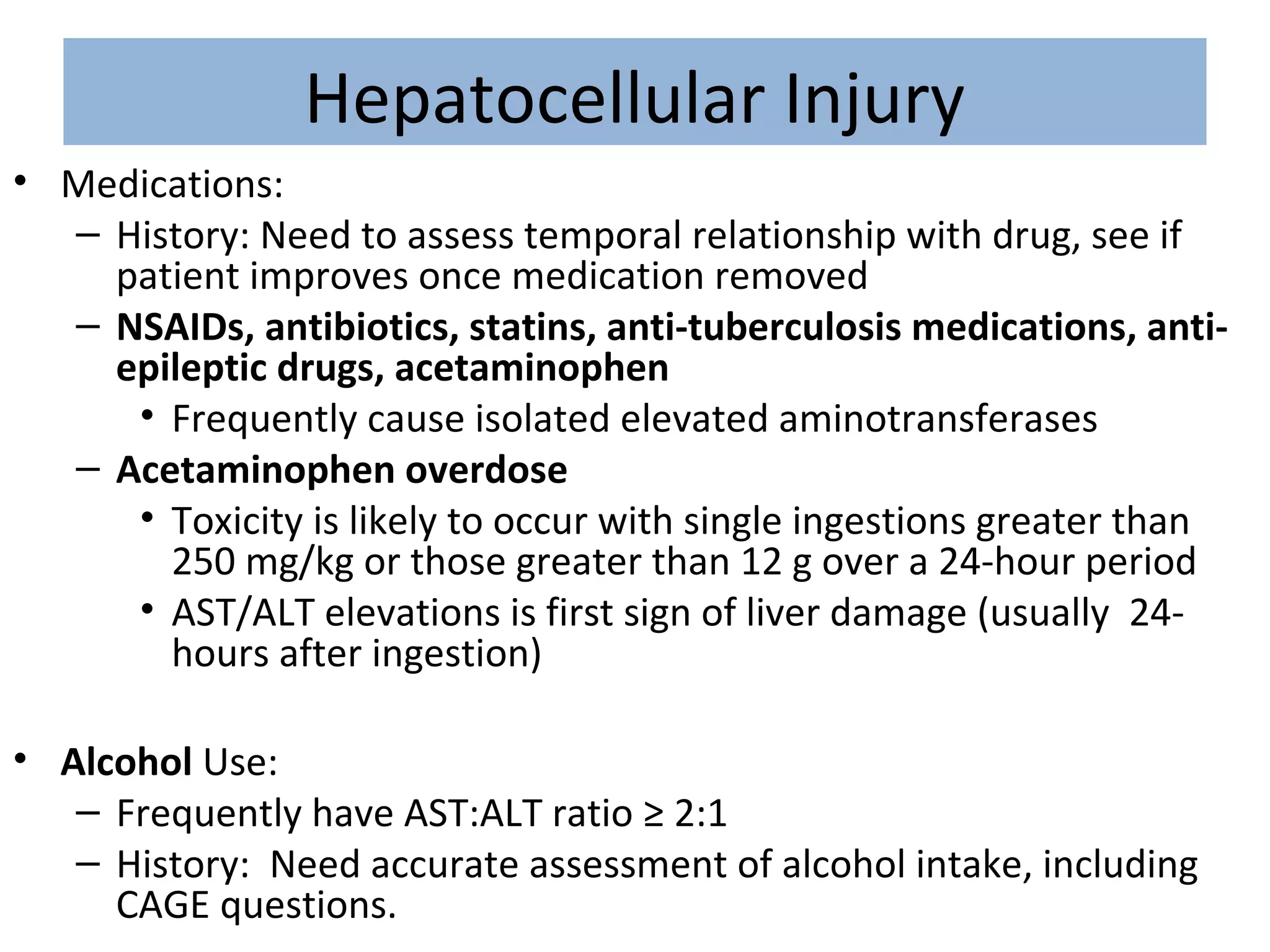
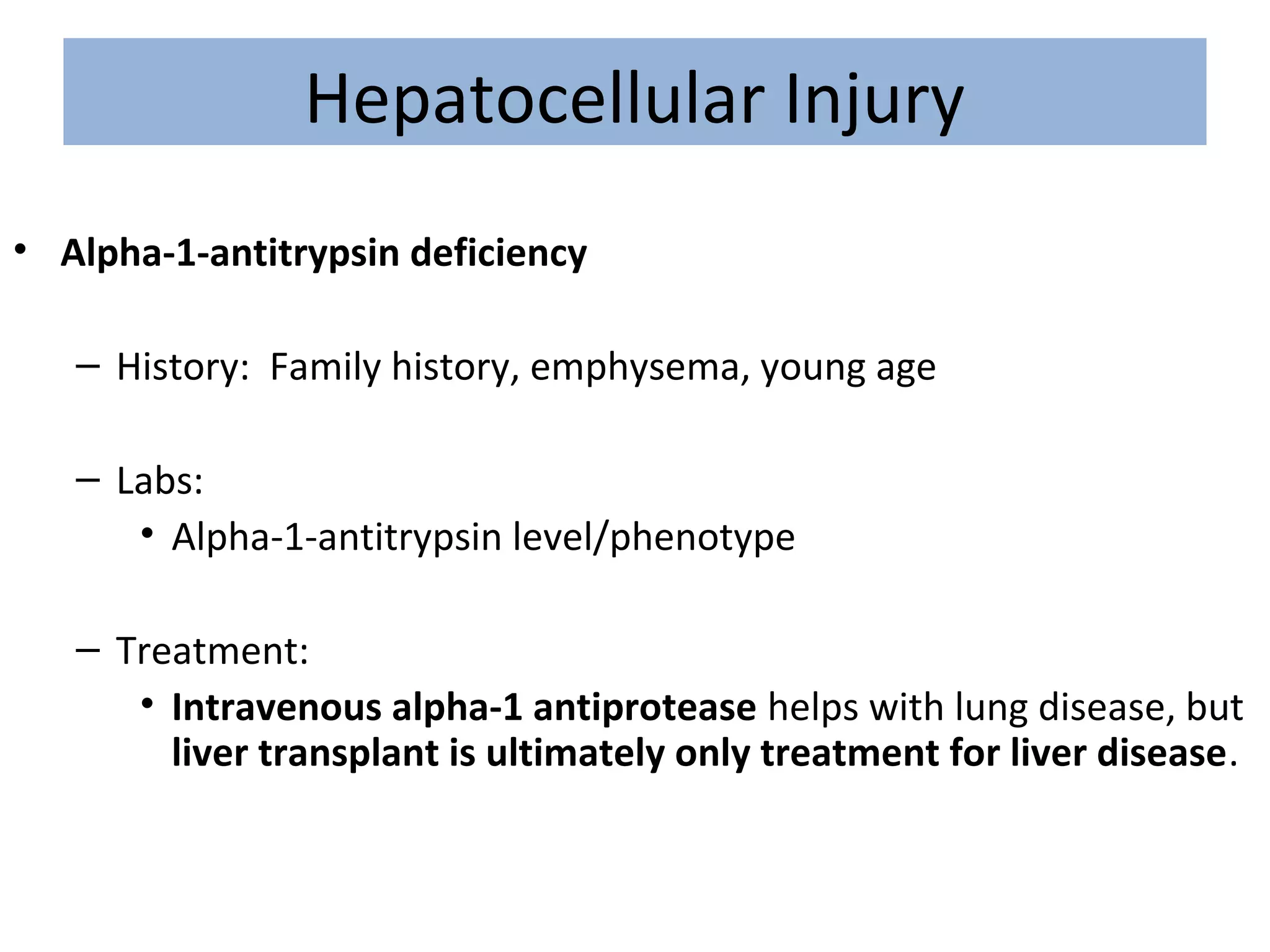
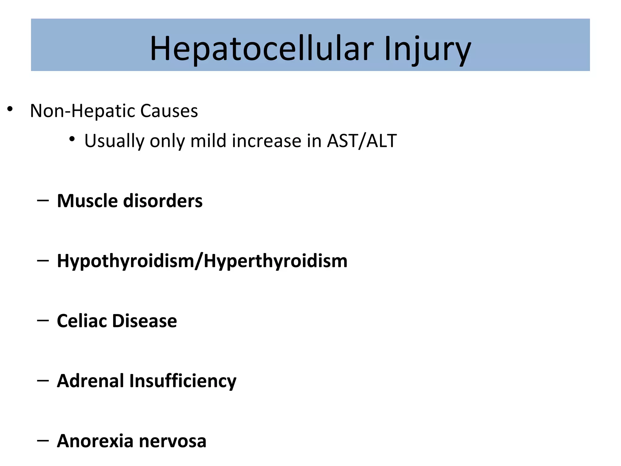
APPROACH TO LIVER FUNCTION
TEST
Dr.ABHINAV KUMAR
Dept. of Gastroenterology
Liver Function Test
• Albumin
• Bilirubin:
– Total Bilirubin
– Direct Bilirubin (conjugated bilirubin)
• Serum aminotransferases
– Aspartate aminotransferase (AST)
– Alanine aminotransferase (ALT)
• Alkaline Phosphatase
• Prothrombin time
Albumin
• Synthesized in the liver
• Production is controlled by multiple factors including nutritional
status, serum oncotic pressure, cytokines, and hormones
• A serum albumin may be reflection of the synthetic function of
the liver.
Bilirubin
• Used to determine liver’s ability to clear endogenous/exogenous
substances from the circulation
• Indirect (unconjugated) bilirubin
– Elevated with hemolysis, hepatic disease
• Direct (conjugated) bilirubin
– Elevated with biliary obstruction and hepatocellular disease.
• Jaundice usually develops with a bilirubin ≥ 3 mg/dL
Aminotransferases
• Hepatic enzymes that are usually intracellular, but are released from
hepatocytes with hepatocellular injury.
• Includes aspartate aminotransferase (AST) and alanine
aminotransferase (ALT)
• AST/ALT ratio
– Normal is 0.8
– In alcoholic hepatitis, is usually > 2
Alkaline Phosphatase
• A group of enzymes that catalyze the hydrolysis of a large number of
organic phosphate esters.
• In liver, believed to play an active role in down-regulating the
secretory activities of the intrahepatic biliary epithelium
• Found in:
• Liver
• Bone
• intestine
• First trimester placenta
• Kidney
– Gamma-glutamyl transpeptidase (GGT):
• Liver origin: Elevated GGT
• Bone origin: Normal GGT
Prothrombin Time (PT)
• Liver is in charge of the synthesis of many clotting factors :
– Factor I (fibrinogen)
– Factor II (prothrombin)
– Factor V
– Factor VII
– Factor IX
– Factor X
– Factors XII and XIII
• Elevated PT may be reflection of decreased synthetic activity of liver.
Assessing the patient with abnormal Liver
Function Tests
• Most of the time, the cause of elevated LFTs can be illicited without
invasive testing (biopsy)
• If no cause of abnormality is found, most frequently the cause is
alcohol liver disease, steatosis, or steatohepatitis
• Certain patterns exist with LFTs
– Hepatocellular Injury: Very high AST, ALT with mild/moderately
elevated alkaline phosphatase.
– Cholestatis: mild/moderately elevated AST/ALT with very high
alkaline phosphatase
Hepatocellular Injury
• Medications:
– History: Need to assess temporal relationship with drug, see if
patient improves once medication removed
– NSAIDs, antibiotics, statins, anti-tuberculosis medications, anti-
epileptic drugs, acetaminophen
• Frequently cause isolated elevated aminotransferases
– Acetaminophen overdose
• Toxicity is likely to occur with single ingestions greater than
250 mg/kg or those greater than 12 g over a 24-hour period
• AST/ALT elevations is first sign of liver damage (usually 24-
hours after ingestion)
• Alcohol Use:
– Frequently have AST:ALT ratio ≥ 2:1
– History: Need accurate assessment of alcohol intake, including
CAGE questions.
Hepatocellular Injury
• Hepatitis A:
– Acute infection
– History: travel, recent outbreak, MSM; nausea, vomiting, jaundice
– Labs: Hepatitis A IgM, frequent elevated bilirubin
• Hepatitis B:
– Can be acute or chronic
– History: See if patient from Asia, Subsaharan Africa; Sexual history,
Drug use
– Labs: Hepatitis B surface antigen, surface antibody, core antibody
• Hepatitis C:
– History: IV drug abuse, blood transfusion prior to 1992, Sexual history,
Tattoos
– Labs: Hepatitis C antibody (Hepatitis C viral load if HIV positive or
immunocompromised)
• HIV:
– Often causes isolated elevated aminotransferases
– History: Sexual History, IV drug use
– Labs: HIV Antibody test (ELISA with reflex Western Blot)
Hepatocellular Injury
• Hereditary Hemochromatosis
– History: Family history of liver disease? Diabetes? Heart Failure?
Bronze skin?
– Labs:
• Serum iron, TIBC
– Calculate iron saturation = serum iron/TIBC
– If iron saturation > 45%, check ferritin
• Ferritin
– If > 400 ng/mL in men, or > 300 ng/mL in women, then
need to check liver biopsy or genetic testing
• Liver biopsy
– Homozygous hereditary hemochromatosis if iron index >
1.9
– If under age 40, and positive genetic testing, no biopsy
needed.
• Genetic Testing
Hepatocellular Injury
• Hepatic steatosis/Non-alcoholic steatohepatitis (NASH)
– Increase in AST/ALT are usually less than 4-fold.
– Ratio of AST/ALT is usually < 1
– History: Female, obesity, diabetes
– Labs:
• Labs to rule out other causes of hepatitis
• Abdominal Ultrasound: look for fatty infiltration of liver
Hepatocellular Injury
• Autoimmune Hepatitis
– History: Young to middle-aged female
– Labs:
• Serum protein electrophoresis (SPEP) – if polyclonal increase
in gamma globulin
• Anti-nucleur antibody: Positive
• Anti-smooth-muscle antibody (SMA)
• Liver biopsy: should be performed if the above are negative,
but autoimmune hepatitis still suspected.
Hepatocellular Injury
• Alpha-1-antitrypsin deficiency
– History: Family history, emphysema, young age
– Labs:
• Alpha-1-antitrypsin level/phenotype
– Treatment:
• Intravenous alpha-1 antiprotease helps with lung disease, but
liver transplant is ultimately only treatment for liver disease.
Hepatocellular Injury
• Wilson’s Disease
– A genetic disorder of biliary copper excretion
– History: Age (usually age 5 – 25, but up to age 40), family history
of liver disease; neuropsychiatric disease
– Evaluation:
• Serum ceruloplasmin: Low
• Opthalmologist: Exam for Kayser-Fleisher rings
• 24-hour urine copper
• Liver biopsy: Evaluate liver copper levels
– Treatment:
• Copper chelating agents
• Zinc
Wilson’s Disease – Kayser-Fleisher Rings
Hepatocellular Injury
• Shock Liver (ischemic hepatitis)
– Etiology: Shock, severe hypotension
– Severely elevated AST/ALT (50 times normal)
– Treatment: Re-establish good blood pressure/perfusion.
– Prognosis: Usually patients recover, but can progress to
fulminant liver failure requiring transplant.
Hepatocellular Injury
• Non-Hepatic Causes
• Usually only mild increase in AST/ALT
– Muscle disorders
– Hypothyroidism/Hyperthyroidism
– Celiac Disease
– Adrenal Insufficiency
– Anorexia nervosa
Hepatocellular Injury
• What if work-up is negative and AST/ALT remain elevated?
– Observe:
• Patients with two-fold or less increase in AST/ALT and no
hyperbilirubinemia
– Liver Biopsy
• Patients with > two-fold increase in AST/ALT, or abnormalities
of other liver function tests.
Cholestatic Pattern
• Predominantly elevated alkaline phosphatase
– Need to check GGT to see if bone or liver in origin
• Blood types O and B: can have elevated serum alkaline phosphatase
after eating a fatty meal due to an influx of intestinal alkaline
phosphatase
• Need to determine if the cholestasis is intrahepatic or extrahepatic
in origin.
Cholestatic Pattern - Intrahepatic
• Drugs:
– Anabolic steroids, contraceptives, antibiotics
• Total parenteral nutrition (TPN)
• Cirrhosis:
– Viral hepatitis (Hepatitis B, C)
– Alcohol hepatitis
Cholestatic Pattern - Intrahepatic
• Primary Biliary Cirrhosis
– Autoimmune disease
– Predominately in women, usually ages 35-65
– May have history of other autoimmune disease
– Symptoms: Pruritis, fatigue, hyperpigmentation, musculoskeletal
complaints
– Labs:
• RUQ Ultrasound
• Anti-mitochondrial antibody
• Liver biopsy to verify diagnosis
Cholestatic Pattern – Both Intrahepatic and
Extrahepatic
• Primary Sclerosing Cholangitis
– chronic progressive disorder of unknown etiology that is
characterized by inflammation, fibrosis, and stricturing of medium
size and large ducts in the intrahepatic and extrahepatic biliary
tree
– ~ 90% have inflammatory bowel disease, especially ulcerative
colitis
– Symptoms: Pruritus, fatigue, RUQ pain
– Diagnosis:
• Ultrasound
• Cholangiogram: multifocal stricturing and dilation of
intrahepatic and/or extrahepatic bile ducts
– Prognosis:
• Poor; average life expectancy after diagnosis is ~12 years
• 10-15% risk of developing cholangiocarcinoma
• Liver transplant is ultimate only treatment
Cholangiogram of Primary Sclerosing
Cholangitis
Cholestatic Pattern - Extrahepatic
• Choledocholithiasis (gall stones!)
– History:
• The 3 F’s
• RUQ colicky abdominal pain
– Diagnosis/Treatment: Ultrasound, ERCP (to remove stones)
• Malignancy
– Cholangiocarcinoma
– Pancreatic
– Metastatic cancer
– Diagnosis: Ultrasound, MRCP
– Treatment: ERCP, biliary stent
Cholestatic Pattern - Extrahepatic
• Chronic Pancreatitis
– History: Recurrent pancreatitis
– Symptoms: Abdominal pain, frequently referred to back
• HIV Cholangiopathy
– Usually seen in AIDS patients with CD4 count well below 100/mm3
– Usually caused by: Cryptosporidium. Microsporidium, CMV
– Symptoms: RUQ pain, Diarrhea, Occassional fever, Occassional
jaundice
– Diagnosis:
• ERCP
• Cholangiography – shows multifocal strictures of extrahepatic
biliary tree
Isolated Hyperbilirubinemia
• indirect hyperbilirubinemia
– Overproduction of bilirubin
• Hemolysis
– Decrease in uptake, conjugation, or excretion of bilirubin
• Increased unconjugated (indirect) bilirubin
• Liver Disease
Isolated Unconjugated Hyperbilirubinemia
• Drugs
– Probenecid, Rifampicin
• Gilbert’s Disease
– Autosomal recessive disorder
– 3 to 7 % of population
– Most common in white males
– Jaundice, increased unconjugated bilirubin (always < 6)
– Occurs when patient under stress/infection
• Crigler-Najjar type II
– Caused by gene mutation
– Reduced activity of Bilirubin UDP glucuronosyl
SUMMARY
• Hepatocellular Injury –
mostly ↑ AST/ALT
– Drugs
– Alcohol hepatitis
– Hepatitis A
– Hepatitis B
– Hepatitis C
– Steatohepatitis (NASH)
– Autoimmune hepatitis
– Wilson’s Disease
– Hereditary
Hemochromatosis
– Alpha-1 antitrypsin
deficiency
SUMMARY
• EXTRAHEPATIC
– Gall stones
– Primary Sclerosing
Cholangitis
– Malignancy
– Chronic pancreatitis
– HIV cholangiopathy
Cholestatic Pattern
• INTRAHEPATIC
– Drugs
– Hepatitis A, B, C
– Alcoholic hepatitis
– TPN
– Primary Sclerosing
Cholangitis
– Primary Biliary Cirrhosis
SUMMARY
• Isolated elevated indirect (unconjugated) bilirubin
– Hemolysis
– Drugs
– Gilbert’s Disease
– Crigler-Najjar type II
Approach to lft

Approach to lft

  • 1.
    APPROACH TO LIVERFUNCTION TEST Dr.ABHINAV KUMAR Dept. of Gastroenterology
  • 2.
    Liver Function Test •Albumin • Bilirubin: – Total Bilirubin – Direct Bilirubin (conjugated bilirubin) • Serum aminotransferases – Aspartate aminotransferase (AST) – Alanine aminotransferase (ALT) • Alkaline Phosphatase • Prothrombin time
  • 3.
    Albumin • Synthesized inthe liver • Production is controlled by multiple factors including nutritional status, serum oncotic pressure, cytokines, and hormones • A serum albumin may be reflection of the synthetic function of the liver.
  • 4.
    Bilirubin • Used todetermine liver’s ability to clear endogenous/exogenous substances from the circulation • Indirect (unconjugated) bilirubin – Elevated with hemolysis, hepatic disease • Direct (conjugated) bilirubin – Elevated with biliary obstruction and hepatocellular disease. • Jaundice usually develops with a bilirubin ≥ 3 mg/dL
  • 5.
    Aminotransferases • Hepatic enzymesthat are usually intracellular, but are released from hepatocytes with hepatocellular injury. • Includes aspartate aminotransferase (AST) and alanine aminotransferase (ALT) • AST/ALT ratio – Normal is 0.8 – In alcoholic hepatitis, is usually > 2
  • 6.
    Alkaline Phosphatase • Agroup of enzymes that catalyze the hydrolysis of a large number of organic phosphate esters. • In liver, believed to play an active role in down-regulating the secretory activities of the intrahepatic biliary epithelium • Found in: • Liver • Bone • intestine • First trimester placenta • Kidney – Gamma-glutamyl transpeptidase (GGT): • Liver origin: Elevated GGT • Bone origin: Normal GGT
  • 7.
    Prothrombin Time (PT) •Liver is in charge of the synthesis of many clotting factors : – Factor I (fibrinogen) – Factor II (prothrombin) – Factor V – Factor VII – Factor IX – Factor X – Factors XII and XIII • Elevated PT may be reflection of decreased synthetic activity of liver.
  • 8.
    Assessing the patientwith abnormal Liver Function Tests • Most of the time, the cause of elevated LFTs can be illicited without invasive testing (biopsy) • If no cause of abnormality is found, most frequently the cause is alcohol liver disease, steatosis, or steatohepatitis • Certain patterns exist with LFTs – Hepatocellular Injury: Very high AST, ALT with mild/moderately elevated alkaline phosphatase. – Cholestatis: mild/moderately elevated AST/ALT with very high alkaline phosphatase
  • 9.
    Hepatocellular Injury • Medications: –History: Need to assess temporal relationship with drug, see if patient improves once medication removed – NSAIDs, antibiotics, statins, anti-tuberculosis medications, anti- epileptic drugs, acetaminophen • Frequently cause isolated elevated aminotransferases – Acetaminophen overdose • Toxicity is likely to occur with single ingestions greater than 250 mg/kg or those greater than 12 g over a 24-hour period • AST/ALT elevations is first sign of liver damage (usually 24- hours after ingestion) • Alcohol Use: – Frequently have AST:ALT ratio ≥ 2:1 – History: Need accurate assessment of alcohol intake, including CAGE questions.
  • 10.
    Hepatocellular Injury • HepatitisA: – Acute infection – History: travel, recent outbreak, MSM; nausea, vomiting, jaundice – Labs: Hepatitis A IgM, frequent elevated bilirubin • Hepatitis B: – Can be acute or chronic – History: See if patient from Asia, Subsaharan Africa; Sexual history, Drug use – Labs: Hepatitis B surface antigen, surface antibody, core antibody • Hepatitis C: – History: IV drug abuse, blood transfusion prior to 1992, Sexual history, Tattoos – Labs: Hepatitis C antibody (Hepatitis C viral load if HIV positive or immunocompromised) • HIV: – Often causes isolated elevated aminotransferases – History: Sexual History, IV drug use – Labs: HIV Antibody test (ELISA with reflex Western Blot)
  • 11.
    Hepatocellular Injury • HereditaryHemochromatosis – History: Family history of liver disease? Diabetes? Heart Failure? Bronze skin? – Labs: • Serum iron, TIBC – Calculate iron saturation = serum iron/TIBC – If iron saturation > 45%, check ferritin • Ferritin – If > 400 ng/mL in men, or > 300 ng/mL in women, then need to check liver biopsy or genetic testing • Liver biopsy – Homozygous hereditary hemochromatosis if iron index > 1.9 – If under age 40, and positive genetic testing, no biopsy needed. • Genetic Testing
  • 12.
    Hepatocellular Injury • Hepaticsteatosis/Non-alcoholic steatohepatitis (NASH) – Increase in AST/ALT are usually less than 4-fold. – Ratio of AST/ALT is usually < 1 – History: Female, obesity, diabetes – Labs: • Labs to rule out other causes of hepatitis • Abdominal Ultrasound: look for fatty infiltration of liver
  • 13.
    Hepatocellular Injury • AutoimmuneHepatitis – History: Young to middle-aged female – Labs: • Serum protein electrophoresis (SPEP) – if polyclonal increase in gamma globulin • Anti-nucleur antibody: Positive • Anti-smooth-muscle antibody (SMA) • Liver biopsy: should be performed if the above are negative, but autoimmune hepatitis still suspected.
  • 14.
    Hepatocellular Injury • Alpha-1-antitrypsindeficiency – History: Family history, emphysema, young age – Labs: • Alpha-1-antitrypsin level/phenotype – Treatment: • Intravenous alpha-1 antiprotease helps with lung disease, but liver transplant is ultimately only treatment for liver disease.
  • 15.
    Hepatocellular Injury • Wilson’sDisease – A genetic disorder of biliary copper excretion – History: Age (usually age 5 – 25, but up to age 40), family history of liver disease; neuropsychiatric disease – Evaluation: • Serum ceruloplasmin: Low • Opthalmologist: Exam for Kayser-Fleisher rings • 24-hour urine copper • Liver biopsy: Evaluate liver copper levels – Treatment: • Copper chelating agents • Zinc
  • 16.
    Wilson’s Disease –Kayser-Fleisher Rings
  • 17.
    Hepatocellular Injury • ShockLiver (ischemic hepatitis) – Etiology: Shock, severe hypotension – Severely elevated AST/ALT (50 times normal) – Treatment: Re-establish good blood pressure/perfusion. – Prognosis: Usually patients recover, but can progress to fulminant liver failure requiring transplant.
  • 18.
    Hepatocellular Injury • Non-HepaticCauses • Usually only mild increase in AST/ALT – Muscle disorders – Hypothyroidism/Hyperthyroidism – Celiac Disease – Adrenal Insufficiency – Anorexia nervosa
  • 19.
    Hepatocellular Injury • Whatif work-up is negative and AST/ALT remain elevated? – Observe: • Patients with two-fold or less increase in AST/ALT and no hyperbilirubinemia – Liver Biopsy • Patients with > two-fold increase in AST/ALT, or abnormalities of other liver function tests.
  • 20.
    Cholestatic Pattern • Predominantlyelevated alkaline phosphatase – Need to check GGT to see if bone or liver in origin • Blood types O and B: can have elevated serum alkaline phosphatase after eating a fatty meal due to an influx of intestinal alkaline phosphatase • Need to determine if the cholestasis is intrahepatic or extrahepatic in origin.
  • 21.
    Cholestatic Pattern -Intrahepatic • Drugs: – Anabolic steroids, contraceptives, antibiotics • Total parenteral nutrition (TPN) • Cirrhosis: – Viral hepatitis (Hepatitis B, C) – Alcohol hepatitis
  • 22.
    Cholestatic Pattern -Intrahepatic • Primary Biliary Cirrhosis – Autoimmune disease – Predominately in women, usually ages 35-65 – May have history of other autoimmune disease – Symptoms: Pruritis, fatigue, hyperpigmentation, musculoskeletal complaints – Labs: • RUQ Ultrasound • Anti-mitochondrial antibody • Liver biopsy to verify diagnosis
  • 23.
    Cholestatic Pattern –Both Intrahepatic and Extrahepatic • Primary Sclerosing Cholangitis – chronic progressive disorder of unknown etiology that is characterized by inflammation, fibrosis, and stricturing of medium size and large ducts in the intrahepatic and extrahepatic biliary tree – ~ 90% have inflammatory bowel disease, especially ulcerative colitis – Symptoms: Pruritus, fatigue, RUQ pain – Diagnosis: • Ultrasound • Cholangiogram: multifocal stricturing and dilation of intrahepatic and/or extrahepatic bile ducts – Prognosis: • Poor; average life expectancy after diagnosis is ~12 years • 10-15% risk of developing cholangiocarcinoma • Liver transplant is ultimate only treatment
  • 24.
    Cholangiogram of PrimarySclerosing Cholangitis
  • 25.
    Cholestatic Pattern -Extrahepatic • Choledocholithiasis (gall stones!) – History: • The 3 F’s • RUQ colicky abdominal pain – Diagnosis/Treatment: Ultrasound, ERCP (to remove stones) • Malignancy – Cholangiocarcinoma – Pancreatic – Metastatic cancer – Diagnosis: Ultrasound, MRCP – Treatment: ERCP, biliary stent
  • 26.
    Cholestatic Pattern -Extrahepatic • Chronic Pancreatitis – History: Recurrent pancreatitis – Symptoms: Abdominal pain, frequently referred to back • HIV Cholangiopathy – Usually seen in AIDS patients with CD4 count well below 100/mm3 – Usually caused by: Cryptosporidium. Microsporidium, CMV – Symptoms: RUQ pain, Diarrhea, Occassional fever, Occassional jaundice – Diagnosis: • ERCP • Cholangiography – shows multifocal strictures of extrahepatic biliary tree
  • 27.
    Isolated Hyperbilirubinemia • indirecthyperbilirubinemia – Overproduction of bilirubin • Hemolysis – Decrease in uptake, conjugation, or excretion of bilirubin • Increased unconjugated (indirect) bilirubin • Liver Disease
  • 28.
    Isolated Unconjugated Hyperbilirubinemia •Drugs – Probenecid, Rifampicin • Gilbert’s Disease – Autosomal recessive disorder – 3 to 7 % of population – Most common in white males – Jaundice, increased unconjugated bilirubin (always < 6) – Occurs when patient under stress/infection • Crigler-Najjar type II – Caused by gene mutation – Reduced activity of Bilirubin UDP glucuronosyl
  • 29.
    SUMMARY • Hepatocellular Injury– mostly ↑ AST/ALT – Drugs – Alcohol hepatitis – Hepatitis A – Hepatitis B – Hepatitis C – Steatohepatitis (NASH) – Autoimmune hepatitis – Wilson’s Disease – Hereditary Hemochromatosis – Alpha-1 antitrypsin deficiency
  • 30.
    SUMMARY • EXTRAHEPATIC – Gallstones – Primary Sclerosing Cholangitis – Malignancy – Chronic pancreatitis – HIV cholangiopathy Cholestatic Pattern • INTRAHEPATIC – Drugs – Hepatitis A, B, C – Alcoholic hepatitis – TPN – Primary Sclerosing Cholangitis – Primary Biliary Cirrhosis
  • 31.
    SUMMARY • Isolated elevatedindirect (unconjugated) bilirubin – Hemolysis – Drugs – Gilbert’s Disease – Crigler-Najjar type II