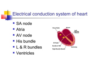
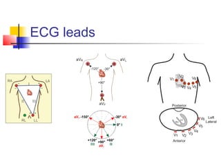
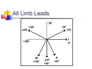
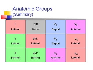
ECG
Heartbeat on a paper
Electrical conduction system of heart
 SA node
 Atria
 AV node
 His bundle
 L & R bundles
 Ventricles
ECG leads
All Limb Leads
Anatomic Groups
(Summary)
A heartbeat
 P- atrial depolarization
 QRS- ventricular depolarization
 T- ventricular repolarization
Interpreting ECG
 Rate-
 <60 bradycardia, >100 tachycardia
 Rhythm- relation of P & QRS
 Regular or irregular
 Axis- use QRS in leads I & aVF
 0-90 normal, <0 LAD, >90 RAD
 P wave- best seen in lead II
 Peaked P- P pulmonale- RAH, wide bifid P- P mitrale- LAH
 PR interval- normal ~0.2 sec.
 Short- WPW syndrome, long- AV blocks
Interpretation- continued
 QRS complex- normal <0.12 sec
 Broad- ectopic, BBB-RSR’ in V1 RBBB, ‘M’ in V1 LBBB
 Q wave >2 mm is pathological, suggesting old MI
 ST segment- difficult to interpret with BBB
 Depressed- ischemia, elevated- infarction
 Saddle shaped across the leads- pericarditis
 QT interval- normal ~0.4 sec
 Prolongation predisposes to ventricular arrythmias
 T wave- upright in all, except III & V1
 Tall tented- hyperkalemia
 Inverted- ischemia, previous infarction, hypokalemia
A normal ECG
Commonest arrythmia
Sinus brady/tachycardia
Ectopics- atrial/ventricular
Majority do not require Rx
Bradycardia- AV blocks
 1st
degree-
prolonged PR
 2nd
degree-
regular P & regular QRS,
uncoordinated
 3rd
degree-
irregular P & wide QRS
Treatment
Atropine
Pacemaker-
(temporary/permanent)
Tachyarrythmia
 Rate >100 bpm
 Types-
 Narrow-complex- supraventricular
Irregular- Atrial fibrillation
Regular at 300/150 bpm- Atrial flutter
Regular >150- AVNRT/AVRT
 Broad-complex- ventricular or SVT with BBB
h/o CAD- VT, unless proved otherwise
Management
 Unstable- DC cardioversion
 Stable-
 Asymptomatic ectopics- don’t treat
 Supraventricular- try carotid massage/valsalva
 Anti-arrythmics- Amiodarone-VT, Adenosine-SVT
 RF ablation, Maze surgery, AICD
 Control precipitating factors
 MI, infection, thyrotoxicosis, alcohol, caffeine,
electrolyte disorders, drug toxicity

Electrocardiogram

  • 1.
  • 2.
    Electrical conduction systemof heart  SA node  Atria  AV node  His bundle  L & R bundles  Ventricles
  • 3.
  • 4.
  • 5.
  • 6.
    A heartbeat  P-atrial depolarization  QRS- ventricular depolarization  T- ventricular repolarization
  • 7.
    Interpreting ECG  Rate- <60 bradycardia, >100 tachycardia  Rhythm- relation of P & QRS  Regular or irregular  Axis- use QRS in leads I & aVF  0-90 normal, <0 LAD, >90 RAD  P wave- best seen in lead II  Peaked P- P pulmonale- RAH, wide bifid P- P mitrale- LAH  PR interval- normal ~0.2 sec.  Short- WPW syndrome, long- AV blocks
  • 8.
    Interpretation- continued  QRScomplex- normal <0.12 sec  Broad- ectopic, BBB-RSR’ in V1 RBBB, ‘M’ in V1 LBBB  Q wave >2 mm is pathological, suggesting old MI  ST segment- difficult to interpret with BBB  Depressed- ischemia, elevated- infarction  Saddle shaped across the leads- pericarditis  QT interval- normal ~0.4 sec  Prolongation predisposes to ventricular arrythmias  T wave- upright in all, except III & V1  Tall tented- hyperkalemia  Inverted- ischemia, previous infarction, hypokalemia
  • 9.
  • 10.
    Commonest arrythmia Sinus brady/tachycardia Ectopics-atrial/ventricular Majority do not require Rx
  • 11.
    Bradycardia- AV blocks 1st degree- prolonged PR  2nd degree- regular P & regular QRS, uncoordinated  3rd degree- irregular P & wide QRS
  • 12.
  • 13.
    Tachyarrythmia  Rate >100bpm  Types-  Narrow-complex- supraventricular Irregular- Atrial fibrillation Regular at 300/150 bpm- Atrial flutter Regular >150- AVNRT/AVRT  Broad-complex- ventricular or SVT with BBB h/o CAD- VT, unless proved otherwise
  • 14.
    Management  Unstable- DCcardioversion  Stable-  Asymptomatic ectopics- don’t treat  Supraventricular- try carotid massage/valsalva  Anti-arrythmics- Amiodarone-VT, Adenosine-SVT  RF ablation, Maze surgery, AICD  Control precipitating factors  MI, infection, thyrotoxicosis, alcohol, caffeine, electrolyte disorders, drug toxicity