Download as PDF, PPTX


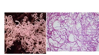
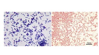
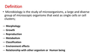
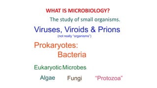
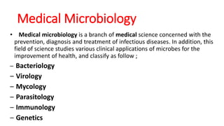
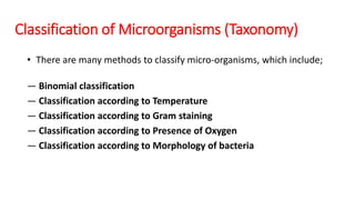
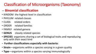
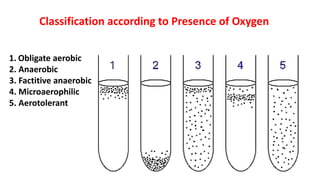
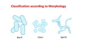
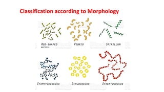
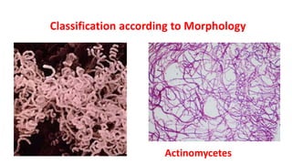
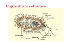
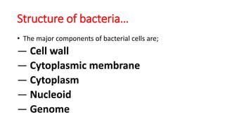
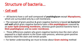
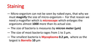
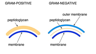
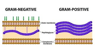

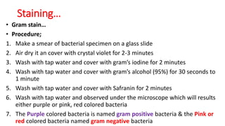
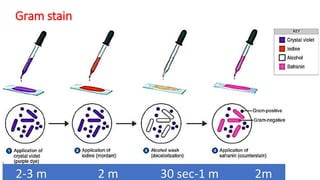
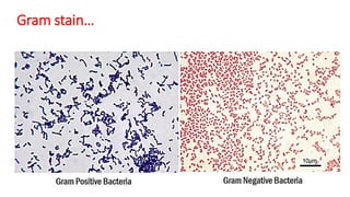
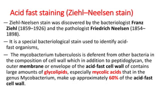
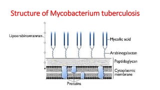

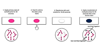
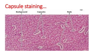


The document provides an introduction to microbiology, defining it as the study of microorganisms, including their morphology, growth, and classification. It highlights the significant role of microbes in various ecosystems and human health, categorizing microbiology into subfields such as medical, industrial, and food microbiology. Additionally, it covers classification methods, staining techniques, and the morphology of microorganisms, essential for their identification and analysis.


![ONFH[AVN HIP] -TRIPLE REGIME -A NOVAL SURGICAL CONCEPT .pptx](https://cdn.slidesharecdn.com/ss_thumbnails/onfhavnhip2026koaconcalicutdrgokuldevdrmashraf-260210064517-213ec005-thumbnail.jpg?width=640&height=640&fit=bounds)


![CTEV [ clubfoot] DR ARUN LAL ,DR MOHAMED ASHRAF travancore medical college k...](https://cdn.slidesharecdn.com/ss_thumbnails/ctevclubfootdrarunlaldrmohamedashraftravancoremedicalcollegekollamkeralaindia-260208063247-18fc466c-thumbnail.jpg?width=640&height=640&fit=bounds)


