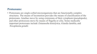
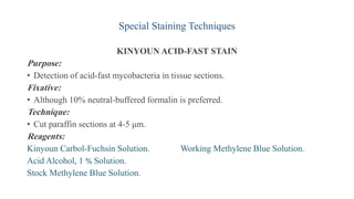
Microorganisms can be classified based on their size, shape, and cellular structure. Bacteria are single-celled organisms that can be further classified as cocci, bacilli, or spirochetes depending on their shape. Special stains like Gram stain and acid-fast stain are used to differentiate bacteria and identify medically important types. Fungi have cell walls containing chitin while viruses are protein-coated genes that need host cells. Protozoa are single-celled organisms that move using pseudopods, flagella, or cilia. A variety of staining techniques exist to identify bacteria, fungi, and other microorganisms in clinical samples and tissue sections.


![• Staphylococcus aureus is the agent of toxic shock syndrome and the cause of
many life-threatening hospital and locker-room infections (eg, methicillin-resistant
Staphylococcus aureus [MRSA]).
• The rod-shaped bacterial organisms are classified as bacilli
• Coccobacilli are rod-shaped, but so short and wide that they resemble cocci.
Examples of coccobacilli are Haemophilus influenzae and Chlamydia trachomatis.
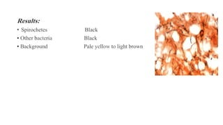
• Bacteria that are spiral or corkscrew-shaped are classified as spirochetes.
Treponema pallidum, the causative organism of syphilis, is a very important
organism in this group.
• Rickettsia, chlamydia, and mycoplasmas are bacteria that do not possess the
typical bacterial attributes.](https://image.slidesharecdn.com/9-230330005744-7b496105/85/9-Microorganisms-pdf-3-320.jpg)


![PERI-PROSTHETIC FRACTURE NAIL-PLATE CONSTRUCT [NPC].pptx](https://cdn.slidesharecdn.com/ss_thumbnails/drarunkumardrmohamedashrafperiprostheticfrasturenail-plateconstructnpc-260209164459-7e9d15a1-thumbnail.jpg?width=640&height=640&fit=bounds)


