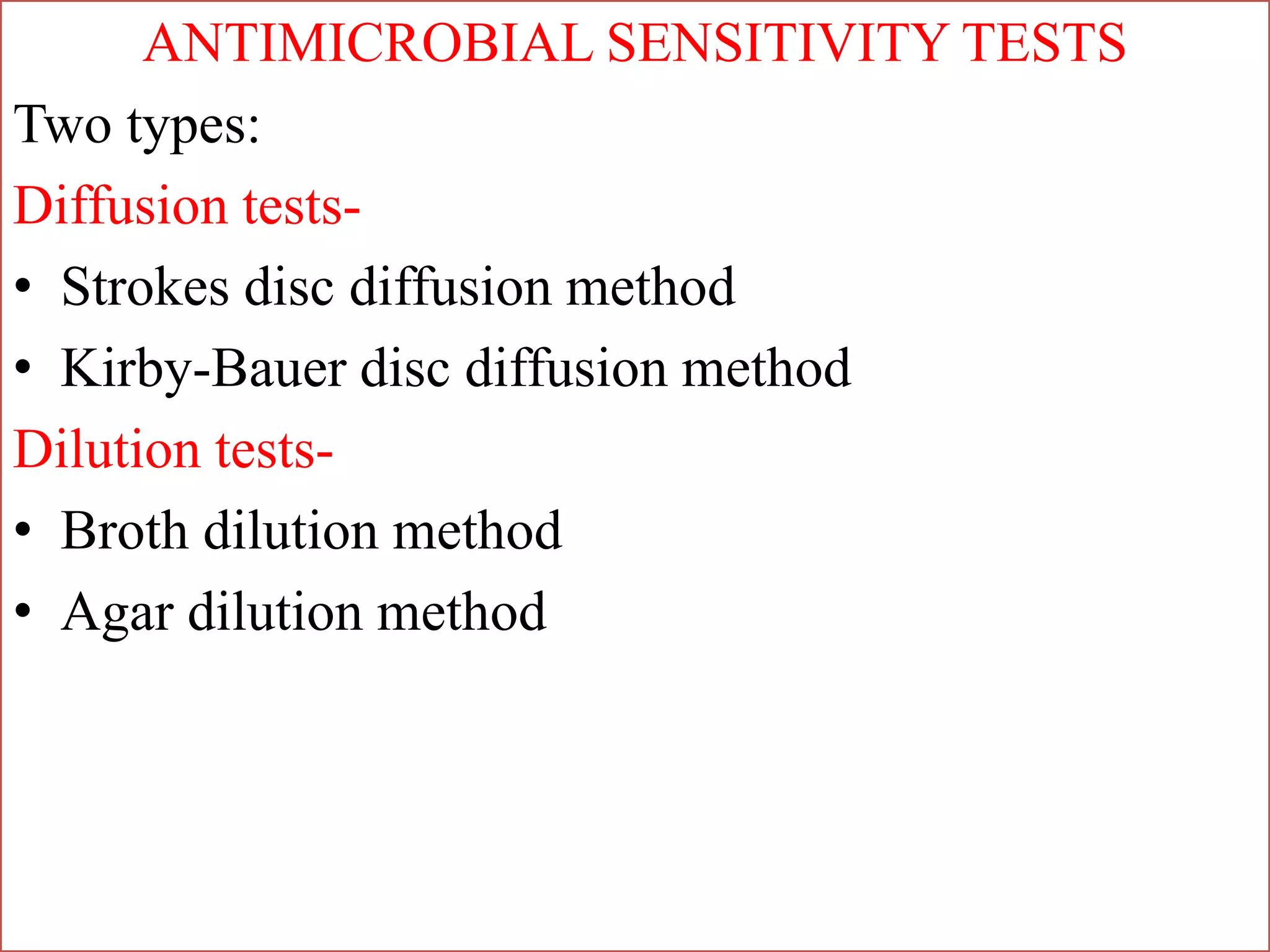
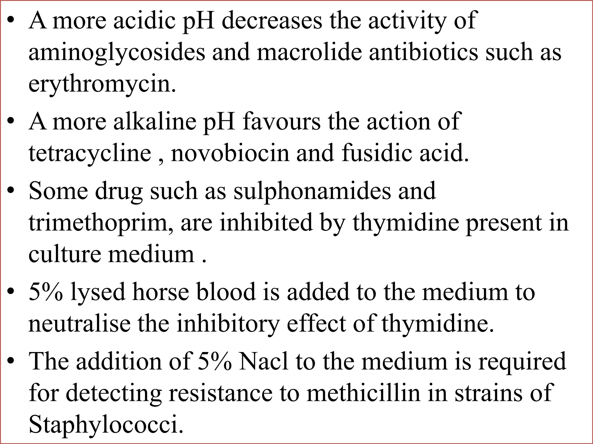
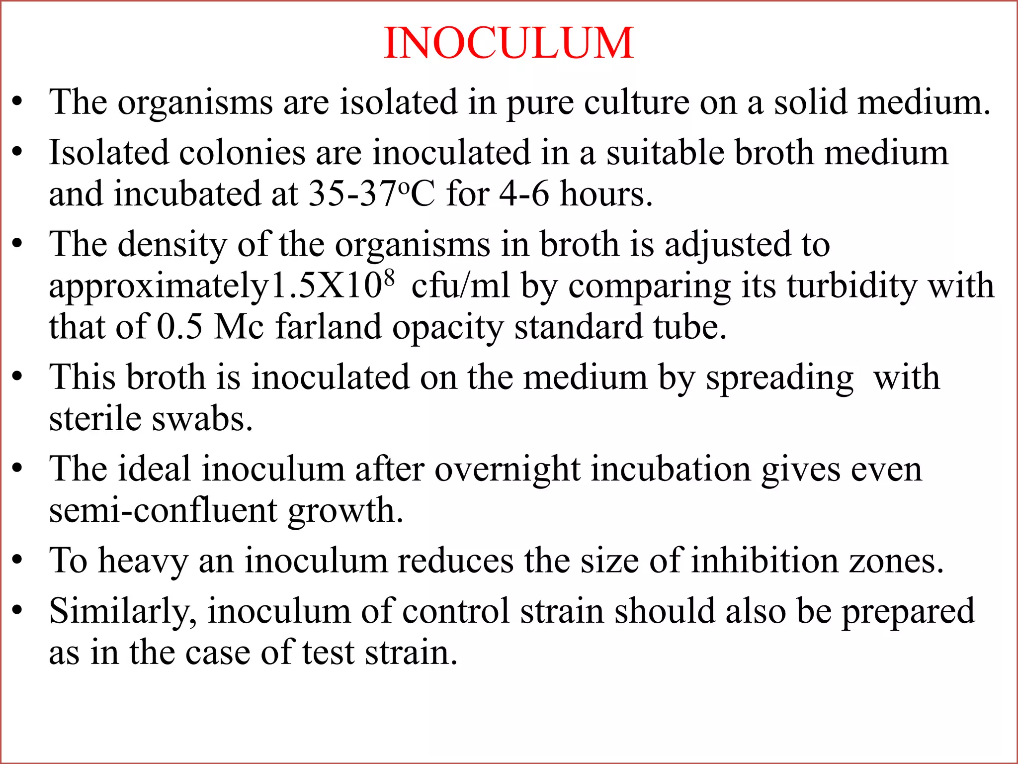
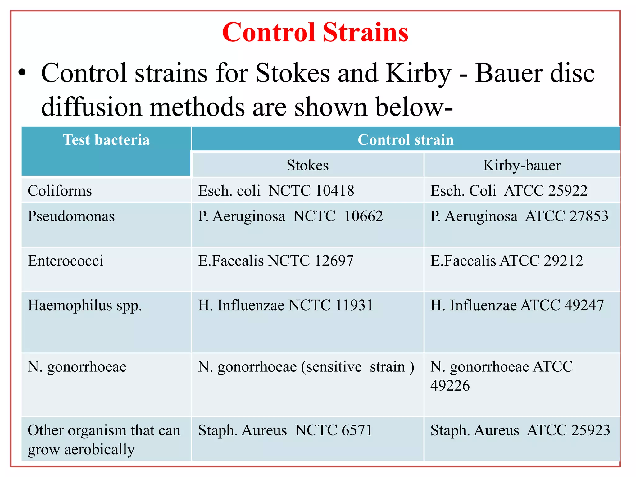
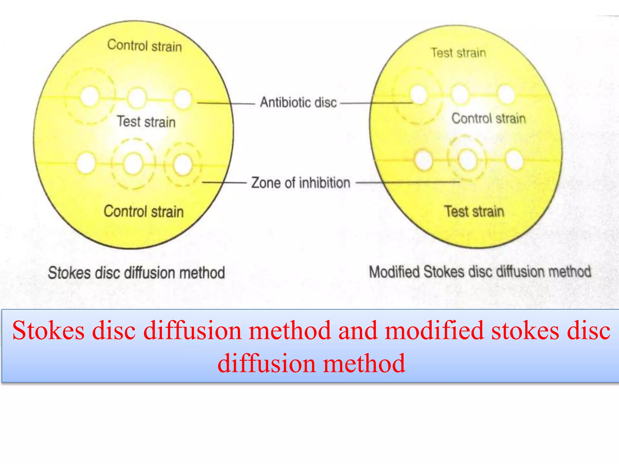
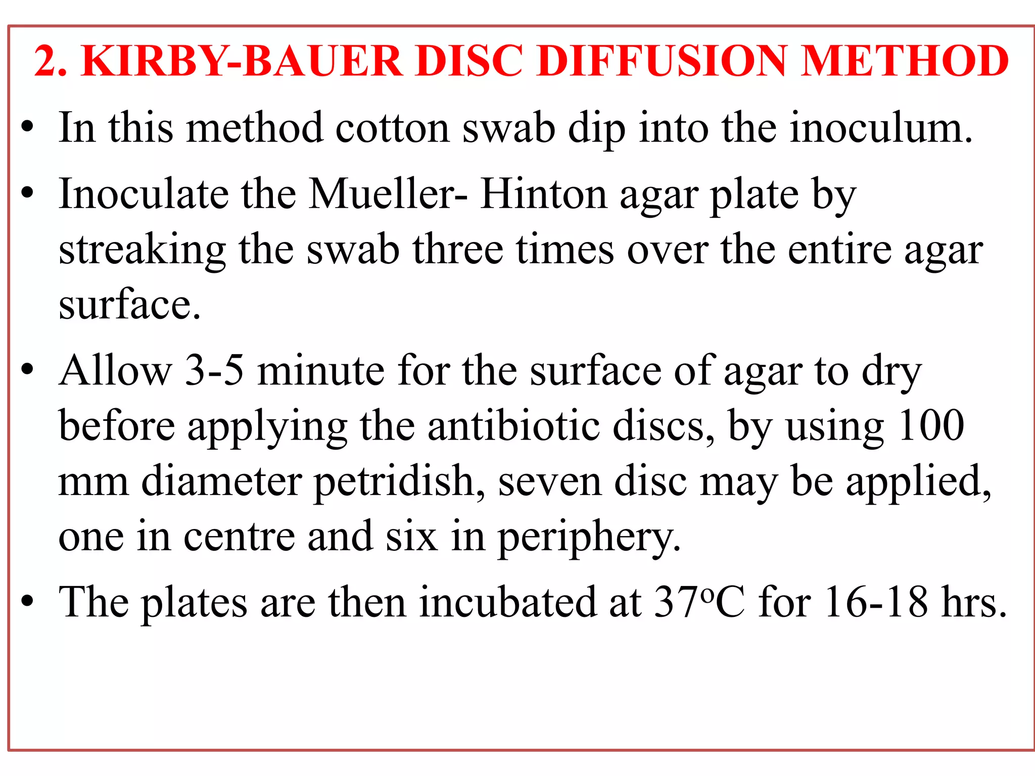
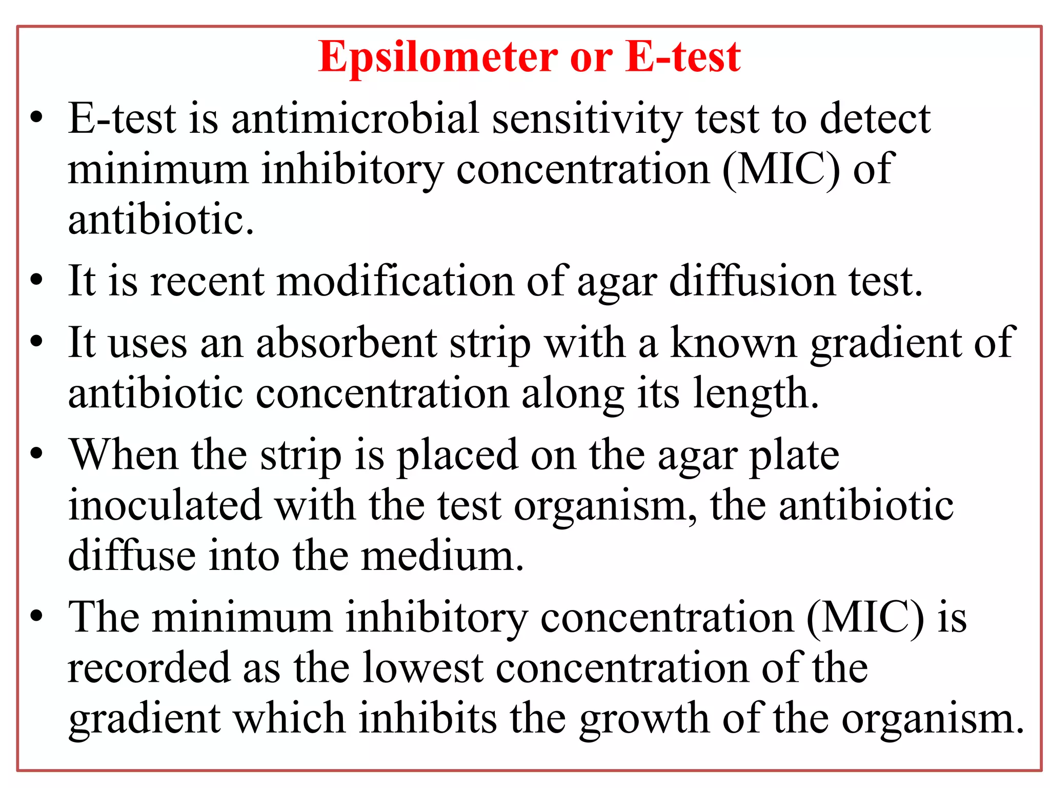
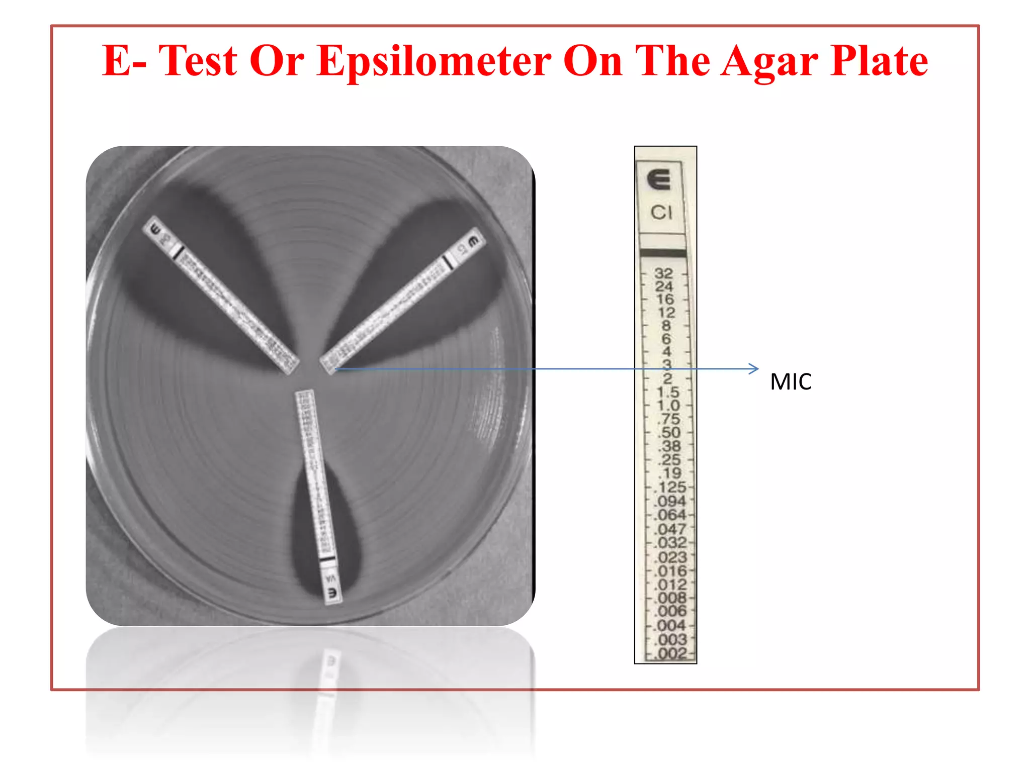
ANTIBIOTIC SENSITIVITY TEST
Antibiotic
• An antibiotic is a substance produced by various species of
living microorganisms.
• (e.g. bacteria and fungi)
Terminology:
Bacteriostatic:
Bactericidal:
Minimum inhibitory concentration:
• MIC is the smallest amount of an agent needed to inhibit growth
of a microorganism.
Minimum bactericidal concentration:
• MBC is the smallest amount of an agent needed to kill the
microorganism.
INTRODUCTION
• Pathogenic bacteria shows great strain wise
variations in susceptibility to antimicrobial agents.
• It is, therefore, essential to determine the
susceptibility of isolates to antibiotics that are likely
to be used in treatment.
• AST is performed only for pathogenic bacteria
isolated from the specimens and not for the
commensals.
ANTIMICROBIAL SENSITIVITY TESTS
Two types:
Diffusion tests-
• Strokes disc diffusion method
• Kirby-Bauer disc diffusion method
Dilution tests-
• Broth dilution method
• Agar dilution method
A. DIFFUSION TESTS
• Disc diffusion tests are most widely used to
determine the susceptibility of isolates of pathogenic
bacteria to antibiotics that are likely to be used in the
treatment.
• Here, the antibiotic is allowed to diffuse through the
solid medium so that concentration is highest near
the site of application of the antibiotic disc and
decreases with the distance.
• These tests is not suitable for slow growing
microbes.
• In the disc diffusion method uses filter paper discs
charged with appropriate concentration of the drugs.
• The test bacterium is inoculated on the medium and
these antibiotic disc are applied.
• Sensitivity to the drug is determined from the
inhibition of bacterial growth around the disc.
Medium:
• The medium should support a good overnight
growth of the test and control organisms.
• Mueller-Hinton agar may be used for the testing
aerobes and facultative anaerobes.
• Nutrient agar is another alternative medium used.
• The medium is prepared in petridish (100 mm in
diameter).
• The depth (thickness) of the medium should be 3-4
mm (around 25ml of the medium is used).
• The plates may be stored at 4oC for up to one week.
• pH - 7.2 to 7.4.
• A more acidic pH decreases the activity of
aminoglycosides and macrolide antibiotics such as
erythromycin.
• A more alkaline pH favours the action of
tetracycline , novobiocin and fusidic acid.
• Some drug such as sulphonamides and
trimethoprim, are inhibited by thymidine present in
culture medium .
• 5% lysed horse blood is added to the medium to
neutralise the inhibitory effect of thymidine.
• The addition of 5% Nacl to the medium is required
for detecting resistance to methicillin in strains of
Staphylococci.
INOCULUM
• The organisms are isolated in pure culture on a solid medium.
• Isolated colonies are inoculated in a suitable broth medium
and incubated at 35-37oC for 4-6 hours.
• The density of the organisms in broth is adjusted to
approximately1.5X108 cfu/ml by comparing its turbidity with
that of 0.5 Mc farland opacity standard tube.
• This broth is inoculated on the medium by spreading with
sterile swabs.
• The ideal inoculum after overnight incubation gives even
semi-confluent growth.
• To heavy an inoculum reduces the size of inhibition zones.
• Similarly, inoculum of control strain should also be prepared
as in the case of test strain.
Control Strains
• Control strains for Stokes and Kirby - Bauer disc
diffusion methods are shown below-
Test bacteria Control strain
Stokes Kirby-bauer
Coliforms Esch. coli NCTC 10418 Esch. Coli ATCC 25922
Pseudomonas P. Aeruginosa NCTC 10662 P. Aeruginosa ATCC 27853
Enterococci E.Faecalis NCTC 12697 E.Faecalis ATCC 29212
Haemophilus spp. H. Influenzae NCTC 11931 H. Influenzae ATCC 49247
N. gonorrhoeae N. gonorrhoeae (sensitive strain ) N. gonorrhoeae ATCC
49226
Other organism that can
grow aerobically
Staph. Aureus NCTC 6571 Staph. Aureus ATCC 25923
ANTIBIOTIC DISCS
• Antibiotic discs may be prepared in the
laboratory or purchased commercially.
• Filter paper discs of 6 mm in diameter are used.
• For preparation of disc in laboratory, pure salt of
antimicrobial agents should be used and not those
use for clinical practice.
• Distilled water serve to dissolve most antibiotic
powders but some antibiotics may require
different diluents:-
Antibiotic Diluent
Chloramphenicol Ethanol
Rifampicin Ethanol
Erythromycin Ethanol
Nitrofurantoin NaOH solution
Sulphonamides NaOH solution
Trimethoprim Acetic acid
Amoxycillin NaHCO3 (Saturated)
ceftazidime NaHCO3 (Saturated)
Table: Diluent of various antibiotics
• Disc concentration of antimicrobial agent are
shown in table:-
Antibiotics Disc concentration
Benzyl penicillin 1.2 µg
Ampicillin 10 µg
Amoxicillin 20 µg
Cephalexin 30 µg
Methicillin 5 µg
Carbenicillin 100 µg
Gentamycin 10 µg
Amikacin 30 µg
Erythromycin 15 µg
Tetracycline 30 µg
Chloramphenicol 30 µg
Nalidixic acid 30 µg
Trimethaprim 5 µg
Ciprofloxacin 1 µg
• The disc are stored at low temperature.
• Hence, disc should be taken out from refrigerator 1-
2 hours before applying on the medium.
1.Stokes Disc Diffusion Method
• The test bacterium is inoculated on the central one
third and control on upper and lower thirds of the
plate.
• However, in Modified Stokes Disc Diffusion Method,
the test bacterium is inoculated over the upper and
lower thirds of the plate and control on central one
third.
• Antibiotic disc are applied between the standard and
test inocula.
• A maximum of six antibiotic discs can be applied on
a 100 mm diameter plate.
• The plates are then incubated at 37oC for 16-18 hrs.
Reporting:
• Plates in which growth is not semi confluent should not be
reported unless the test is repeated.
• The reporting of results is done by comparing the zones of
inhibition of control and test bacterium.
• Measure the zone of size i.e. the distance in mm from edge
of the disc to the zone edge.
Interpretation:-
Sensitive:
• The zone of test bacterium is equal to, or larger than that of
control strain.
• If zone size of the test bacterium is smaller than that of
control, the difference between two should not be at least 3
mm
Intermediate sensitive:
• The zone size of the test bacterium should be
at least 2 mm and the difference between the
zone of test and control strain should be at
least 3 mm.
Resistant:
• The zone size of the test bacterium is smaller
than 2 mm.
Stokes disc diffusion method and modified stokes disc
diffusion method
2. KIRBY-BAUER DISC DIFFUSION METHOD
• In this method cotton swab dip into the inoculum.
• Inoculate the Mueller- Hinton agar plate by
streaking the swab three times over the entire agar
surface.
• Allow 3-5 minute for the surface of agar to dry
before applying the antibiotic discs, by using 100
mm diameter petridish, seven disc may be applied,
one in centre and six in periphery.
• The plates are then incubated at 37oC for 16-18 hrs.
• The zones of complete growth inhibition around
each of the disc is included in this measured.
• The diameter of disc is included in this
measurement.
• The interpretation of zone size into sensitive,
intermediate or resistant is based on interpretation
chart.
• Control strains of Staph. aureus , Esch. Coli, Ps.
aeruginosa etc. should be tested each time when a
new batch of agar or discs is used.
Kirby- Bauer Disc Diffusion Method
• Interpretation Chart Used In Kirby – Bauer Disc
Diffusion Method
Antibiotic Zone size in mm
S I R
Ampicillin(AMP) 17 ─ 16
Chloramphenicol (C) 18 13-17 12
Ciprofloxacin (CIP) 21 16-20 15
Doxycycline (DO) 16 13-15 12
Fosfomycin (FO) 16 13-15 12
High level Gentamycin (HLG) 10 7-9 6
High level Streptomycin (HLS) 10 7-9 6
Levofloxacin (LE) 17 14-16 13
Linezolid (LZ) 23 21-22 20
Nitrofurantion (NIT) 17 15-16 14
Norfloxacin (NX) 17 13-16 12
Not for UTI Erythromycin 23 14-22 13
Penicillin (PN) 15 ─ 14
Teicoplanin (TEI) 14 11-13 10
Tetracycline (TE) 19 15-18 14
Vancomycin (VA) 17 15-16 14
Zone Diameter for Enterococcus spp.
Antibiotic Zone size in mm
S I R
Amikacin (AK) 17 15-16 14
Azithromycin (AZM) 18 14-17 13
Cefoxitin (CX) for CONS 25 ─ 24
Cefoxitin (CX)for S.aureus ≥ 22 ─ ≤ 21
Co- Trimoxazole (COT) 16 11-15 10
Doxycycline (DO) 16 13-15 12
Gentamycin (GEN) 15 13-14 12
Linozolid (LZ) 21 ─ 20
Netilmycin (NET) 15 13-14 12
Nitrofrunction (NIT) 17 15-16 14
Norfloxacin (NX) 17 13-16 12
Not for UTI Erythromycin(E)
Clindamycin (CD)
23 14-22 13
Penicillin (P) ≥ 29 ─ ≤ 28
Sulfonamides (SF) 17 13-16 12
Teicoplanin (TEI) 14 11-13 10
Tetracyclin (TE) 19 15-18 14
Tobramycin (TOB) 15 13-14 12
Trimethoprim (TR) 16 11-15 10
Zone Diameter for Staphylococcus spp.
Antibiotic Zone size in mm
S I R
Amikacin (AK) 17 15-16 14
Aztreonam (AZT) 22 16-21 15
Cefexime (CXM) 18 15-17 14
Ceftazidime (CAZ) 18 15-17 14
Ciprofloxacin (CIP) 21 16-20 15
Colistin (CL) 11 ─ 10
Gentamycin (GEN) 15 13-14 12
Jmipenem (IMP) 19 16-18 15
Levofloxacin (LE) 17 14-16 13
Meropenem (MRP) 19 16-18 15
Netilmycin (NET) 15 13-14 12
Piperacillin (PI) 21 15-20 14
Piperacillin(PI)-Tazobactum 21 15-20 14
Polymyxin B (PB) 12 ─ 11
Tobramycin (TOB) 15 13-14 12
Zone Diameter for Pseudomonas (NLF, Oxidase Positive)
I Line Drugs
Antibiotic Zone size in mm
S I R
Amikacin (AK) 17 15-16 14
Amoxycillin(AMP) - Clavulanate 18 14-17 13
Ampicillin (AMP) 17 14-16 13
Ampicillin(AMP) - Sulbactum 15 12-14 11
Cefotaxime (CTX) 26 23-25 22
Cefoxitin (CX) 18 15-17 14
Ceftazidime (CAZ) 21 18-20 17
Ceftriaxone (CTR) 22 20-22 19
Ciprofloxacin (CIP)for other enterobacteriaceae 21 16-20 15
Co-Trimoxazole (COT) 16 11-15 10
Gentamycin (GEN) 15 13-14 12
Tobramycin (TOB) 15 13-14 12
II Line Drugs
Aztreonam (AZT) 21 18-20 17
Cefepime (CPM) 25 ─ 18
Chloramphenicol (C) 18 13-17 12
Doxycyclin (DO) 14 11-13 10
Imipenem (IMP) 23 20-22 19
Levofloxacin (LE) 17 14-16 13
Meropenem (MRP) 23 20-22 19
Nalidixic Acid 19 14-18 13
Netilmycin (NET) 15 13-14 12
Piperacillin (P) 21 18-20 17
Piperacillin- Tazobactum (PIT) 21 18-20 17
Zone Diameter for Enterobacteriaceae
PRIMARY DISC DIFFUSION TEST
• The disc diffusion methods, as described
above, are done after the pathogenic bacteria
are isolated from the clinical specimens.
• When results are required urgently, the
‘primary disc diffusion test’ may be performed.
• Here, the clinical specimen is directly
inoculated uniformly on the surface of a plate
and the antibiotic discs are applied.
• The results of the primary test should be
verified by testing the isolates subsequently.
Epsilometer or E-test
• E-test is antimicrobial sensitivity test to detect
minimum inhibitory concentration (MIC) of
antibiotic.
• It is recent modification of agar diffusion test.
• It uses an absorbent strip with a known gradient of
antibiotic concentration along its length.
• When the strip is placed on the agar plate
inoculated with the test organism, the antibiotic
diffuse into the medium.
• The minimum inhibitory concentration (MIC) is
recorded as the lowest concentration of the
gradient which inhibits the growth of the organism.
E- Test Or Epsilometer On The Agar Plate
MIC
B. DILUTION TESTS
1.Broth dilution method:
• Serial dilution of the drug in Mueller-Hinton broth
are taken in tubes and a standardised suspension of
the test bacterium inoculated.
• An organism of known sensitivity should also be
titrated.
• Incubate at 37oC for 16-18 hrs.
• For determination of MIC of methicillin incubate at
30oC.
• The minimum inhibitory concentration (MIC) is
ready by noting the lowest concentration of the drug
at which there is no visible growth.
• The minimum bactericidal concentration (MBC) is
determined by subculturing from each tube showing
no growth on nutrient agar plate without any
antimicrobial agent.
• Incubate the plates and examine them for growth.
• The tube containing the lowest concentration of the
drug that fails to show growth, on sub culture, is the
MBC of the drug for that test strain.
Uses of MIC determination:
• When the therapeutic dose is to be regulated
accurately as in the treatment of bacterial
endocarditis.
• For testing antimicrobial sensitivities of slow
growing bacteria such tubercle bacilli.
• When small degree of resistance are to be
demonstrated.
Antibiotic
concentration
in (/ml)
Mueller Hinton
broth
No Growth Turbidity (growth)
No Growth Growth
MIC=8µg /ml
MBC=32µg /ml
Broth Dilution Method Showing MIC And MBC
2.Agar dilution method:
• Serial dilutions of the drug are prepared in agar
and poured into plates.
• Many strains can be inoculated on each plate
containing an antibiotic dilution.
• This method is more convenient when several
strains are to be tested at the same time.
Automated antimicrobial susceptibility
tests
• Several automated systems are available now,
such as,
 VITEK 2 bacterial identification and
antimicrobial sensitivity system (bioMerieux)
 Phoenix System

Antimicrobial sensitivity test

  • 1.
  • 2.
    Antibiotic • An antibioticis a substance produced by various species of living microorganisms. • (e.g. bacteria and fungi) Terminology: Bacteriostatic: Bactericidal: Minimum inhibitory concentration: • MIC is the smallest amount of an agent needed to inhibit growth of a microorganism. Minimum bactericidal concentration: • MBC is the smallest amount of an agent needed to kill the microorganism.
  • 3.
    INTRODUCTION • Pathogenic bacteriashows great strain wise variations in susceptibility to antimicrobial agents. • It is, therefore, essential to determine the susceptibility of isolates to antibiotics that are likely to be used in treatment. • AST is performed only for pathogenic bacteria isolated from the specimens and not for the commensals.
  • 4.
    ANTIMICROBIAL SENSITIVITY TESTS Twotypes: Diffusion tests- • Strokes disc diffusion method • Kirby-Bauer disc diffusion method Dilution tests- • Broth dilution method • Agar dilution method
  • 5.
    A. DIFFUSION TESTS •Disc diffusion tests are most widely used to determine the susceptibility of isolates of pathogenic bacteria to antibiotics that are likely to be used in the treatment. • Here, the antibiotic is allowed to diffuse through the solid medium so that concentration is highest near the site of application of the antibiotic disc and decreases with the distance. • These tests is not suitable for slow growing microbes.
  • 6.
    • In thedisc diffusion method uses filter paper discs charged with appropriate concentration of the drugs. • The test bacterium is inoculated on the medium and these antibiotic disc are applied. • Sensitivity to the drug is determined from the inhibition of bacterial growth around the disc.
  • 7.
    Medium: • The mediumshould support a good overnight growth of the test and control organisms. • Mueller-Hinton agar may be used for the testing aerobes and facultative anaerobes. • Nutrient agar is another alternative medium used. • The medium is prepared in petridish (100 mm in diameter). • The depth (thickness) of the medium should be 3-4 mm (around 25ml of the medium is used). • The plates may be stored at 4oC for up to one week. • pH - 7.2 to 7.4.
  • 8.
    • A moreacidic pH decreases the activity of aminoglycosides and macrolide antibiotics such as erythromycin. • A more alkaline pH favours the action of tetracycline , novobiocin and fusidic acid. • Some drug such as sulphonamides and trimethoprim, are inhibited by thymidine present in culture medium . • 5% lysed horse blood is added to the medium to neutralise the inhibitory effect of thymidine. • The addition of 5% Nacl to the medium is required for detecting resistance to methicillin in strains of Staphylococci.
  • 9.
    INOCULUM • The organismsare isolated in pure culture on a solid medium. • Isolated colonies are inoculated in a suitable broth medium and incubated at 35-37oC for 4-6 hours. • The density of the organisms in broth is adjusted to approximately1.5X108 cfu/ml by comparing its turbidity with that of 0.5 Mc farland opacity standard tube. • This broth is inoculated on the medium by spreading with sterile swabs. • The ideal inoculum after overnight incubation gives even semi-confluent growth. • To heavy an inoculum reduces the size of inhibition zones. • Similarly, inoculum of control strain should also be prepared as in the case of test strain.
  • 10.
    Control Strains • Controlstrains for Stokes and Kirby - Bauer disc diffusion methods are shown below- Test bacteria Control strain Stokes Kirby-bauer Coliforms Esch. coli NCTC 10418 Esch. Coli ATCC 25922 Pseudomonas P. Aeruginosa NCTC 10662 P. Aeruginosa ATCC 27853 Enterococci E.Faecalis NCTC 12697 E.Faecalis ATCC 29212 Haemophilus spp. H. Influenzae NCTC 11931 H. Influenzae ATCC 49247 N. gonorrhoeae N. gonorrhoeae (sensitive strain ) N. gonorrhoeae ATCC 49226 Other organism that can grow aerobically Staph. Aureus NCTC 6571 Staph. Aureus ATCC 25923
  • 11.
    ANTIBIOTIC DISCS • Antibioticdiscs may be prepared in the laboratory or purchased commercially. • Filter paper discs of 6 mm in diameter are used. • For preparation of disc in laboratory, pure salt of antimicrobial agents should be used and not those use for clinical practice. • Distilled water serve to dissolve most antibiotic powders but some antibiotics may require different diluents:-
  • 12.
    Antibiotic Diluent Chloramphenicol Ethanol RifampicinEthanol Erythromycin Ethanol Nitrofurantoin NaOH solution Sulphonamides NaOH solution Trimethoprim Acetic acid Amoxycillin NaHCO3 (Saturated) ceftazidime NaHCO3 (Saturated) Table: Diluent of various antibiotics
  • 13.
    • Disc concentrationof antimicrobial agent are shown in table:- Antibiotics Disc concentration Benzyl penicillin 1.2 µg Ampicillin 10 µg Amoxicillin 20 µg Cephalexin 30 µg Methicillin 5 µg Carbenicillin 100 µg Gentamycin 10 µg Amikacin 30 µg Erythromycin 15 µg Tetracycline 30 µg Chloramphenicol 30 µg Nalidixic acid 30 µg Trimethaprim 5 µg Ciprofloxacin 1 µg
  • 14.
    • The discare stored at low temperature. • Hence, disc should be taken out from refrigerator 1- 2 hours before applying on the medium.
  • 15.
    1.Stokes Disc DiffusionMethod • The test bacterium is inoculated on the central one third and control on upper and lower thirds of the plate. • However, in Modified Stokes Disc Diffusion Method, the test bacterium is inoculated over the upper and lower thirds of the plate and control on central one third. • Antibiotic disc are applied between the standard and test inocula. • A maximum of six antibiotic discs can be applied on a 100 mm diameter plate. • The plates are then incubated at 37oC for 16-18 hrs.
  • 16.
    Reporting: • Plates inwhich growth is not semi confluent should not be reported unless the test is repeated. • The reporting of results is done by comparing the zones of inhibition of control and test bacterium. • Measure the zone of size i.e. the distance in mm from edge of the disc to the zone edge. Interpretation:- Sensitive: • The zone of test bacterium is equal to, or larger than that of control strain. • If zone size of the test bacterium is smaller than that of control, the difference between two should not be at least 3 mm
  • 17.
    Intermediate sensitive: • Thezone size of the test bacterium should be at least 2 mm and the difference between the zone of test and control strain should be at least 3 mm. Resistant: • The zone size of the test bacterium is smaller than 2 mm.
  • 18.
    Stokes disc diffusionmethod and modified stokes disc diffusion method
  • 19.
    2. KIRBY-BAUER DISCDIFFUSION METHOD • In this method cotton swab dip into the inoculum. • Inoculate the Mueller- Hinton agar plate by streaking the swab three times over the entire agar surface. • Allow 3-5 minute for the surface of agar to dry before applying the antibiotic discs, by using 100 mm diameter petridish, seven disc may be applied, one in centre and six in periphery. • The plates are then incubated at 37oC for 16-18 hrs.
  • 20.
    • The zonesof complete growth inhibition around each of the disc is included in this measured. • The diameter of disc is included in this measurement. • The interpretation of zone size into sensitive, intermediate or resistant is based on interpretation chart. • Control strains of Staph. aureus , Esch. Coli, Ps. aeruginosa etc. should be tested each time when a new batch of agar or discs is used.
  • 21.
    Kirby- Bauer DiscDiffusion Method
  • 22.
    • Interpretation ChartUsed In Kirby – Bauer Disc Diffusion Method Antibiotic Zone size in mm S I R Ampicillin(AMP) 17 ─ 16 Chloramphenicol (C) 18 13-17 12 Ciprofloxacin (CIP) 21 16-20 15 Doxycycline (DO) 16 13-15 12 Fosfomycin (FO) 16 13-15 12 High level Gentamycin (HLG) 10 7-9 6 High level Streptomycin (HLS) 10 7-9 6 Levofloxacin (LE) 17 14-16 13 Linezolid (LZ) 23 21-22 20 Nitrofurantion (NIT) 17 15-16 14 Norfloxacin (NX) 17 13-16 12 Not for UTI Erythromycin 23 14-22 13 Penicillin (PN) 15 ─ 14 Teicoplanin (TEI) 14 11-13 10 Tetracycline (TE) 19 15-18 14 Vancomycin (VA) 17 15-16 14 Zone Diameter for Enterococcus spp.
  • 23.
    Antibiotic Zone sizein mm S I R Amikacin (AK) 17 15-16 14 Azithromycin (AZM) 18 14-17 13 Cefoxitin (CX) for CONS 25 ─ 24 Cefoxitin (CX)for S.aureus ≥ 22 ─ ≤ 21 Co- Trimoxazole (COT) 16 11-15 10 Doxycycline (DO) 16 13-15 12 Gentamycin (GEN) 15 13-14 12 Linozolid (LZ) 21 ─ 20 Netilmycin (NET) 15 13-14 12 Nitrofrunction (NIT) 17 15-16 14 Norfloxacin (NX) 17 13-16 12 Not for UTI Erythromycin(E) Clindamycin (CD) 23 14-22 13 Penicillin (P) ≥ 29 ─ ≤ 28 Sulfonamides (SF) 17 13-16 12 Teicoplanin (TEI) 14 11-13 10 Tetracyclin (TE) 19 15-18 14 Tobramycin (TOB) 15 13-14 12 Trimethoprim (TR) 16 11-15 10 Zone Diameter for Staphylococcus spp.
  • 24.
    Antibiotic Zone sizein mm S I R Amikacin (AK) 17 15-16 14 Aztreonam (AZT) 22 16-21 15 Cefexime (CXM) 18 15-17 14 Ceftazidime (CAZ) 18 15-17 14 Ciprofloxacin (CIP) 21 16-20 15 Colistin (CL) 11 ─ 10 Gentamycin (GEN) 15 13-14 12 Jmipenem (IMP) 19 16-18 15 Levofloxacin (LE) 17 14-16 13 Meropenem (MRP) 19 16-18 15 Netilmycin (NET) 15 13-14 12 Piperacillin (PI) 21 15-20 14 Piperacillin(PI)-Tazobactum 21 15-20 14 Polymyxin B (PB) 12 ─ 11 Tobramycin (TOB) 15 13-14 12 Zone Diameter for Pseudomonas (NLF, Oxidase Positive)
  • 25.
    I Line Drugs AntibioticZone size in mm S I R Amikacin (AK) 17 15-16 14 Amoxycillin(AMP) - Clavulanate 18 14-17 13 Ampicillin (AMP) 17 14-16 13 Ampicillin(AMP) - Sulbactum 15 12-14 11 Cefotaxime (CTX) 26 23-25 22 Cefoxitin (CX) 18 15-17 14 Ceftazidime (CAZ) 21 18-20 17 Ceftriaxone (CTR) 22 20-22 19 Ciprofloxacin (CIP)for other enterobacteriaceae 21 16-20 15 Co-Trimoxazole (COT) 16 11-15 10 Gentamycin (GEN) 15 13-14 12 Tobramycin (TOB) 15 13-14 12 II Line Drugs Aztreonam (AZT) 21 18-20 17 Cefepime (CPM) 25 ─ 18 Chloramphenicol (C) 18 13-17 12 Doxycyclin (DO) 14 11-13 10 Imipenem (IMP) 23 20-22 19 Levofloxacin (LE) 17 14-16 13 Meropenem (MRP) 23 20-22 19 Nalidixic Acid 19 14-18 13 Netilmycin (NET) 15 13-14 12 Piperacillin (P) 21 18-20 17 Piperacillin- Tazobactum (PIT) 21 18-20 17 Zone Diameter for Enterobacteriaceae
  • 26.
    PRIMARY DISC DIFFUSIONTEST • The disc diffusion methods, as described above, are done after the pathogenic bacteria are isolated from the clinical specimens. • When results are required urgently, the ‘primary disc diffusion test’ may be performed. • Here, the clinical specimen is directly inoculated uniformly on the surface of a plate and the antibiotic discs are applied. • The results of the primary test should be verified by testing the isolates subsequently.
  • 27.
    Epsilometer or E-test •E-test is antimicrobial sensitivity test to detect minimum inhibitory concentration (MIC) of antibiotic. • It is recent modification of agar diffusion test. • It uses an absorbent strip with a known gradient of antibiotic concentration along its length. • When the strip is placed on the agar plate inoculated with the test organism, the antibiotic diffuse into the medium. • The minimum inhibitory concentration (MIC) is recorded as the lowest concentration of the gradient which inhibits the growth of the organism.
  • 28.
    E- Test OrEpsilometer On The Agar Plate MIC
  • 29.
    B. DILUTION TESTS 1.Brothdilution method: • Serial dilution of the drug in Mueller-Hinton broth are taken in tubes and a standardised suspension of the test bacterium inoculated. • An organism of known sensitivity should also be titrated. • Incubate at 37oC for 16-18 hrs. • For determination of MIC of methicillin incubate at 30oC. • The minimum inhibitory concentration (MIC) is ready by noting the lowest concentration of the drug at which there is no visible growth.
  • 30.
    • The minimumbactericidal concentration (MBC) is determined by subculturing from each tube showing no growth on nutrient agar plate without any antimicrobial agent. • Incubate the plates and examine them for growth. • The tube containing the lowest concentration of the drug that fails to show growth, on sub culture, is the MBC of the drug for that test strain.
  • 31.
    Uses of MICdetermination: • When the therapeutic dose is to be regulated accurately as in the treatment of bacterial endocarditis. • For testing antimicrobial sensitivities of slow growing bacteria such tubercle bacilli. • When small degree of resistance are to be demonstrated.
  • 32.
    Antibiotic concentration in (/ml) Mueller Hinton broth NoGrowth Turbidity (growth) No Growth Growth MIC=8µg /ml MBC=32µg /ml Broth Dilution Method Showing MIC And MBC
  • 33.
    2.Agar dilution method: •Serial dilutions of the drug are prepared in agar and poured into plates. • Many strains can be inoculated on each plate containing an antibiotic dilution. • This method is more convenient when several strains are to be tested at the same time.
  • 34.
    Automated antimicrobial susceptibility tests •Several automated systems are available now, such as,  VITEK 2 bacterial identification and antimicrobial sensitivity system (bioMerieux)  Phoenix System