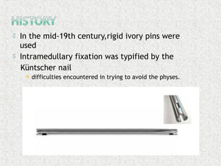
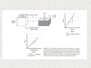
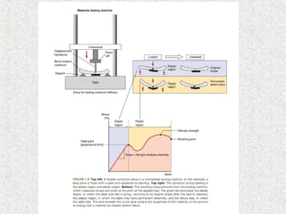
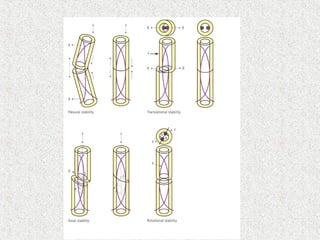
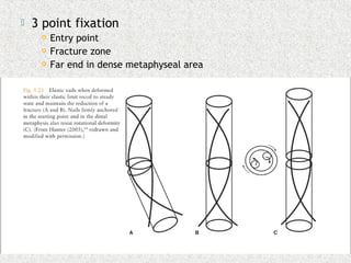
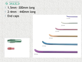
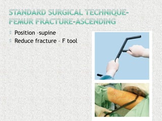
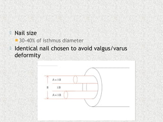
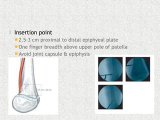
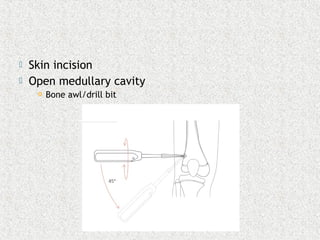
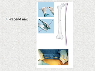
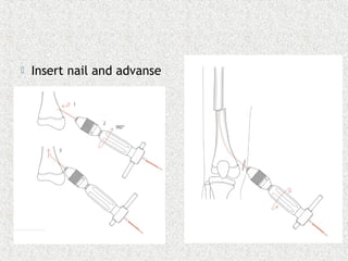
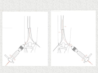
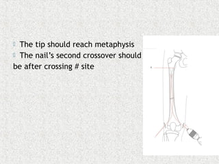
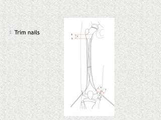
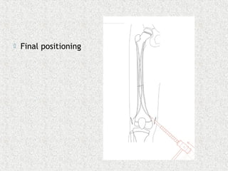
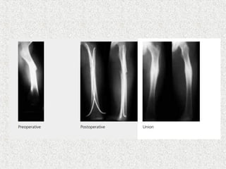
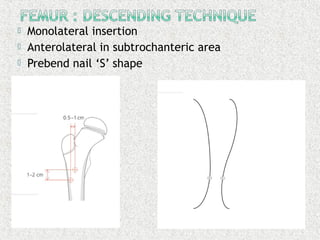
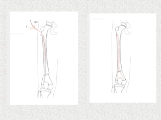
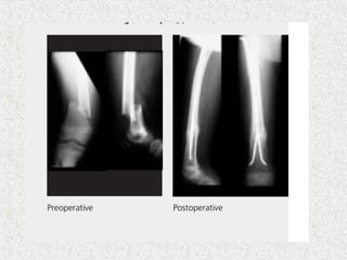
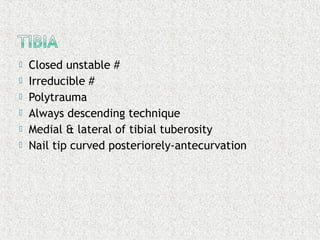
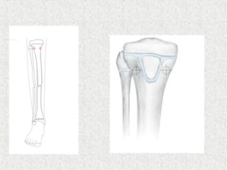
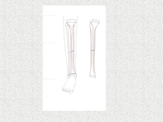
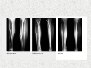
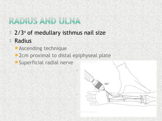
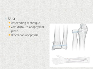
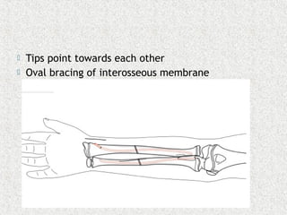
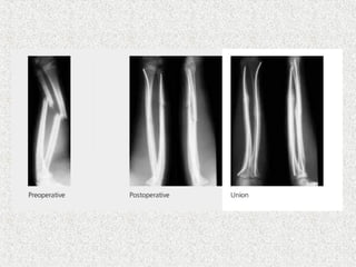
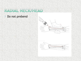
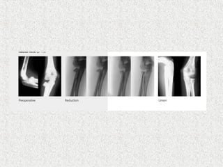
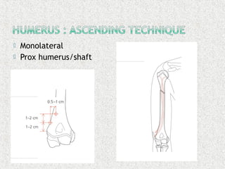
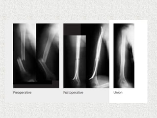
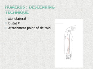
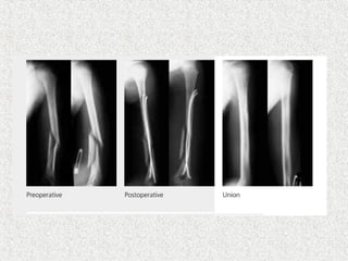
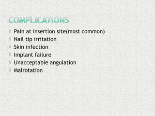
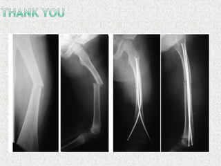
Dr (Major) Parthasarathy S
Pg Resident,MS Orthopaedics
Stanley Medical College,Chennai
Ref : Rockwood & Wilkin’s fractures in children 8th
edition
The elements of fracture fixation 3rd
edition
 Primary definitive fracture care
 Thick periosteum
 Biological healing
 Minimal soft tissue insult
 Callus promoting micro movements
 Maintains length,rotation,alignment
 In the mid-19th century,rigid ivory pins were
used
 Intramedullary fixation was typified by the
Küntscher nail
 difficulties encountered in trying to avoid the physes.
 The Rush nail
 forerunner of modern elastic intramedullary fixation
 three-point fixation
 slightly flexible
 pre-bent
 rotational stability was poor
 flexibility was insufficient
 Hackethal Marchetti
 bundles of thinner wires which filled the medullary cavity
 Stabilisation achieved by splaying the ends of the wires
 Ender
 safely inserted into the metaphysis
 In the early 1980s, surgeons in Nancy,France
 Developed an elastic stable intramedullary nail based
on a theoretical concept by Firica.
 elasticity and stability combined in one construct
 two pre-tensioned nails inserted from opposite sides
of the bone
 Metazieau, Ligier et al were able to show that
titanium nails which were accurately contoured and
properly inserted could impart excellent axial and
lateral stability to diaphyseal fractures in long bones.
 Rotational stability weakest point of the technique
 Young’s/elastic/tensile modulus-object’s
resistance to being deformed elastically
 Young’s modulus=stress/strain
 Stress-forcing causing deformation/area
 Strain-change in length
 Precurved-3 times the narrowest diameter of
bone
 Maximum curvature in fracture zone
 2 nails inserted often
 Opposite to each other
 4 properties
 Flexural,axial,rotational,transitional stability
 3 point fixation
 Entry point
 Fracture zone
 Far end in dense metaphyseal area
 Intraarticular fracture
 Complex fracture particularly in connection
with overweight & age >15
 1.5mm -300mm long
 2-4mm – 440mm long
 End caps
 Position –supine
 Reduce fracture – F tool
 Nail size
 30-40% of isthmus diameter
 Identical nail chosen to avoid valgus/varus
deformity
 Insertion point
 2.5-3 cm proximal to distal epiphyeal plate
 One finger breadth above upper pole of patella
 Avoid joint capsule & epiphysis
 Skin incision
 Open medullary cavity
 Bone awl/drill bit
 Prebend nail
 Insert nail and advanse
 The tip should reach metaphysis
 The nail’s second crossover should
be after crossing # site
 Trim nails
 Final positioning
 Monolateral insertion
 Anterolateral in subtrochanteric area
 Prebend nail ‘S’ shape
 Closed unstable #
 Irreducible #
 Polytrauma
 Always descending technique
 Medial & lateral of tibial tuberosity
 Nail tip curved posteriorely-antecurvation
 2/3rd
of medullary isthmus nail size
 Radius
 Ascending technique
 2cm proximal to distal epiphyseal plate
 Superficial radial nerve
 Ulna
 Descending technique
 2cm distal to apophyseal
plate
 Olecranon apophysis
 Tips point towards each other
 Oval bracing of interosseous membrane
 Do not prebend
 Monolateral
 Prox humerus/shaft
 Monolateral
 Distal #
 Attachment point of deltoid
 Pain at insertion site(most common)
 Nail tip irritation
 Skin infection
 Implant failure
 Unacceptable angulation
 Malrotation
TENS

TENS

  • 1.
    Dr (Major) ParthasarathyS Pg Resident,MS Orthopaedics Stanley Medical College,Chennai Ref : Rockwood & Wilkin’s fractures in children 8th edition The elements of fracture fixation 3rd edition
  • 2.
     Primary definitivefracture care  Thick periosteum  Biological healing  Minimal soft tissue insult  Callus promoting micro movements  Maintains length,rotation,alignment
  • 3.
     In themid-19th century,rigid ivory pins were used  Intramedullary fixation was typified by the Küntscher nail  difficulties encountered in trying to avoid the physes.
  • 4.
     The Rushnail  forerunner of modern elastic intramedullary fixation  three-point fixation  slightly flexible  pre-bent  rotational stability was poor  flexibility was insufficient
  • 5.
     Hackethal Marchetti bundles of thinner wires which filled the medullary cavity  Stabilisation achieved by splaying the ends of the wires  Ender  safely inserted into the metaphysis
  • 6.
     In theearly 1980s, surgeons in Nancy,France  Developed an elastic stable intramedullary nail based on a theoretical concept by Firica.  elasticity and stability combined in one construct  two pre-tensioned nails inserted from opposite sides of the bone  Metazieau, Ligier et al were able to show that titanium nails which were accurately contoured and properly inserted could impart excellent axial and lateral stability to diaphyseal fractures in long bones.  Rotational stability weakest point of the technique
  • 7.
     Young’s/elastic/tensile modulus-object’s resistanceto being deformed elastically  Young’s modulus=stress/strain  Stress-forcing causing deformation/area  Strain-change in length
  • 11.
     Precurved-3 timesthe narrowest diameter of bone  Maximum curvature in fracture zone  2 nails inserted often  Opposite to each other  4 properties  Flexural,axial,rotational,transitional stability
  • 13.
     3 pointfixation  Entry point  Fracture zone  Far end in dense metaphyseal area
  • 16.
     Intraarticular fracture Complex fracture particularly in connection with overweight & age >15
  • 17.
     1.5mm -300mmlong  2-4mm – 440mm long  End caps
  • 18.
     Position –supine Reduce fracture – F tool
  • 19.
     Nail size 30-40% of isthmus diameter  Identical nail chosen to avoid valgus/varus deformity
  • 20.
     Insertion point 2.5-3 cm proximal to distal epiphyeal plate  One finger breadth above upper pole of patella  Avoid joint capsule & epiphysis
  • 21.
     Skin incision Open medullary cavity  Bone awl/drill bit
  • 22.
  • 23.
     Insert nailand advanse
  • 25.
     The tipshould reach metaphysis  The nail’s second crossover should be after crossing # site
  • 26.
  • 27.
  • 29.
     Monolateral insertion Anterolateral in subtrochanteric area  Prebend nail ‘S’ shape
  • 32.
     Closed unstable#  Irreducible #  Polytrauma  Always descending technique  Medial & lateral of tibial tuberosity  Nail tip curved posteriorely-antecurvation
  • 36.
     2/3rd of medullaryisthmus nail size  Radius  Ascending technique  2cm proximal to distal epiphyseal plate  Superficial radial nerve
  • 37.
     Ulna  Descendingtechnique  2cm distal to apophyseal plate  Olecranon apophysis
  • 38.
     Tips pointtowards each other  Oval bracing of interosseous membrane
  • 40.
     Do notprebend
  • 42.
  • 44.
     Monolateral  Distal#  Attachment point of deltoid
  • 46.
     Pain atinsertion site(most common)  Nail tip irritation  Skin infection  Implant failure  Unacceptable angulation  Malrotation