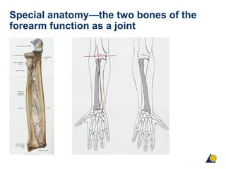
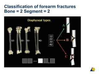
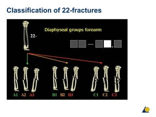
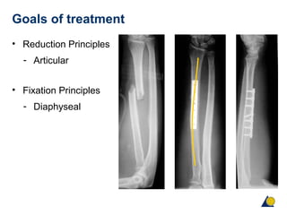
The document discusses fractures of the forearm and their treatment. It summarizes that the forearm functions as a joint with six articulations. Forearm fractures can result in deformities like shortening, angulation, and loss of alignment if not treated properly. Treatment goals are anatomical reduction, restoration of length and rotation, and early return of function. Plate fixation is the gold standard and provides stable fixation, allowing early motion to restore function with high union rates over 95%.
2. Objectives
• Review the concept of the forearm as a joint
• Discuss the assessment, problems and options for
treatment of forearm fractures
• Review the options for surgical approaches
• Discuss complications and outcomes
8. Investigations
• X-ray in two planes—must include both joints
• CT—rarely indicated
• MRI—useful for assessing damage to articular cartilage
of distal radioulnar joint
• Angiography—vital in cases involving vascular trauma
11. Fracture with joint disruption
Monteggia fractures
• Ulnar shaft fracture with
- Dislocation of radial head
- Types I – IV depending on direction of radial head
dislocation
12. Fracture with joint disruption
Monteggia fractures
Line drawn through radial head
and shaft should line up with the
capitellum in all views
13. Fracture with joint disruption
Galeazzi fractures
• Radial shaft fracture with:
- Dislocation of distal ulna
- Multiple variants in
location of radius fracture
14. Personality of fracture
• Soft-tissue damage
• Degree of fracture displacement
• Degree of comminution
• Degree of joint involvement
• Osteoporosis
• Nerve/blood vessel injury
15. Goals of treatment
• Anatomical reduction
• Restore ulnar & radial length
• Reduce/stabilize joints
• Restore rotational alignment
• Repair soft-tissue injuries
• Restore normal function
18. Indications for operative treatment
• Combined fractures of ulna and radius
• Displaced isolated fracture of either bone
• Monteggia and Galeazzi
• Every open fracture
23. Operative treatment—implant
• LC-DCP 3.5 (limited-contact dynamic compression plate)
- Anatomical reduction
• LCP 3.5 (locking compression plate)
- Restoration of length, axis, and alignment
• At least 3 screws in each main fragment!
24. Plate fixation
• The “Gold Standard” treatment for most diaphyseal
forearm fractures
• Stable, strong, anatomical fixation
• Union rates > 95%
25. Tips for operative surgery
• Begin with easiest fracture
• Look for elbow and wrist joint
• Be aware of rotation of radius
• Test function of forearm joint after fixation
26. Plate fixation—reduction preserving soft tissue
• Minimize periosteal stripping
• Narrow retractors placed to
- avoid penetration of interosseous
- membrane
• Extreme care with butteryfly
- fragments
27. Timing of surgery
• Splint application with elective scheduling for
uncomplicated closed fractures
• Immediate fixation for:
- Open fractures
- Impending open fractures
- Compartment syndrome
- Unreducable dislocations
• Easier fracture reduction with early surgery, especially if
shortening is present
28. Postoperative care
• Avoid prolonged immobilization
• If you fix it internally, do not fix it externally
• Immobilize the minimum time needed to protect the soft
tissues
• Dislocations may require immobilization
• Begin early active ROM
29. Problems
• Soft-tissue loss
• Infection
• Synostosis 2,6–6,6%
• Nonunion 3,7–10,3%
• Refracture after implant removal up to 25%
30. Results of plate fixation
MW Chapman et al (1989) J Bone Joint Surg
• Healing 98%
• Excellent to satisfactory 92%
• Infection rate 2.3%
• Refracture rate 2.3%
31. External fixator
• Indications
- Bridging fixation
- Types IIIB & C open fractures
- Later revision to internal
fixation when soft tissues
allow
- NOT definitive fixation
32. Intramedullary nailing
Indications
• Not routinely used
• Problems of rotational instability, loss of radial bow,
shortening, and nonunion
• May be useful when soft tissues compromised
• Children
• Pathologic fractures or impending fractures where the
device protects the whole bone
34. Summary
• Analysis of fracture mechanism and associated soft-
tissue lesions are vital to allow adequate treatment
planning
• Complete reconstruction of anatomy is essential to
restore normal function
• Stable fixation with long plates and early movement are
the keys to success
• Plate fixation gives good results