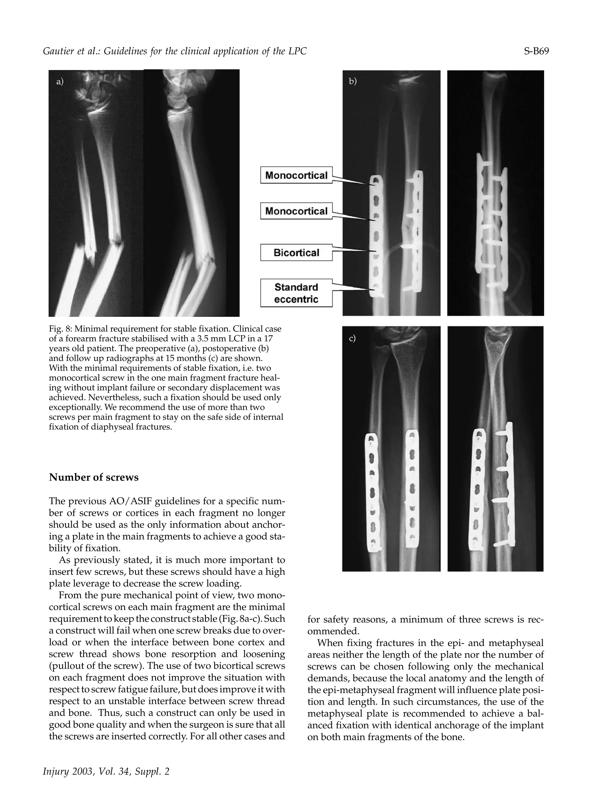
The document provides guidelines for using the Locking Compression Plate (LCP) internal fixation device. It discusses that the LCP requires an adapted surgical technique and new thinking about plate fixation concepts. Key points include:
- Reduction technique and minimal-invasive plate insertion are important to preserve bone viability.
- Proper implant length, screw type, number and placement are essential for sound fixation and low stress on screws.
- Knowledge of screw length selection and bicortical versus monocortical placement is important depending on bone quality.
- Rules are provided for compression plating, bridge plating, and combination techniques according to fracture type and bone quality. Guidance is given on plate and screw
![Injury, Int. J. Care Injured 34 (2003) S-B63–S-B76
Summary1
The Locking Compression Plate (LCP), in combination
with the LISS and the PHILOS, is part of a new plate
generation requiring an adapted surgical technique and
new thinking about commonly used concepts of inter-
nal fixation using plates. The following guidelines are
needed to avoid failures and possible complications in
the hands of surgeons not yet confident with the new
implant philosophy.
The importance of the reduction technique and min-
imal-invasive plate insertion and fixation is addressed
to keep bone viability undisturbed. Understanding of
the mechanical background for choosing the proper
implant length and the type and number of screws is
essential to obtain a sound fixation with a high plate
span ratio and a low plate screw density. A high plate
span ratio decreases the load onto the plate. A high
working length of the plate in turn reduces the screw
loading, thus fewer screws need to be inserted and the
plate screw density can be kept low. Knowledge of the
working length of the screw is helpful for the proper
choice of monocortical or bicortical screws. Selection is
done according to the quality of the bone structure and
it is important to avoid problems at the screw thread
bone interface with potential pullout of screws and sec-
ondary displacement. Conclusive rules are given at the
end of this chapter.
Keywords:Internalfixator,plateosteosynthesis,lock-
ing compression plate, bridging plate, minimal-inva-
sive internal fixation
Injury 2003, Vol. 34, Suppl. 2
Introduction
The recent evolution in reduction and internal fixation
of fractures is based on an improved understanding of
the biology of bone, of the biomechanics of fracture fix-
ation and fracture healing and on the analysis of previ-
ous failures [10, 12, 17, 21, 24, 32, 34]. Improvements in
implant designs [15, 37] play an important role in avoid-
ingpossiblecomplicationsandinachievingtheprimary
goalsofoperativefracturetreatment[29,35],i.e.restora-
tion of the overall function of the extremity involved
and recovery of the biological and mechanical integrity
of the osseous tissue with return of the prefracture tis-
sue vitality and structure as well as the prefracture stiff-
ness and strength of the injured bone segment [4, 38].
New implants can never be regarded as isolated from
the surgical action. New, minimally invasive techniques
have had to be developed to optimise the potential of a
specific implant able to fulfil the mechanical demands of
the fracture and to preserve the biological competence of
the involved tissues [1, 2, 5, 6, 8, 9, 14, 18-20, 25, 26, 33, 39,
40]. Such modifications have influenced our thinking
about everything that is currently valid [28, 30]. This
process requires a careful analysis of each step of a surgi-
cal procedure and in many cases previously used opera-
tive techniques and strategies judged to be sound cannot
bevalidatedanymoreandthereforehavetobeabandoned.
The Locking Compression Plate (LCP) is such a type
of new implant that is revolutionising internal fixation
using plates. Our task is to give some guidelines to the
current state of the art in plating techniques, in the full
knowledge that very soon these recommendations will
be under criticism and will need to be re-evaluated and
re-validated.
In our daily life, guidelines allow the safe application
of a device and help avoid potential complications and
1 Abstracts in German, French, Italian, Spanish, Japanese and
Russian are printed at the end of this supplement.
Guidelines for the clinical application of the LCP
Emanuel Gautier1, Christoph Sommer2
1 Department of Orthopaedic Surgery, Kantonsspital Fribourg, 1708 Fribourg, Switzerland
2 Department of Surgery, Kantonsspital Chur, 7000 Chur, Switzerland
0020–1383/$ – see front matter # 2003 Published by Elsevier Ltd.
doi:10.1016/j.injury.2003.09.026](https://image.slidesharecdn.com/lcpgautier-160323191152/75/Lcp-gautier-1-2048.jpg)
![dangers due to its inappropriate use. The following
chapter describes technical details on the use of the
internal fixator. The mechanical and biological back-
groundinformationwillhelpprovidemechanicallyand
biologically sound constructs to achieve fracture heal-
ing.
Even when incorrect from the mechanical point of
view, the terms “plate” and “screw” are used as syn-
onyms for the terms “internal fixator” and “bolt”.
Concepts of fixation
Generally, there are two basic principles of internal frac-
turefixation,:interfragmentarycompressionandsplint-
ing. Both are useful and have their places in the reper-
toire of the orthopaedic trauma surgeon.
Compression is a safe, high rigidity method of fixa-
tion suitable for simple fracture patterns in each seg-
ment of a bone. Splinting is the more flexible method of
fixation that should be used mainly in complex or com-
minutedfracturesofthemeta-anddiaphysealsegments
of a long bone [3, 16, 22, 36].
Due to the specific design of the plate hole, the locked
compression plate can be used as a standard plate with
standard screws and as an internal fixator using lock-
ing screws. The simultaneous use of both concepts is
called combination fixation. The mechanical concept of
the internal fixator is more or less identical to the con-
cept of the external fixator. The Locking Compression
Plate (LCP) allows the following mechanically different
internal fixations to be performed (Table 1):
Compression plating.
This can be accomplished either with the tension device
or the dynamic compression principle of the plate with
eccentric screw insertion. The good indication for this
mechanical concept is the simple transverse or oblique
fracture in the meta- or diaphyseal segments of a long
bone with low soft tissue compromise.
Bridge plating technique or non-gliding splint
technique.
Splinting consists of the connection of an implant to the
broken bone. The stability of this composite system
depends on the stiffness of the splint and the quality of
anchorage of the splint to the bone. This technique can
be performed with both types of screws for fixation of
a comminuted fracture of the metaphysis or diaphysis.
The advantage of the use of locked screws is the possi-
blereductionofthescrewlengthtomonocorticaldimen-
sions and the use of self-drilling screws, which do not
Concept Technique Screws
Type
Fracture Type Bone
Quality
Compression plating
technique
DC-principle Standard eccentric
and standard neutral
Standard eccentric
and locked
Full or partial contact
between main
fragments
Normal
Tension device Standard alone,
Locked alone
Standard and locked
Full or partial contact
between main
fragments
Normal
Bridge plating
technique
Distractor, Fracture
table
Standard neutral No contact between
main fragments
Normal
Distractor, Fracture
table
Locked No contact between
main fragments
Poor or normal
Combination
technique
DC-principle and
bridge plate technique
Standard eccentric and
locked
Segmental fracture one
level simple, one level
comminuted fracture
pattern
Normal or poor
Plate lagging screw in
meta- or diaphyseal
bone segment
Standard and locked simple oblique
fracture
Normal or poor
Plate lagging screws
for articular fragment
Standard and locked Articular fracture Normal or poor
Hybrid use of both
types of screws
Reduction
onto the plate
Locked and standard Diaphyseal or
metaphyseal fracture
Normal or poor
Malalignment of the
plate on bone axis
Locked and standard Diaphyseal or
metaphyseal fracture
Normal or poor
Table 1: Biomechanical concepts of internal fixation using plates
S-B64](https://image.slidesharecdn.com/lcpgautier-160323191152/75/Lcp-gautier-2-2048.jpg)

![by inserting a long selftapping screw or using an angu-
lated standard screw (Fig. 6a, b). The problem can be
recognised in an earlier stage of the procedure and
avoided by using the drill bit to centre the screw and to
feel the bone cortex before inserting the monocortical
selfdrilling screw.
Position of the implant
The standard positions of the LC-DCP can be adopted
for the positioning of the LCP. The function as an inter-
nalfixatorwithlockedscrewshasnotyetneededamod-
ification of our standard approaches.
Length of the LCP
The choice of the appropriate length of the LCP is one
of the most important steps in internal fixation using
plates. It depends on the fracture pattern and the
mechanical concept used for fixation.
In intramedullary nailing, the length of the nail to be
used is not under debate. The nail length more or less
equals the complete length of the fractured bone from
one epiphysis to the other. In contrast to intramedullary
nailing, the length of the plate remained controversial
inplateosteosynthesisforalongtime.Inthepast,ashort
(or too short) plate was often chosen to avoid a long skin
incision and extensive soft tissue dissection. With the
newer techniques of indirect reduction, subcutaneous
or submuscular implant insertion, the plate length can
be increased without additional soft tissue dissection.
Thus, no or not much additional damage on the bio-
logical side is created and plate length can be chosen
according to the pure mechanical demands of the spe-
cific fracture that has to be stabilised. From the mechan-
ical point of view, we should keep the plate loading and
the screw loading as low as possible to avoid fatigue
failure due to cyclic loading.
Three segments of the plate can be distinguished: the
middle segment at the fracture site between the two
innermost screws, and the proximal and the distal plate
segments anchoring the implant onto the proximal and
distal main fragments. The length of the plate and the
positioning of the screws influence the loading condi-
tion of the plate itself, as well as the screws. In the mid-
dle segment, spanning the fracture, is the local mechan-
ical environment responsible for the biological response
of fracture healing (indirect healing, direct healing, non-
healing).
The ideal length of the internal fixator can be deter-
mined by means of two values: the plate span width and
the plate screw density [34]. The plate span width is the
quotient of plate length and overall fracture length.
Empirically, we found that the plate span width should
behigherthan2to3incomminutedfracturesandhigher
than 8 to 10 in simple fractures. The plate screw density
is the quotient formed by the number of screws inserted
and the number of plate holes. Empirically, we recom-
mend values below 0.5 to 0.4, indicating that less than
half of the plate holes are occupied by screws (Fig. 7).
come the pr
Fig. 6: Screw insertion in eccentric plate position. To over-
oblem of insufficient anchorage of a monocorti-
cal selfdrilling screw in case of eccentric plate position either
insertion of a long bicortical selftapping screw (a) or of a
standard screw allowing angulation in the plate hole (b) is
recommended.
Fig. 7: Importance of the plate span ratio and plate screw
density in bridge plate technique. The schematic drawing
shows a mechanically sound fixation of a comminuted dia-
physeal fracture of the lower leg. The ratio between the frac-
ture length and the plate length is called plate span ratio. In
this case this ratio is high enough, i.e. about 3 indicating that
the plate is three times longer than the overall fracture area.
On the other side the plate screw density is shown for all the
three bone segments: the proximal main fragment has a
plate screw density of 0.5 (3 hole out of 6 occupied), the seg-
ment over the fracture of 0 (0 holes out of 4 occupied), and
the distal main fragment of 0.75 (3 holes out of 4). The
higher plate screw density in the distal main fragment has
to be accepted because for anatomical reasons there is no
possibility to decrease it. The overall plate screw density of
the construct in this example is 0.43 (6 screws for a 14-hole
plate).
S-B68](https://image.slidesharecdn.com/lcpgautier-160323191152/75/Lcp-gautier-6-2048.jpg)
![as close as possible to the fracture, with the peripheral
screws inserted at each plate end. For a comminuted dia-
physeal fracture spanned with the internal fixator as a
non-gliding splint, a longer distance between the two
screws adjacent to the fracture is needed to obtain a
longer distance and a lower elastic deformation of the
plate [7, 11] and also of the interfragmentary tissues.
Effect of plate length on bone healing
Flexible fixation allows the fracture fragments to dis-
place in relation to each other when load is applied. The
external load results only in reversible deformation of
the splint.After unloading, the fracture fragments move
back into their former relative position. When the load
results in an irreversible deformation of the splint, the
fragments remain permanently displaced. Such a situ-
ation with plastic deformation of the implant is called
unstable fixation.
It appears likely that some flexibility of fixation is the
mostimportantmechanismtriggeringandinducingcal-
lus. A maintained low tissue strain allows the safe dif-
ferentiationofthegranulationtissueintocallus[31].Sta-
bilityitselfseemstobeofsecondaryimportanceforbone
healing. The most critical precondition for sound repair
consists of an unimpaired viability of the involved tis-
sues. Avascularity of bone fragments prevents callus
bridging of the fracture gaps.
Effect of the internal fixator concept on bone healing
The internal fixator leaves a distance underneath the
implant; thus, the hole circumference of the bone shows
Fig. 10: Plate strain in three point bending. A bending
moment leads depending on the amount of the moment to
reversible deformation (i.e. reversible angulation) of the
implant. When the segment to be bent is short (a, b) the rela-
tive deformation (strain) is high and the implant is prone to
undergo very soon fatigue failure. When the plate spans a
longer comminuted fracture area (c, d) the same three point
bending leads to an equal absolute deformation (angulation)
of the plate. But, the deformation is distributed over a
longer distance leading to low implant strain and higher
resistance against fatigue.
Fig. 11: Callus healing underneath the internal fixator.
Clinical case of a lower leg fracture in a 19 years old patient
treated with a 4.5/5.0 mm LCP. The postoperative antero-
posterior radiograph shows the no contact situation
between plate and bone (a). The enlargement of the area at
the fracture site shows callus formation underneath the
plate at 4 months (b) and after implant removal at 2 years
(c) proving vital bone and periosteum underneath the
implant.
Injury 2003, Vol. 34, Suppl. 2
Gautier et al.: Guidelines for the clinical application of the LPC S-B71](https://image.slidesharecdn.com/lcpgautier-160323191152/75/Lcp-gautier-9-2048.jpg)
![identical callus formation even directly underneath the
implant. Callus formation starts at areas with undis-
turbed vitality of the bone and the periosteum with
direct subperiosteal bone apposition. This is in contrast
to the standard plating technique with load transfer by
friction where the bone underneath the implant shows
a slow healing capacity with a long lasting weakness of
the cortex due to the initial implant-induced avascular-
ity.Thiseffectcanleadtoastressrisereffectwithapoten-
tial danger for refracture after implant removal [23].
Therefore, for biological reasons, the concept of the
locked internal fixator should be preferred whenever it
is technically feasible (Fig. 11a-c).
Technique of reduction
With the new implant system the main goal of internal
fixation remains unchanged, i.e. anatomical reduction
and stable fixation of fractures of the articular surface,
and correct restoration of axial alignment, rotational
alignment and length of the bone. To achieve these aims,
reduction can be performed directly or indirectly. For
biological reasons, the indirect approach should be con-
sidered whenever it is technically feasible [13, 27].
In the submuscular plating technique of the femur
and subcutaneous plating technique of the tibia for
meta-diaphyseal fractures, the correction of the length
is mainly achieved by traction (manual, fracture table,
distractor). The axial alignment needs to be controlled
with intraoperative radiographs or an image intensifier
in both planes; the rotational alignment is mainly con-
trolled clinically. The advantages of the indirect reduc-
tion method include minimal soft tissue compromise
and negligible devascularisation of the fracture frag-
ments during surgery. These advantages in turn result
in a closer approximation of the natural course of bone
healingwithrapidintegrationofthevitalfragmentsinto
the fracture callus buttressing the fracture area opposite
the implant and preventing fatigue failure of the
implant.
Indirect reduction and closed fixation techniques are
technically much more demanding than an open pro-
cedure; thus, accurate preoperative planning is needed
to choose the appropriate implant size and length, shap-
ing of the plate and the number, position and order of
insertion of the screws (standard or locked) [27].
Shaping of the LCP
Conventional plating calls for the exact adaptation of
the implant to the bone to maintain precise reduction.
The screws serve to apply a compressive preload at the
interface between plate and bone. The bone fragments
are pulled towards the implant. Thus, when using the
LCP like a conventional LC-DC-Plate, accurate shaping
of the implant is needed. Load transfer from one to the
other main fragment is by friction. Most of the implant-
induced damage to the bone has been traced to the areas
of direct contact between the implant and the bone, and
attributed primarily to the disruption of the periosteal
vascular supply.
Using the LCP as an internal fixator, the exact adap-
tation of the implant to the bony surface is not manda-
tory. Load transfer occurs by locking. Once the fracture
fragmentsareproperlyaligned,tighteningofthescrews
in the conical threaded plate holes does not lead to a sec-
ondary displacement and no compression of the soft tis-
sues in the interface plate bone occurs.
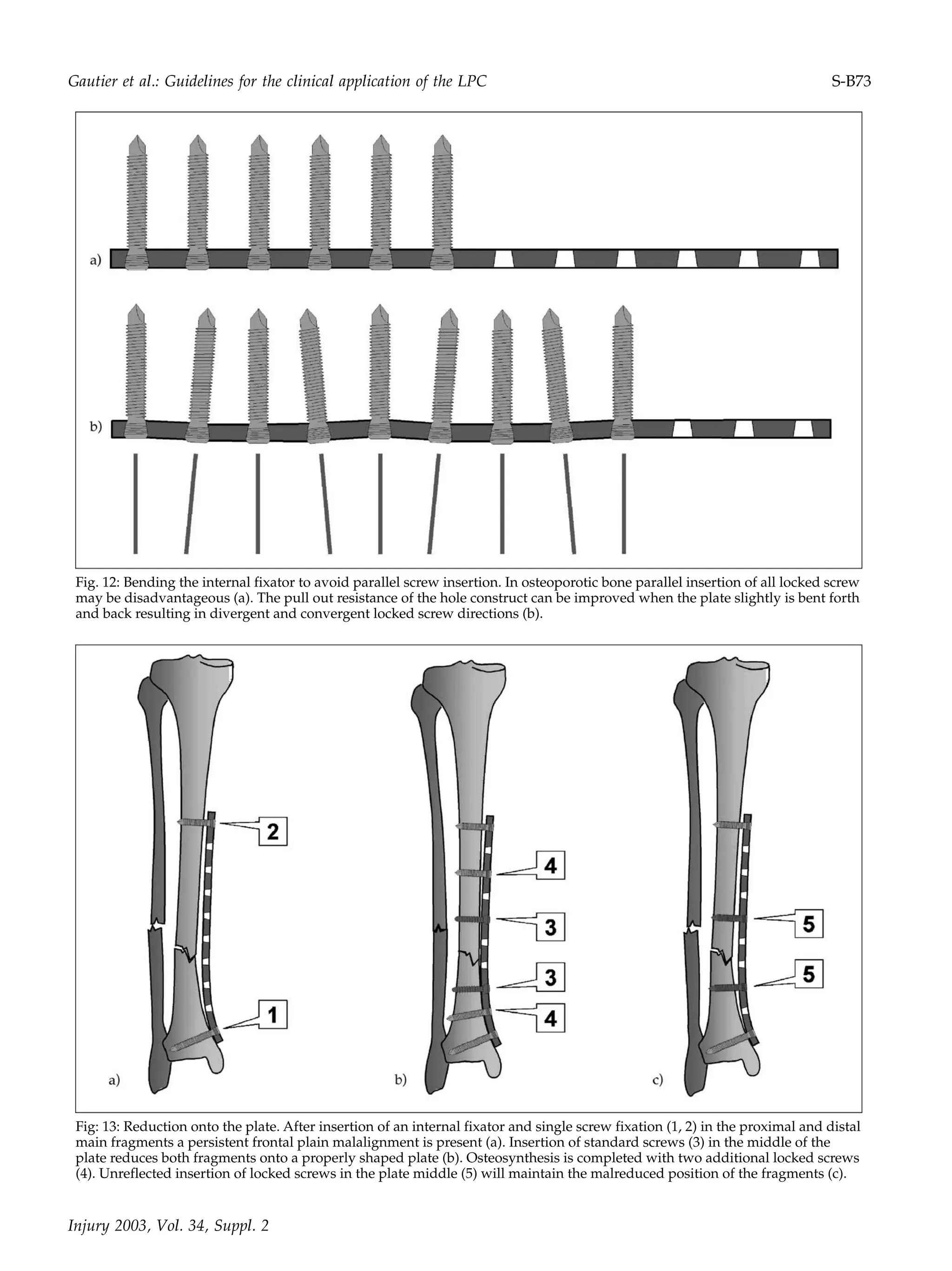
The straight and the bent internal fixator
To stabilise a straight segment of a diaphysis a straight
internalfixatorcanbeused.Insuchasituationallscrews
have the same direction and the screw loading is iden-
tical. In very osteoporotic bone, this can be dangerous.
To avoid identical screw directions for all the screws,
slight bending (multiple waves forth and back) of the
internal fixator offers an advantage because it results in
divergent and convergent screw direction enhancing
the pullout strength of the screws, comparable to the
PHILOS plate. This slight shaping can be used in so-
calledeggshellboneincasesofsevereosteoporosis(Fig.
12a, b). Other bone segments, such as the olecranon,
need a bent plate from the beginning onwards. The
shaping of the plate avoids the screws being parallel to
each other.
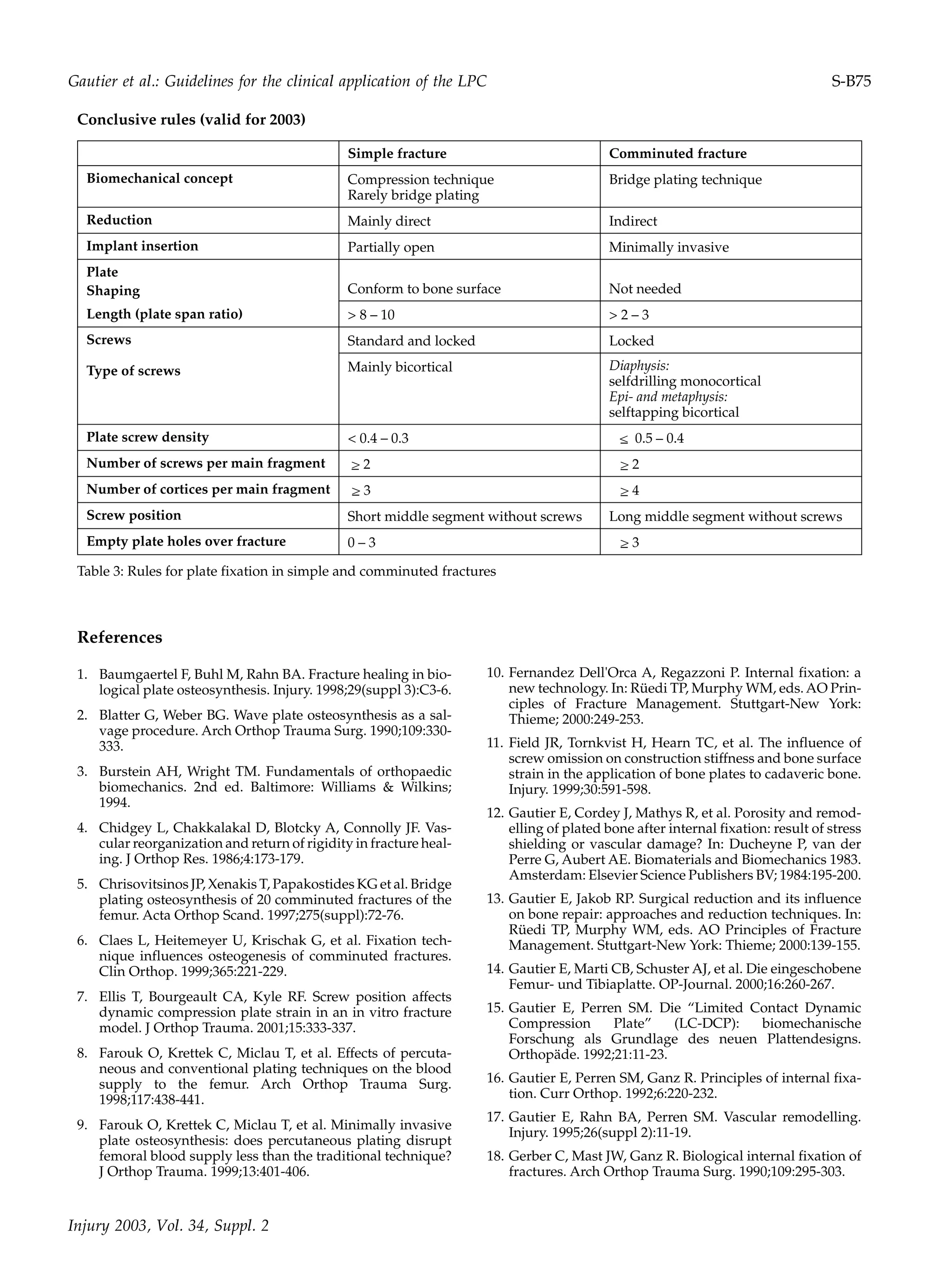
Reduction onto the plate
The first screw insertion (locked or standard) is in the
shorter metaphyseal fragment close to the joint line.
Then the screw at the other plate end is inserted and the
reduction is checked with the image intensifier or an
intraoperative radiograph in both planes. With a flex-
ion or extension malalignment the fragments are
reduced with pointed reduction forceps through stab
incisions. Where the malalignment is in the frontal
plane, introducing the standard screws nearer the frac-
ture can help to reduce the fracture onto the plate. The
fixation is completed either with locked or standard
screws, depending on the bone quality (Fig. 13a, b).
Insertion of locked screws in the middle of the plate
maintains the malreduction (Fig. 13c).
Dislocation onto the plate
The inappropriate use of either standard or locked
screwscanabolishthepreviouseffortstoachieveproper
axial alignment. Thus, the remaining axial malalign-
S-B72](https://image.slidesharecdn.com/lcpgautier-160323191152/75/Lcp-gautier-10-2048.jpg)