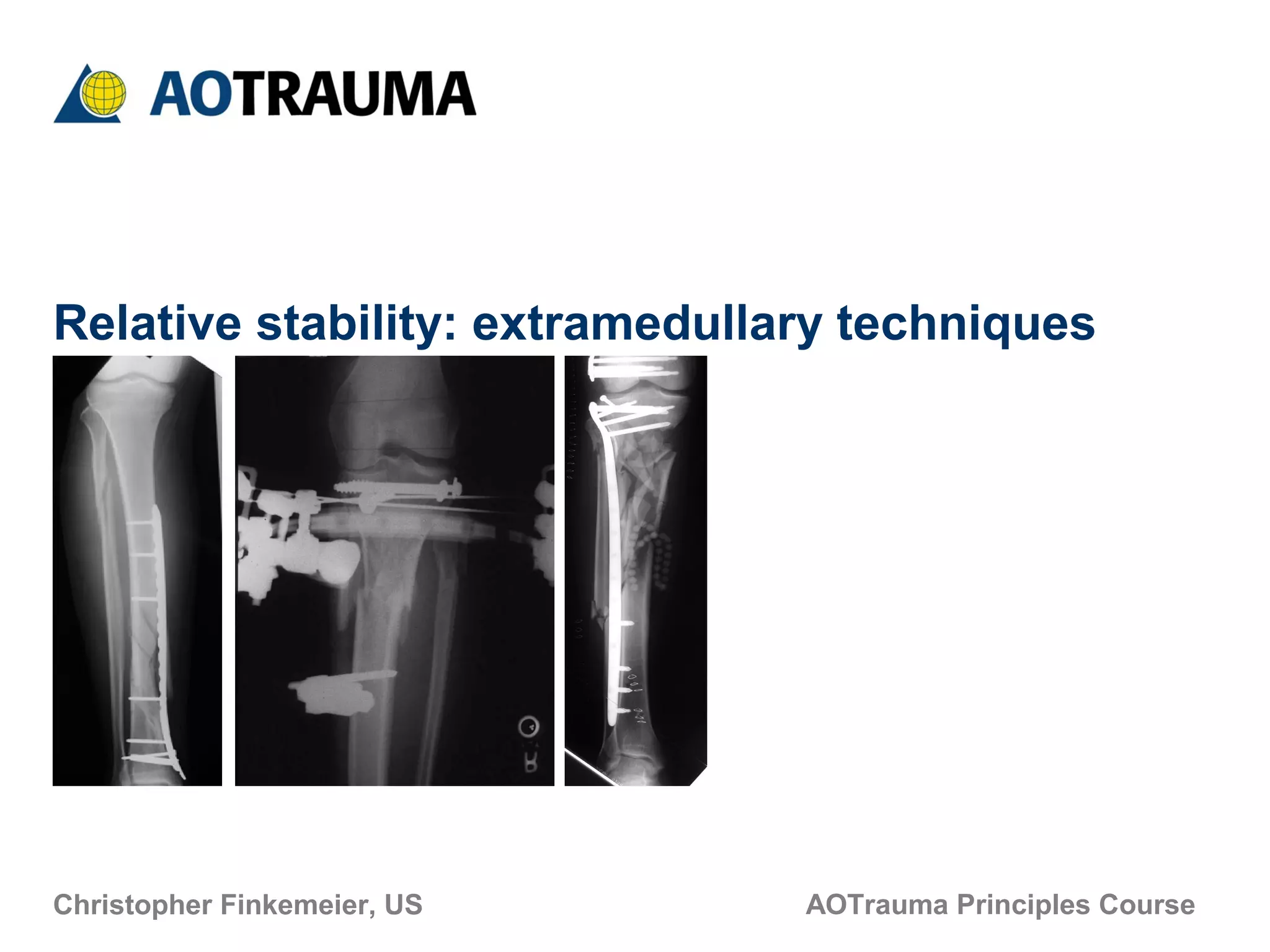
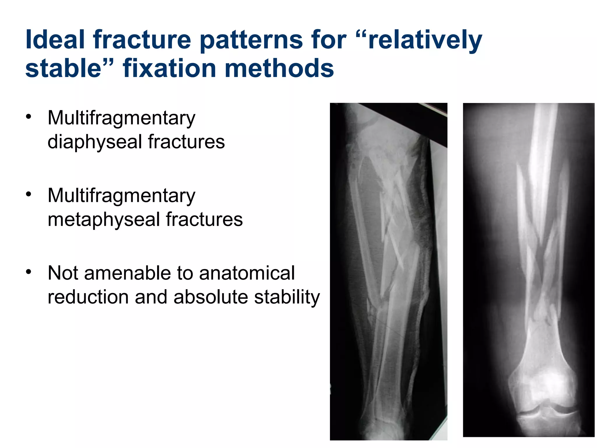
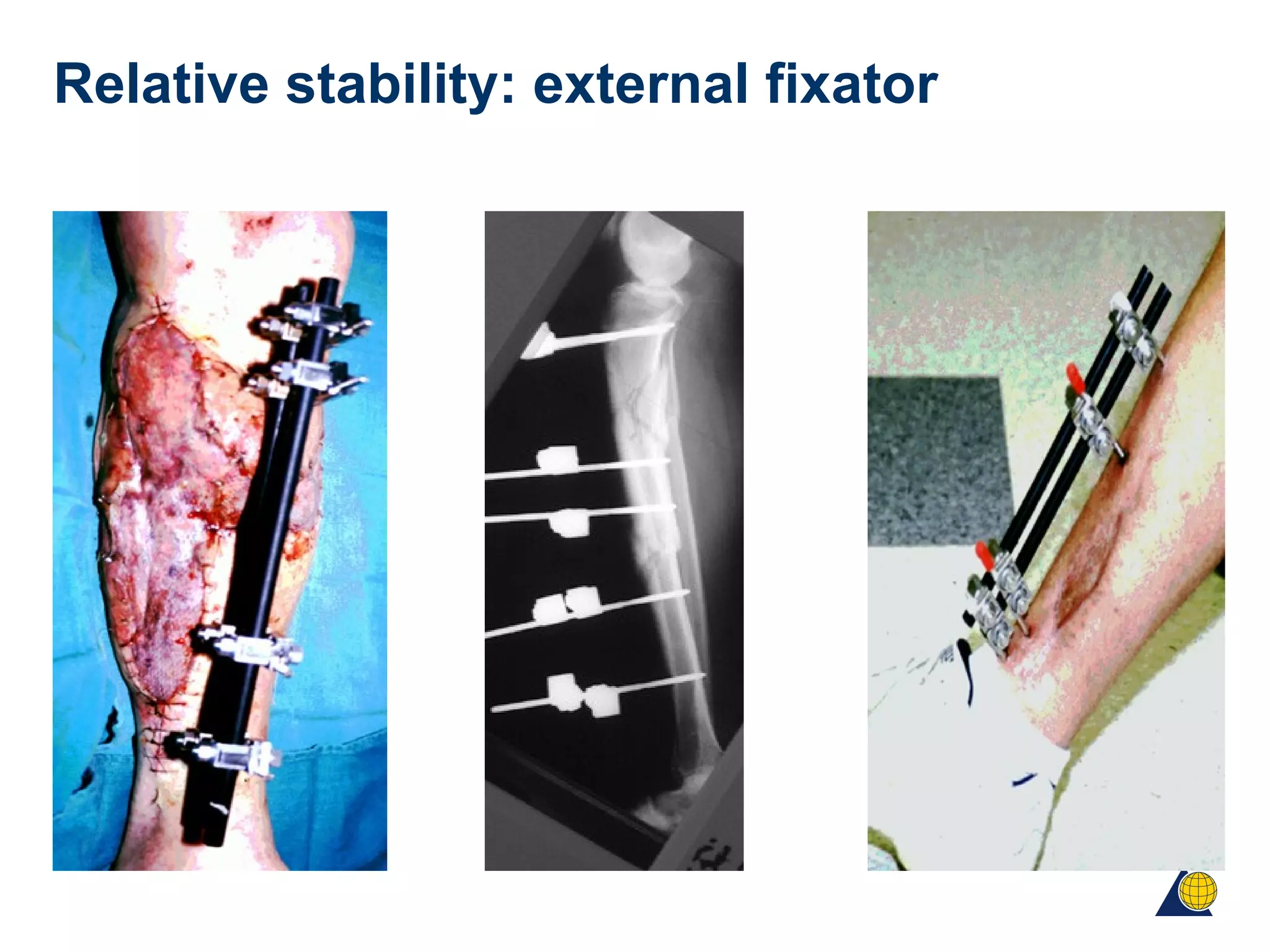
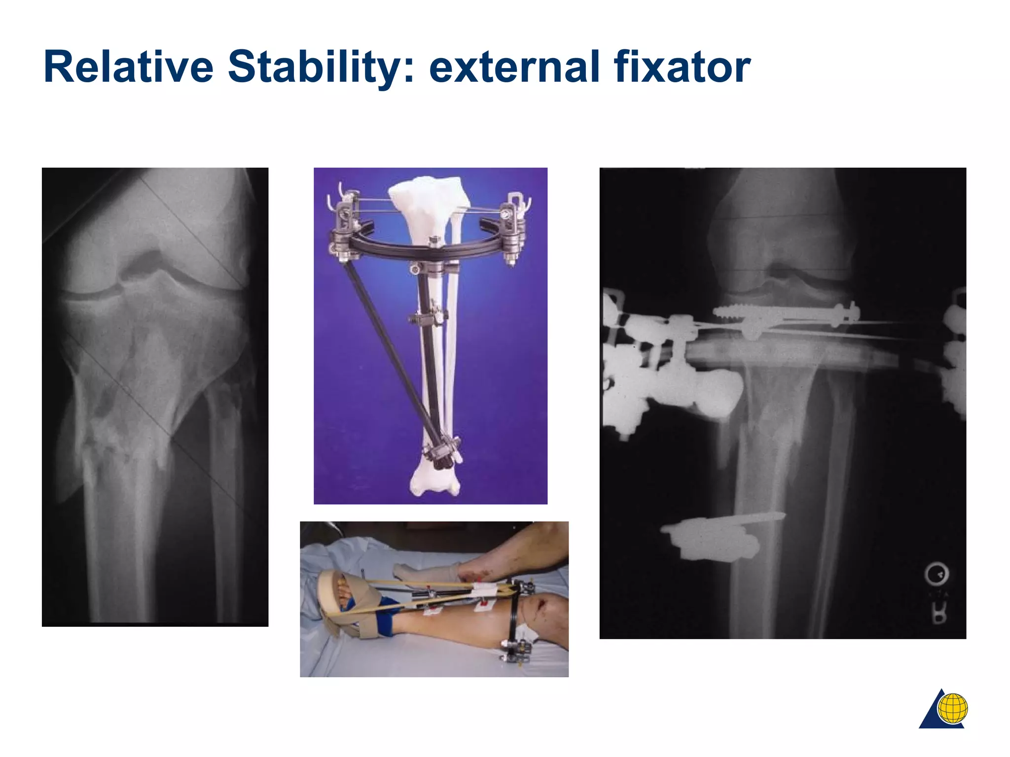
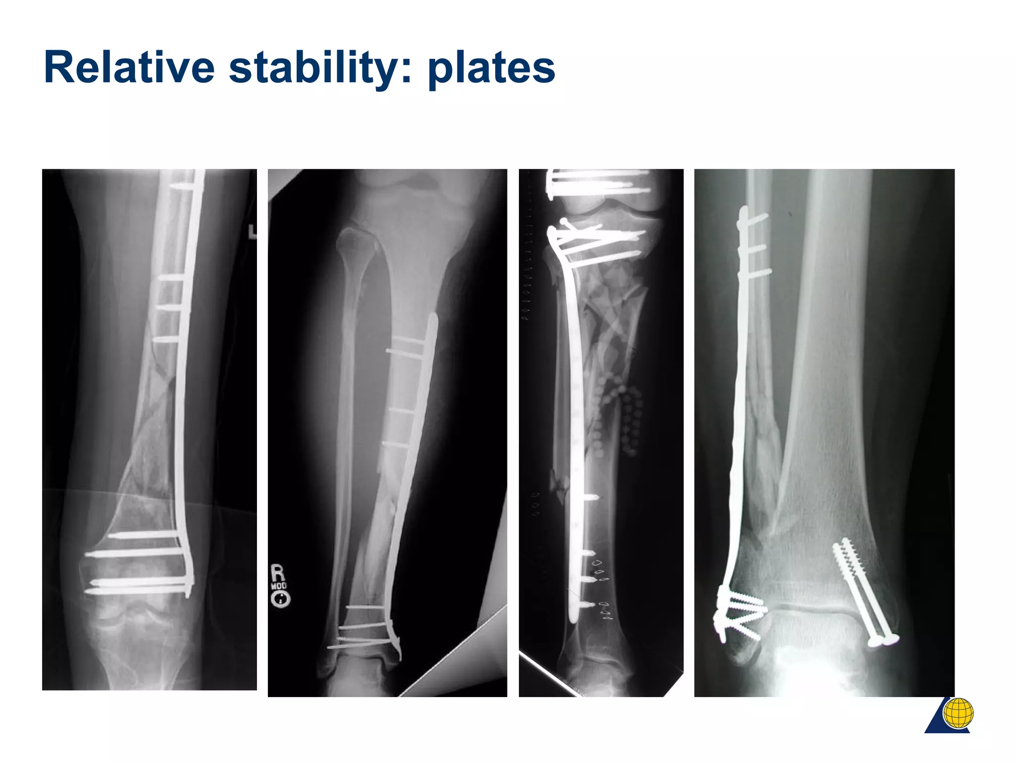
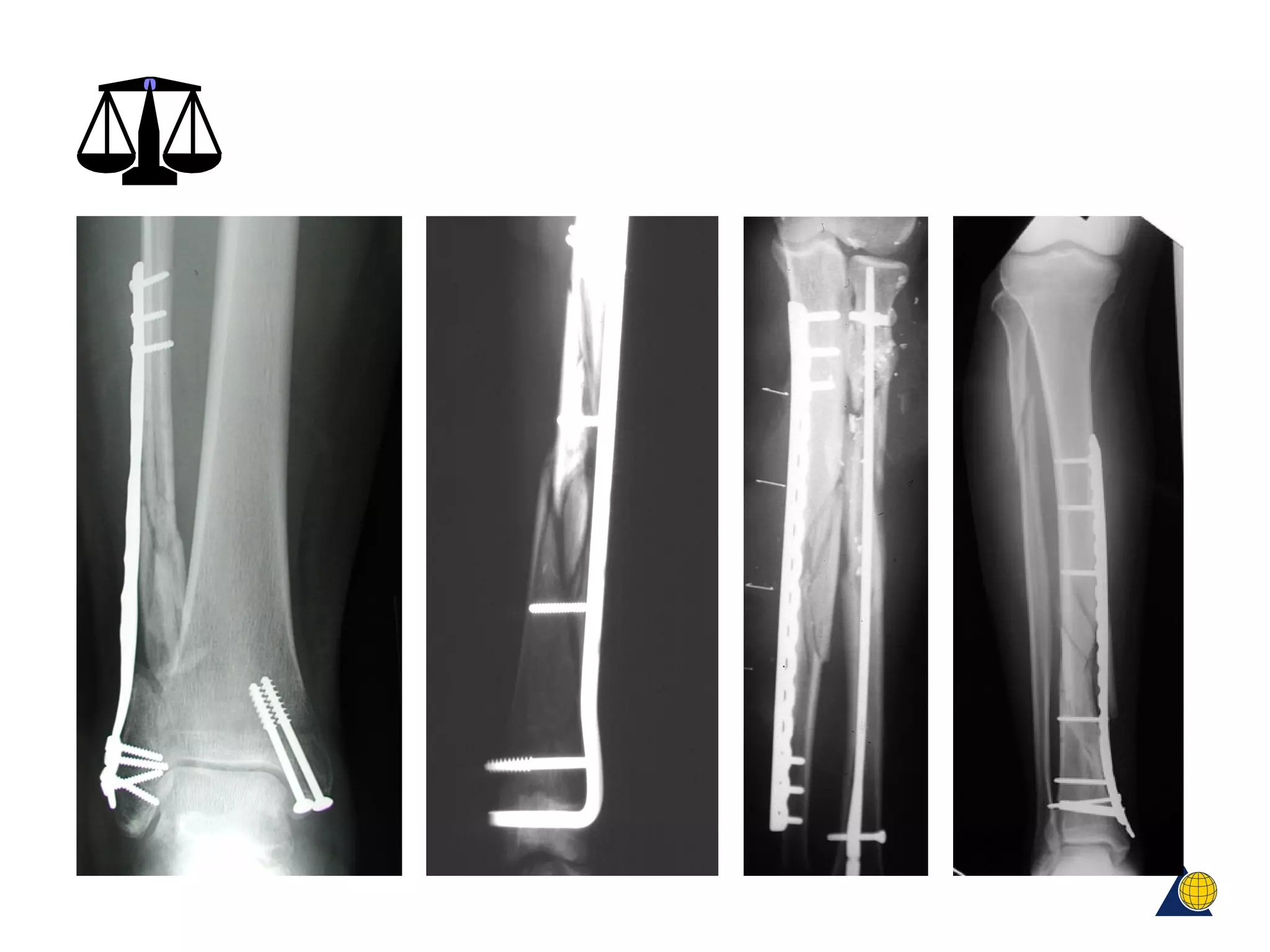
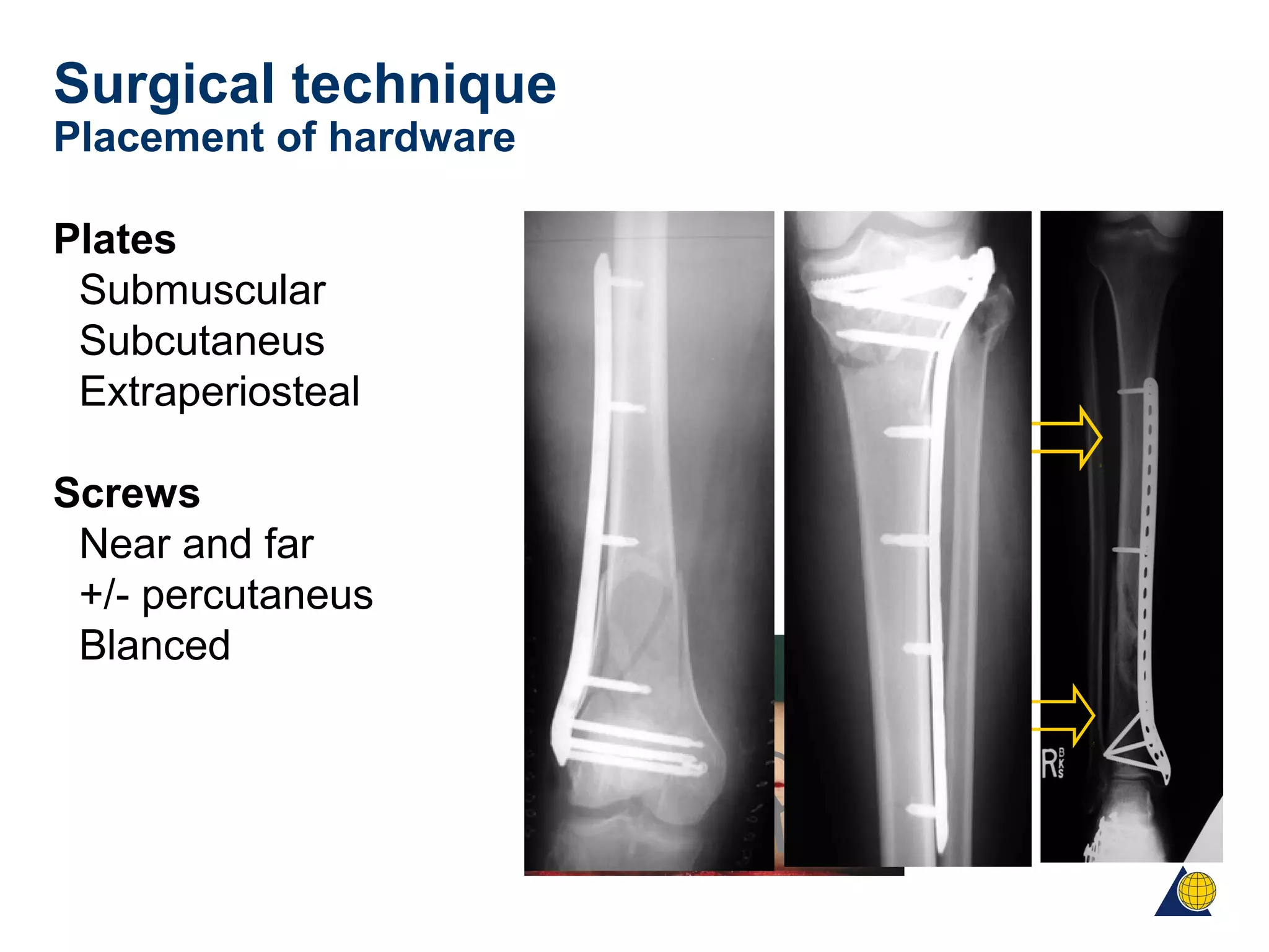
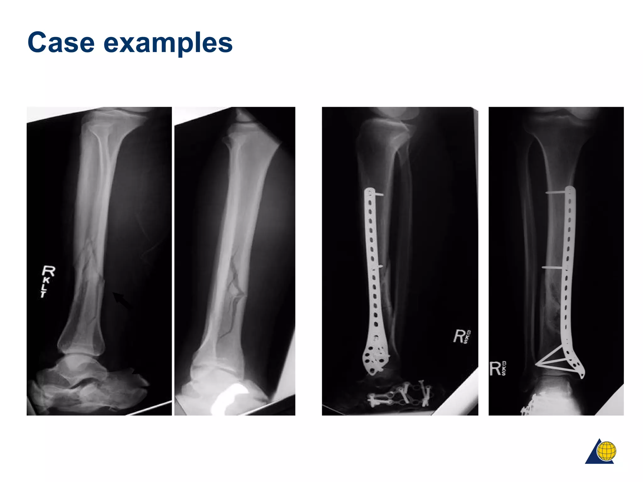
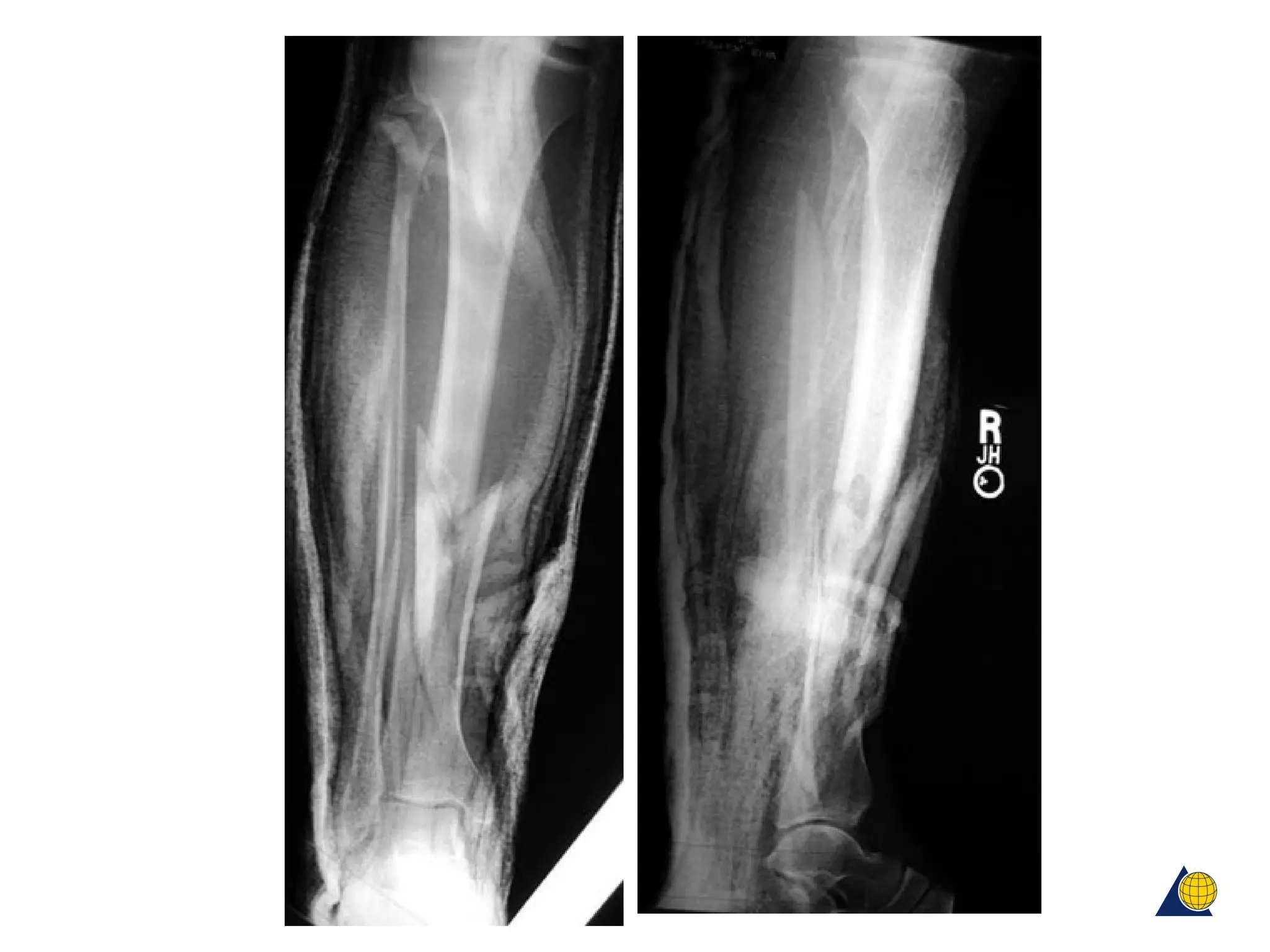
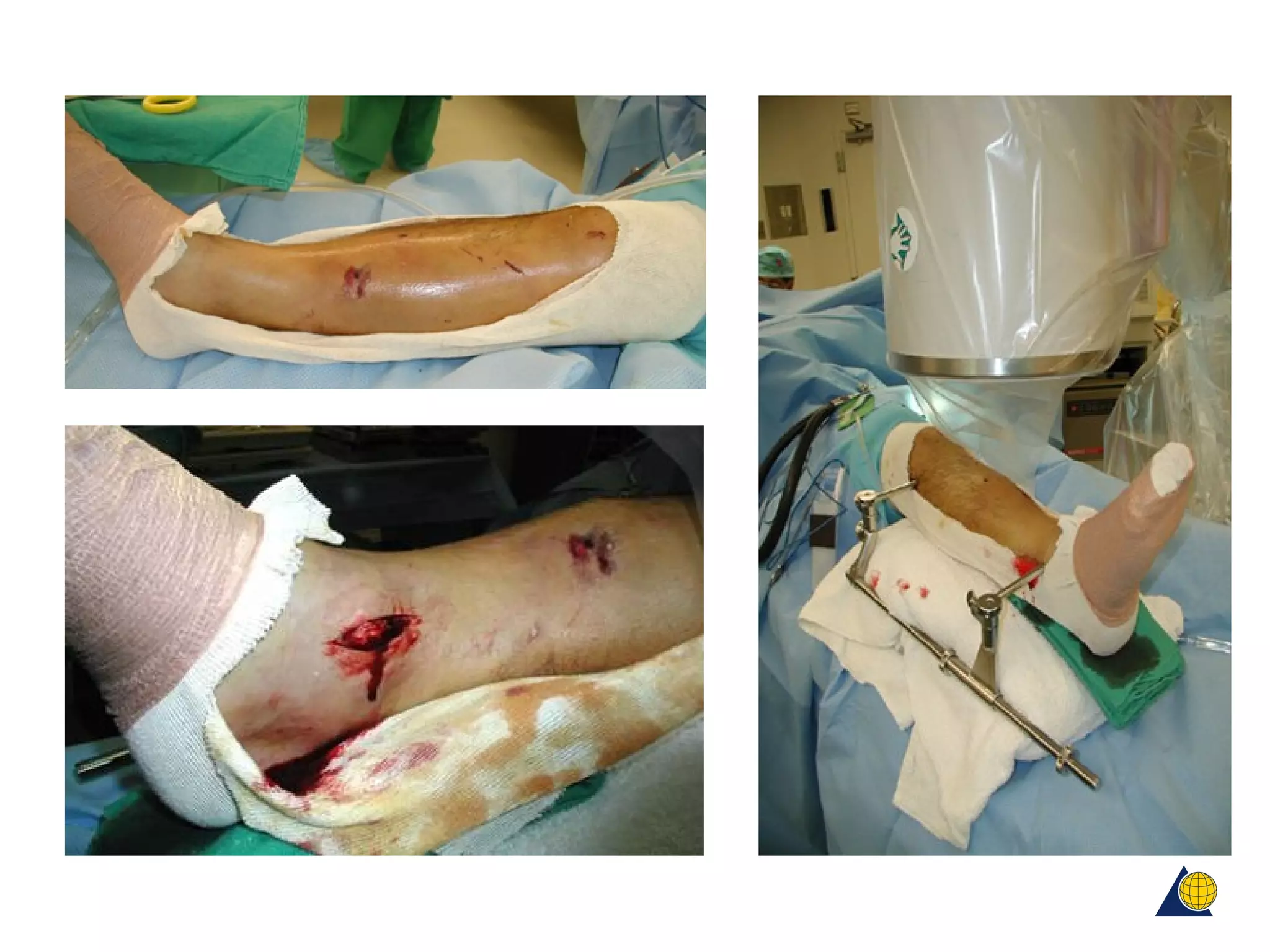
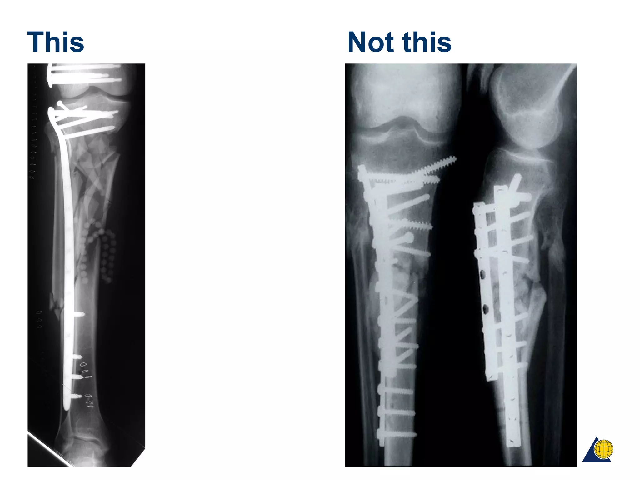
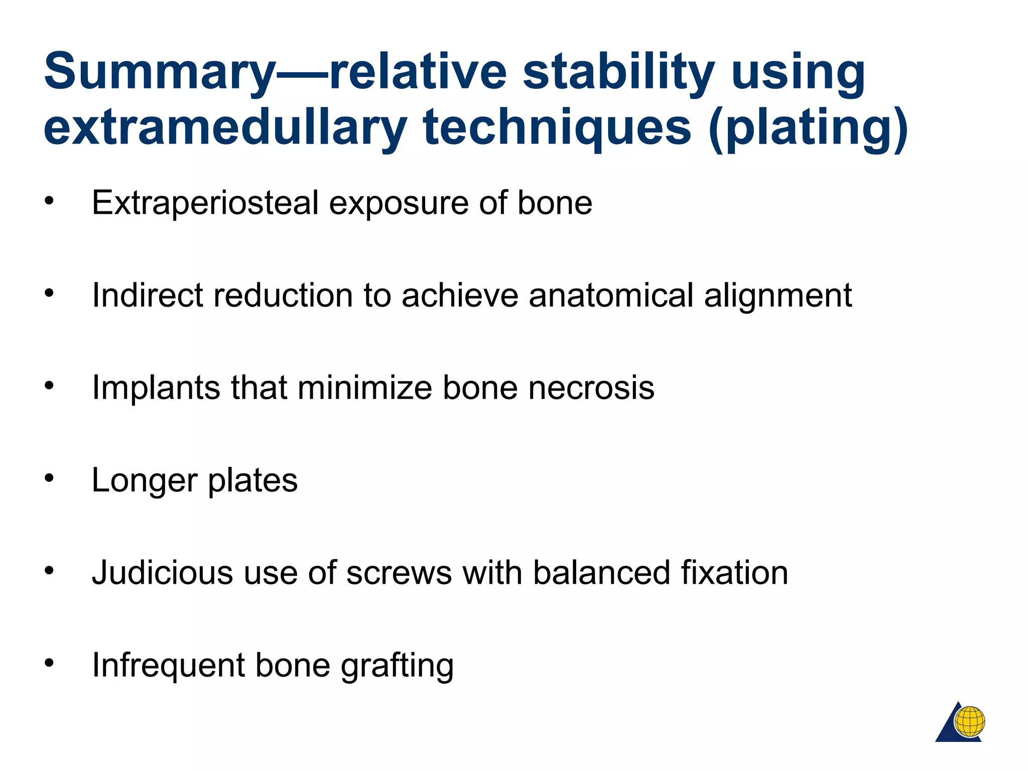
AOTrauma Principles Course
Relative stability: extramedullary techniques
Christopher Finkemeier, US
Objectives
• Identify what fracture patterns are best suited for
relatively stable fixation techniques
• Identify two common extramedullary techniques for
obtaining relative stability
• Identify several fundamental features of bridge plating
• Understand several surgical techniques unique to bridge
plating
Relative stability—review
• Applied load to fracture exceeds the preload
• Small amount of motion between fragments leads to
callus formation and “indirect bone healing”
• Healing occurs if the interfragmental strain remains
below the critical strain level for the repair tissue
• The more fragments present, the less strain between
fragments and the less rigid the construct requirement
Ideal fracture patterns for “relatively
stable” fixation methods
• Multifragmentary
diaphyseal fractures
• Multifragmentary
metaphyseal fractures
• Not amenable to anatomical
reduction and absolute stability
Extramedullary techniques
• External fixation
• Plate fixation:
- Bridge plating
- “Internal fixator”
Aim of technique
• To preserve the vascularity of the fracture site and
fracture fragments
• To provide sufficient stabilization to promote union
External fixation
• External fixator may be used as provisional or definitive
management of fracture
• Provisional fixators are used mainly to treat the soft
tissues
• Definitive fixators treat both bone and soft tissues
Relative stability: external fixator
Relative Stability: external fixator
Current trend in external fixation
• Provisional external fixation
Provisional external fixation—
eg, femoral fractures
IMN
(intramedullary nail)
4 days later
Unstable
multitrauma
patient
Immediate
provisional
external fixator
“Damage Control”
High-energy
mechanism with severe
depression
Provisional external fixation—eg, tibial
plateau fractures
Provisional external fixator
restores length
and alignment
Definitive ORIF (open
reduction and internal
fixation) when soft tissues
ready
Articular segment
remains depressed
Relative stability: plates
• Extraperiosteal exposure of bone
• Indirect reduction to achieve anatomic alignment
• Implants that minimize bone necrosis
• Longer plates
• Judicious use of screws with balanced fixation
• Infrequent bone grafting
Epiperiosteal exposure of bone
• Fractures disrupt the normal blood
supply to bone (predominantly
intramedullary via nutrient artery)
Epiperiosteal exposure of bone
• After fracture the surrounding soft tissues provide an
• extraosseous blood supply
• Proliferation of periosteal osteoblasts occur when
vessels
• grow from the musculature to the periosteum
Epiperiosteal exposure of bone
• Damage to the periosteum:
• Escape of hematoma
• Diffusion of pluripotent mesenchymal cells
• Necrosis at the fracture site
Indirect reduction to achieve anatomical
alignment
Ligamentotaxis
Length
Rotation
axis
Indirect reduction to achieve anatomical
alignment (slide 1 of 2)
Indirect reduction to achieve anatomical
alignment (slide 2 of 2)
Implants that minimize bone necrosis
Limited contact dynamic compression plate
LC-DCP
Less invasive stabilization system LISS
Fewer screws/longer plates
• Longer plates improve the construct by increasing the
lever arm of the plate
• Longer plates require fewer screws to achieve optimal
fixation (near fracture and farthest from fracture)
• The strain on longer plates is reduced as is the strain on
the screws
• Fewer screws minimize damage to the bone
• A tensioned plate without lag screws acts as an elastic
but rigid spring
=
Gotzen et al, 1983
Mini—max fixation
Bone grafting is unnecessary
Rozbruch et al, 1998
• Incidence of primary bone grafting femoral
• Shaft fractures: 16% to 4%
Krettek et al, 1997
• 92% union in proximal and distal femur fractures without
bone grafting
Kregor et al, 1999
• 97% union rate (Type A and C supracondylar femoral
fractures) without bone grafting
Planning and reduction technique in
fracture surgery
• Fracture configuration
• Implant templates
• Plan fixation construct
• Step-by-step operative plan (open vs MIPO)
Surgical technique
• Equipment
• Radiolucent OR table
• Image intensifier
Surgical technique
• External fixator or femoral distractor (2)
• Sterile towels (to make bumps if needed)
Surgical technique
Implants (precontoured)
Surgical technique
Reduction instruments: pointed clamps, lamina spreader, dental pick
Surgical technique
Minimize use of clamps that disrupt soft tissues and blood
supply
Lohman worst
Locking Lane
Verbrugge
Weber tenaculum least
Surgical technique
• Anatomical reduction
• Anatomical reduction of articular surfaces
Surgical technique
• Indirect reduction
• Restore length, axial alignment, rotation
Surgical technique
• Exposure
• Open, periosteal preserving
Surgical technique
• Exposure
• Percutaneous
Surgical technique
Placement of hardware
Plates
Submuscular
Subcutaneus
Extraperiosteal
Screws
Near and far
+/- percutaneus
Blanced
Case examples
Postoperatively 2 weeks 4 months
This Not this
Summary—relative stability using
extramedullary techniques (plating)
• Extraperiosteal exposure of bone
• Indirect reduction to achieve anatomical alignment
• Implants that minimize bone necrosis
• Longer plates
• Judicious use of screws with balanced fixation
• Infrequent bone grafting

Relative extramedullary stability