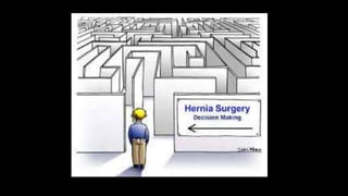
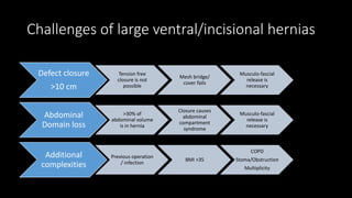
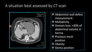
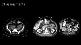
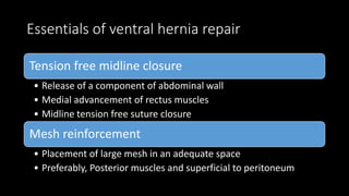
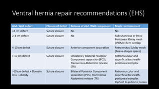
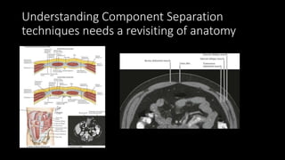
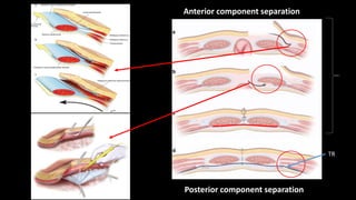
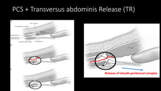
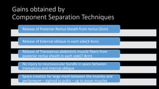
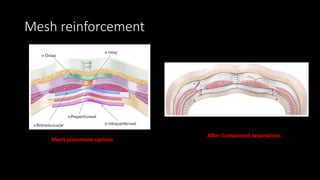
Component separation for ventral hernias prof. ahm shamsul alam
1. Abdominal wall Component
Separation Techniques for large
Ventral Hernia Repair
Prof. AHM Shamsul Alam
Bangabandhu Sheikh Mujib Medical University, 14th September, 2019
5. Challenges of large ventral/incisional hernias
Defect closure
>10 cm
Tension free
closure is not
possible
Mesh bridge/
cover fails
Musculo-fascial
release is
necessary
Abdominal
Domain loss
>30% of
abdominal volume
is in hernia
Closure causes
abdominal
compartment
syndrome
Musculo-fascial
release is
necessary
Additional
complexities
Previous operation
/ infection
BMI >35
COPD
Stoma/Obstruction
Multiplicity
6. A situation best assessed by CT scan
Abdominal wall defect
measurement
Multiplicity
Domain loss: >30% of
abdominal volume in
hernia
Previous mesh
position
Obesity
Stoma position
8. Essentials of ventral hernia repair
Tension free midline closure
• Release of a component of abdominal wall
• Medial advancement of rectus muscles
• Midline tension free suture closure
Mesh reinforcement
• Placement of large mesh in an adequate space
• Preferably, Posterior muscles and superficial to peritoneum
9. Ventral hernia repair recommendations (EHS)
Abd. Wall defect Closure of defect Release of abd. Wall component Mesh reinforcement
<2 cm defect Suture closure No No
2-4 cm defect Suture closure No Subcutaneous or Intra-
Peritoneal Onlay mesh
(IPOM) >5cm overlap
4-10 cm defect Suture closure Anterior component separation Retro rectus Sublay mesh
(Reeve-stoppa space)
> 10 cm defect Suture closure Unilateral / Bilateral Posterior
Component separation (PCS),
Transversus Abdominis release
(TR)
Retromuscular and
superficial to sheath-
peritoneal complex
>10 cm defect + Domain
loss + obesity
Suture closure Bilateral Posterior Component
separation (PCS), Transversus
Abdominis release (TR)
Retro-muscular and
superficial to sheath-
peritoneal complex
Xiphoid to pubis to psosas
13. Gains obtained by
Component Separation Techniques
Release of Posterior Rectus Sheath from rectus (2cm)
Release of External oblique in each side(3-4cm)
Release of Transversus abdominis muscle fibers from
posterior rectus sheath in each side(7-8cm)
No injury to neurovascular bundle in space between
transversus and internal oblique
Space creation for large mesh between the muscles and
peritoneum – xiphoid to pubis – up to psoas muscles
17. Minimal access abdominal Component
Separation
• e-TEP laparoscopic and Robotic approach is currently in
practice for posterior component separation (PCS) and
Transversus Release (TAR)
• Principals and dissection is not different from that of open
operations
• Retro-rectus space (Reeve-Stoppa) created by balloon / air
pump/ telescopic stretching on making port 1cm medial to
linea semilunaris
18. Outcome of Component separation
techniques
Rafat Y Afifi Hernia 9(4),310-315, 2005
Massive ventral hernias are difficult to repair, numerous methods has been
described with overall recurrence rate up to 33% after first repair and 44%
after second repair, mostly occurring within 3 years of repair.
Novitsky, Yuri W. et al: Annals of Surgery, Volume 264, Number 2, August
2016, pp. 226-232(7)
December 2006 to December 2014. 428 consecutive PCS-TAR procedures
were analyzed.
Eighty (18.7%) surgical-site events occurred, of which 39 (9.1%) were
surgical-site infections. Three patients required mesh debridement; however,
no instances of mesh explantation occurred. Of the 347 (81%) patients with
at least 1-year follow-up (mean 31.5 mo), there were 13 (3.7%) recurrences.
19. Complications Open CST and TR (%) MICST and TR (%)
Superficial infections 8.9 3.5
Skin dehiscence 8.2 5.3
Skin necrosis 6.8 2.1
Haematoma/seroma 7.4 4.6
Fistul 1 .4
Fascial dehiscence .4 0
Intra-abdominal abscess 3.8 4.6
Mortality .4 .6
Recurrence 11.1 15.1
Meta analysis of 63 primary studies (3055 patients) by
Noah J Switzer et al, Surgical endoscopy 29(4),787-795,2015
Outcome of Component separation
techniques
20. Outcome of Component separation
techniques compared
Issues Onlay open IPOM OPEN CST MIS CST
Defect closure <5cm <4cm >10 cm >10 cm
Operating hazards
and time
+ + ++ ++
Post-op wound
events
++ + + +
Recurrences +++ + + +
Cosmetic outcome + +++ ++ +++
Patient satisfaction + +++ ++ +++
Cost + +++ ++ ++++
21. Summary
• Midline fascial closure is an essential component of ventral hernia repair
• Large defect needs abdominal wall musculo-fascial release in order to
prevent
• Tension in suture line
• To prevent abdominal compartment syndrome
• Anterior component separation could be sufficient to provide musculo-
fascial release <10 cm of defect and allows mesh placement in retro rectus
space(Reeve-Stopa)
• Posterior component separation + TR provides sufficient musculo-fascial
release for > 10 cm defect and in complex cases and allows large mesh
under cover of sheath-peritoneal complex avoiding gut contact.
• Outcome in large series and meta analysis shows lower rates of wound
related events and recurrences in both open and minimal access
approaches.
22. Conclusion
• Component separation techniques are powerful tools in
order to overcome challenges of complex large ventral
hernias
• Anatomical orientation of abdominal wall layers is the key to
dissect in correct plane.
• Use of MIS is promising in this field
• Outcome of surgery depends on correct decision for correct
techniques.
23.
24. Next Scientific meeting proposals of
Hernia Society of Bangladesh (Monthly)
Date Topic Institute
October 2019 Open groin hernia repair- am I doing it correctly? Sir Salimullah MC
November 2019 Prevention of chronic pain after hernia surgeries Dhaka Medical College
December 2019 Guidelines for groin hernia repairs (EHS, AHS, APHS) BIRDEM
January 2020 Mesh fixation Shahid Shohruardy MC
February 2020 Training module of Minimally Invasive Hernia
surgeries
BCPS
Visit Facebook page of “Hernia Society Of Bangladesh” and click Like button for regular updates
Editor's Notes
The “classic” anterior component separation technique is well described by Clarke
“Midline scar excision is followed by extensive skin flap mobilization. The lateral border of the rectus muscle is located, as well as a point 1 cm lateral to the rectus, the external oblique aponeurosis and muscle are divided from the inguinal region to the costal margin. Lateral dissection deep to the external oblique allows creation of a “sliding myofascial flap” consisting of internal oblique and transversus muscles. Cephalad to the costal margin, where the rib cage protects against herniation, the lateral border of the rectus may be released to allow these muscles to be mobilized from the chest wall and apposed in the midline to “fill” the epigastrium. Attenuated tissue around the hernia is resected, and the posterior rectus sheath may also be incised longitudinally, if additional mobilization is desirable. The midline is then closed with a single layer of heavy monofilament suture” (7).
Novitsky et al. (9) have described the novel technique of transversus abdominis release (TAR) (Figure 1B):
“The retromuscular plane is developed toward the linea similunaris, visualizing the junction between the posterior and anterior rectus sheaths. The perforators to the rectus muscle (branches of the thoracoabdominal nerves, penetrating the lateral edge of the posterior rectus sheath) are visualized and preserved. Starting in the upper third of the abdomen, about 0.5 cm medial to the anterior/posterior rectus sheath junction, the posterior rectus sheath is incised to expose the underlying transversus abdominis muscle. The muscle is then divided along its entire medial edge using electrocautery. This step is initiated in the upper third of the abdomen where medial fibers of the transversus abdominis muscle are easiest to identify and separate from the underlying fascia. This step allows entrance to the space between the transversalis fascia and the divided transversus abdominis muscle. Once similar release is performed on both sides, the posterior rectus sheaths are reapproximated in the midline with a running monofilament suture. The mesh is placed as a sublay in the retromuscular space and secured with sutures. The anterior rectus sheaths are then reapproximated in the midline to restore the linea alba ventral to the mesh”(9).