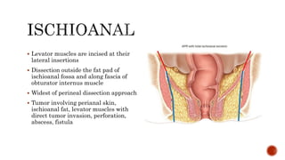
Presenter: Dr. Mohammad Masoom Parwez
Moderator: Prof. Dr. Pradeep Saxena
ď‚§ Historical Background
ď‚§ Indications
ď‚§ Preoperative Planning
ď‚§ Surgical Technique
ď‚§ Post operative care
ď‚§ Management of complications
ď‚§ Outcomes
ď‚§ W. Ernest Miles, 1908 : credited with describing modern APR
ď‚§ Single stage surgery incorporating both abdominal and perineal procedure
ď‚§ Creation of abdominal colostomy
ď‚§ Removal of entire pelvic colon and meso colon below the common iliac artery
ď‚§ Lymph node at the bifurcation of common iliac artery
ď‚§ Levator muscles at their origin, ischiorectal fat and anus
ď‚§ O.V Lloyd Davis popularized synchronous two team approach with patient in
lithotomy position
ď‚§ Bill Heald, 1982: revolutionized the technique of entire mesorectum excision via
precise and meticulous dissection
 Adenocarcinoma lower rectum – for negative circumferential resection margin
(not distal margin)
 Anal SCC – that persists or recurs after chemoRT
ď‚§ Anal melanoma
ď‚§ Anal sarcoma
ď‚§ GIST
 Extended APR – vulvar, vaginal or prostate ca involving distal rectum/anal
spinchter
 Benign – as a part of total proctocolectomy in FAP or IBD
Intersphincteric
Extralevator
Ischioanal
ď‚§ Creates narrowest perineal wound
ď‚§ For patients without levator
involvement, but incontinent
ď‚§ Patients at high risk for anastomotic
leak – poor wound healing, inadequate
physiological reserve
 ELAPE – Extralevator abdominoperineal
excision
ď‚§ Levator muscles are removed en bloc with
the specimen
ď‚§ Tumor within a cm from the dentate line
ď‚§ Low rectal tumor with threatened CRM
ď‚§ Where a sphincter saving procedure is
oncologically impossible
ď‚§ Levator muscles are incised at their
lateral insertions
ď‚§ Dissection outside the fat pad of
ischioanal fossa and along fascia of
obturator internus muscle
ď‚§ Widest of perineal dissection approach
ď‚§ Tumor involving perianal skin,
ischioanal fat, levator muscles with
direct tumor invasion, perforation,
abscess, fistula
ď‚§ Significant morbidity
ď‚§ Medical clearance
ď‚§ Marking of stoma site
ď‚§ Counselling by stoma therapist
ď‚§ Bowel cleansing/enema (original Nichols and Condon oral antibiotic regime, 1970
of Neomycin and Erythromycin base)
Extent of resection:
ď‚§ Physical examination and review of imaging
ď‚§ Multidisciplinary approach with specialists
 Anterior structures – prostate, post wall of vagina
ď‚§ Posterior structures - sacrum
 Pelvic sidewall lymphadenopathy – extended lymphadenectomy
 Dr. Solomon – extended resections and pelvic exenteration
Stoma marking:
ď‚§ Outer third of rectus abdominis
ď‚§ Abdomen characteristics: Protuberant abdomen, folds, scars, costal margin and
iliac crest, pendulous breasts an hernias
ď‚§ Patient characteristics: mobility (wheelchair bound), posture (kyphosis), dexterity,
vision
ď‚§ Patient preference for location (belt line)
ď‚§ Surgical considerations: type of stoma (loop vs end), segment of intestine, need for
both urinary and fecal stoma, continence
Workup:
ď‚§ History - baseline urinary and sexual function, continence
 DRE – relationship of tumor to surrounding structures
 Staging workup – CT scan of chest, abdomen and pelvis, baseline CEA, endorectal
US, MRI of pelvis (T and N stage)
 MERCURY Trial – 91% accuracy of MRI in predicting negative resection margin
in patients going for primary surgery and 77% accuracy among [patients who have
received NACT
 Rectal MRI – invasion of levator muscle and sphincter complex relation
Abdominal dissection
Perineal dissection
 Lloyd Davis position – both procedures can be performed without repositioning
the patient
ď‚§ Prone jack knife position
ď‚§ Lithotomy position
Abdominal dissection:
ď‚§ Lower midline or low transverse laparotomy
ď‚§ Thorough examination of abdomen and pelvis
ď‚§ Sigmoid colon is mobilized by dividing the attachments and adhesions to lateral
abdominal and pelvic sidewall
ď‚§ Incision is extended cephalad upto splenic flexure and distally towards pelvis
ď‚§ Mesentery is lifted from RP attachment exposing the left gonadal vessels and left
ureter
 Stay in retromesocolic plane. Deeper dissection – damage to left ureter and
gonadal vessels
ď‚§ Sigmoid colon is retracted anteriorly
and laterally exposing the root of the
sigmoid mesentery
ď‚§ Incision is made to right of sigmoid
mesocolon, proximal to promontory
ď‚§ Plane is developed underneath the
superior rectal vessels in the loose
areolar tissue, between the origin of
inferior mesenteric artery and
promontory
 Avoid injuring the superior hypogastric plexus – situated between the sup rectal
vessels and bifurcation of aorta
ď‚§ Hypogastric nerves can be seen as two band like structures just lateral to the
midline, as the dissection proceeds inferior to the sacral promontory
ď‚§ Identify and secure the ureter in the retroperitoneum, isolate and ligate the
superior rectal vessels between the origin of left colic and first sigmoid vessel
ď‚§ Left colic artery is not routinely divided, but any enlarged LN around the
bifurcation of IMA should be dissected
ď‚§ High ligation of IMA carries the risk of damaging the hypogastric plexus
ď‚§ The mesentery of the sigmoid colon is divided at the point chosen to create the end
colostomy
ď‚§ The colon itself is divided using a linear stapler after ensuring sufficient
mobilisation to ensure a tension free, well vascularized colostomy
ď‚§ Areolar plane behind the fascia proper of the rectum is opened by anterior
reflection of the stump of the superior rectal vessels and the proximal rectum
away from the promontory
ď‚§ Identify and protect B/L hypogastric nerves present in the lateral aspect of the
areolar space
ď‚§ Inferior dissection is carried along the
concavity of the sacrum, till the
sacrococcygeal junction and extended
from the midline, laterally
ď‚§ Distally, preserve the pelvic splanchnic
nerve coursing from the lateral pelvic
sidewall near the anterior sacral
foramina to join the pelvic plexus
ď‚§ Division of the areolar connective
tissue should be done using
electrocautery or other sharp
dissection
ď‚§ The lateral stalks are exposed and divided using electrocautery by providing
traction on the rectum to the opposite side of the pelvis
ď‚§ Forceful retraction can damage the pelvic plexus
ď‚§ Dissection is continued laterally until the origin of levator muscle is reached
ď‚§ Finally, the peritoneum is opened anteriorly in the cul de sac
ď‚§ Dissection is carried to the level of prostate in men, and halfway down the vagina
in women
ď‚§ Placement of pelvic drain, closure of midline incision and maturing of stoma
Perineal dissection:
ď‚§ Position: lithotomy or prone jack
knife
ď‚§ Elliptical incision made outside the
lateral edge of external sphincter and
medial to ischial tuberosity
ď‚§ Posteriorly, incision should be
midway between anus and coccyx
ď‚§ Anteriorly, it should divide the
perineal body
ď‚§ Can be extended
ď‚§ The dissection is done using electrocautery, controlling the perforating vessels as
well as the inferior rectal arteries
ď‚§ Palpate the coccyx and aim the posterior dissection to join with the posterior
mesorectal dissection
ď‚§ Dividing the anococcygeal ligament at the tip of the coccyx provides access to the
posterior pelvis for joining the mesorectal dissection
ď‚§ Once the two dissection planes have been connected posteriorly, divide the levator
muscles close to their insertion on the tendinous arch covering the obturator
internus
ď‚§ Placing the index finger of the non dominant hand into the pelvis and hooking the
levator muscles facilitates this division
ď‚§ Puborectalis should be divided anteriorly before the transverse perineal muscle is
reached
ď‚§ The rectum is still attached anteriorly to the prostate or vagina
ď‚§ At this point, it should be delivered through the wound to facilitate exposure for
anterior dissection
ď‚§ This is best performed from proximal to distal following the anterior surface of the
prostate/vagina and from medial to lateral
ď‚§ This approach provides better visualization of the neurovascular bundle (pelvic
plexus) and avoids injury to the urethra
ď‚§ In males, palpating the urethra and the urinary catheter can help avoid injury
ď‚§ In females, placing a finger in the vagina helps define the plane of the
rectovaginal septum
ď‚§ Once the specimen is removed, the pelvic cavity is irrigated
ď‚§ Pelvic drains placed previously are repositioned as required and perineal defect is
closed
ď‚§ For primary closure, meticulous multi-layered closure of the ischiorectal fat with
absorbable sutures is done, and skin is approximated in a vertical mattress
pattern using nylon
ď‚§ In patients with large perineal defects, reinforcement with a biological absorbable
mesh anchored to the coccyx, coccygeus muscle and pelvic sidewall is done to
prevent wound dehiscence and perineal hernia
ď‚§ Other options
ď‚§ VRAM flap
ď‚§ B/L V-Y advancement gluteal flaps
ď‚§ Gracilis muscle flaps
 ERAS –
ď‚§ Early ingestion of clear liquids advancing to low residue diet as tolerated
ď‚§ Limited IV fluid resuscitation
ď‚§ Early ambulation
ď‚§ Non narcotic pain control
ď‚§ Urinary catheter at least till third post op day
ď‚§ Post APR drain management
 Primary closure – remove prior to discharge
 Rectus flap – as long as 2 weeks or till output diminishes
ď‚§ Restrict physical activity to protect the perineal wound
 For primary closure, avoid sitting for 4 – 5 weeks
ď‚§ Perineal sutures to remain in place for 3-4 weeks
ď‚§ For flap closure, avoid bending at the waist for 4-5 weeks
 Most common source of morbidity – perineal wound
ď‚§ Perineal wound infection as high as 40%, high risk of evisceration
ď‚§ Wound is irrigated, necrotic tissue removed, additional drain placed and wound
reclosed if possible
ď‚§ Otherwise, negative pressure dressing has good success rate
ď‚§ Synthetic prosthetic devices (silicone breast implants) can also fill the defect,
though not recommended in contaminated fields
 Genitourinary and sexual dysfunction – up to 50%
 Prevention –
ď‚§ Surgical technique
ď‚§ Identification and protection of pelvic nerve
ď‚§ Minor dysfunction improves 12 months after surgery
ď‚§ However, a significant minority of patients sustain permanent dysfunction
 Stoma related complication – parastomal hernia – significant long term
consequence
ď‚§ Studies support the use of preperitoneal prosthetic mesh for constructing
permanent ostomies
 APR performed in lithotomy – risk of intraoperative bowel perforation and
positive CRM – higher recurrence rate and mortality compared to LAR
ď‚§ With neoadjuvant chemoradiation, local recurrence as high as 30%
 Prone positioning for cylindrical APR – reduction in local recurrence rate to 6%
and perineal wound infection to 11%, decreased rate of positive CRM
ď‚§ Patient with clinical stage 2 or 3 treated with neoadjuvant chemoradiotherapy
and optimal surgery, 5 year survival rate after APR is around 70% compared to
85% after sphincter preserving surgery
ď‚§ Fischers Mastery of Surgery, 7th international edition (2019)
 Maingot’s Abdominal Operations, 13th edition (2019)
Abdominoperineal resection.pptx

Abdominoperineal resection.pptx

  • 1.
    Presenter: Dr. MohammadMasoom Parwez Moderator: Prof. Dr. Pradeep Saxena
  • 3.
    ď‚§ Historical Background ď‚§Indications ď‚§ Preoperative Planning ď‚§ Surgical Technique ď‚§ Post operative care ď‚§ Management of complications ď‚§ Outcomes
  • 5.
    ď‚§ W. ErnestMiles, 1908 : credited with describing modern APR ď‚§ Single stage surgery incorporating both abdominal and perineal procedure ď‚§ Creation of abdominal colostomy ď‚§ Removal of entire pelvic colon and meso colon below the common iliac artery ď‚§ Lymph node at the bifurcation of common iliac artery ď‚§ Levator muscles at their origin, ischiorectal fat and anus ď‚§ O.V Lloyd Davis popularized synchronous two team approach with patient in lithotomy position ď‚§ Bill Heald, 1982: revolutionized the technique of entire mesorectum excision via precise and meticulous dissection
  • 9.
     Adenocarcinoma lowerrectum – for negative circumferential resection margin (not distal margin)  Anal SCC – that persists or recurs after chemoRT  Anal melanoma  Anal sarcoma  GIST  Extended APR – vulvar, vaginal or prostate ca involving distal rectum/anal spinchter  Benign – as a part of total proctocolectomy in FAP or IBD
  • 10.
  • 11.
     Creates narrowestperineal wound  For patients without levator involvement, but incontinent  Patients at high risk for anastomotic leak – poor wound healing, inadequate physiological reserve
  • 12.
     ELAPE –Extralevator abdominoperineal excision  Levator muscles are removed en bloc with the specimen  Tumor within a cm from the dentate line  Low rectal tumor with threatened CRM  Where a sphincter saving procedure is oncologically impossible
  • 13.
    ď‚§ Levator musclesare incised at their lateral insertions ď‚§ Dissection outside the fat pad of ischioanal fossa and along fascia of obturator internus muscle ď‚§ Widest of perineal dissection approach ď‚§ Tumor involving perianal skin, ischioanal fat, levator muscles with direct tumor invasion, perforation, abscess, fistula
  • 15.
    ď‚§ Significant morbidity ď‚§Medical clearance ď‚§ Marking of stoma site ď‚§ Counselling by stoma therapist ď‚§ Bowel cleansing/enema (original Nichols and Condon oral antibiotic regime, 1970 of Neomycin and Erythromycin base)
  • 16.
    Extent of resection: Physical examination and review of imaging  Multidisciplinary approach with specialists  Anterior structures – prostate, post wall of vagina  Posterior structures - sacrum  Pelvic sidewall lymphadenopathy – extended lymphadenectomy  Dr. Solomon – extended resections and pelvic exenteration
  • 18.
    Stoma marking: ď‚§ Outerthird of rectus abdominis ď‚§ Abdomen characteristics: Protuberant abdomen, folds, scars, costal margin and iliac crest, pendulous breasts an hernias ď‚§ Patient characteristics: mobility (wheelchair bound), posture (kyphosis), dexterity, vision ď‚§ Patient preference for location (belt line) ď‚§ Surgical considerations: type of stoma (loop vs end), segment of intestine, need for both urinary and fecal stoma, continence
  • 19.
    Workup:  History -baseline urinary and sexual function, continence  DRE – relationship of tumor to surrounding structures  Staging workup – CT scan of chest, abdomen and pelvis, baseline CEA, endorectal US, MRI of pelvis (T and N stage)  MERCURY Trial – 91% accuracy of MRI in predicting negative resection margin in patients going for primary surgery and 77% accuracy among [patients who have received NACT  Rectal MRI – invasion of levator muscle and sphincter complex relation
  • 20.
  • 21.
     Lloyd Davisposition – both procedures can be performed without repositioning the patient  Prone jack knife position  Lithotomy position
  • 22.
    Abdominal dissection:  Lowermidline or low transverse laparotomy  Thorough examination of abdomen and pelvis  Sigmoid colon is mobilized by dividing the attachments and adhesions to lateral abdominal and pelvic sidewall  Incision is extended cephalad upto splenic flexure and distally towards pelvis  Mesentery is lifted from RP attachment exposing the left gonadal vessels and left ureter  Stay in retromesocolic plane. Deeper dissection – damage to left ureter and gonadal vessels
  • 24.
    ď‚§ Sigmoid colonis retracted anteriorly and laterally exposing the root of the sigmoid mesentery ď‚§ Incision is made to right of sigmoid mesocolon, proximal to promontory ď‚§ Plane is developed underneath the superior rectal vessels in the loose areolar tissue, between the origin of inferior mesenteric artery and promontory
  • 25.
     Avoid injuringthe superior hypogastric plexus – situated between the sup rectal vessels and bifurcation of aorta  Hypogastric nerves can be seen as two band like structures just lateral to the midline, as the dissection proceeds inferior to the sacral promontory  Identify and secure the ureter in the retroperitoneum, isolate and ligate the superior rectal vessels between the origin of left colic and first sigmoid vessel  Left colic artery is not routinely divided, but any enlarged LN around the bifurcation of IMA should be dissected  High ligation of IMA carries the risk of damaging the hypogastric plexus
  • 26.
    ď‚§ The mesenteryof the sigmoid colon is divided at the point chosen to create the end colostomy ď‚§ The colon itself is divided using a linear stapler after ensuring sufficient mobilisation to ensure a tension free, well vascularized colostomy ď‚§ Areolar plane behind the fascia proper of the rectum is opened by anterior reflection of the stump of the superior rectal vessels and the proximal rectum away from the promontory ď‚§ Identify and protect B/L hypogastric nerves present in the lateral aspect of the areolar space
  • 27.
    ď‚§ Inferior dissectionis carried along the concavity of the sacrum, till the sacrococcygeal junction and extended from the midline, laterally ď‚§ Distally, preserve the pelvic splanchnic nerve coursing from the lateral pelvic sidewall near the anterior sacral foramina to join the pelvic plexus ď‚§ Division of the areolar connective tissue should be done using electrocautery or other sharp dissection
  • 29.
    ď‚§ The lateralstalks are exposed and divided using electrocautery by providing traction on the rectum to the opposite side of the pelvis ď‚§ Forceful retraction can damage the pelvic plexus ď‚§ Dissection is continued laterally until the origin of levator muscle is reached ď‚§ Finally, the peritoneum is opened anteriorly in the cul de sac ď‚§ Dissection is carried to the level of prostate in men, and halfway down the vagina in women ď‚§ Placement of pelvic drain, closure of midline incision and maturing of stoma
  • 34.
    Perineal dissection: ď‚§ Position:lithotomy or prone jack knife ď‚§ Elliptical incision made outside the lateral edge of external sphincter and medial to ischial tuberosity ď‚§ Posteriorly, incision should be midway between anus and coccyx ď‚§ Anteriorly, it should divide the perineal body ď‚§ Can be extended
  • 35.
    ď‚§ The dissectionis done using electrocautery, controlling the perforating vessels as well as the inferior rectal arteries ď‚§ Palpate the coccyx and aim the posterior dissection to join with the posterior mesorectal dissection ď‚§ Dividing the anococcygeal ligament at the tip of the coccyx provides access to the posterior pelvis for joining the mesorectal dissection ď‚§ Once the two dissection planes have been connected posteriorly, divide the levator muscles close to their insertion on the tendinous arch covering the obturator internus
  • 36.
    ď‚§ Placing theindex finger of the non dominant hand into the pelvis and hooking the levator muscles facilitates this division ď‚§ Puborectalis should be divided anteriorly before the transverse perineal muscle is reached
  • 41.
    ď‚§ The rectumis still attached anteriorly to the prostate or vagina ď‚§ At this point, it should be delivered through the wound to facilitate exposure for anterior dissection ď‚§ This is best performed from proximal to distal following the anterior surface of the prostate/vagina and from medial to lateral ď‚§ This approach provides better visualization of the neurovascular bundle (pelvic plexus) and avoids injury to the urethra ď‚§ In males, palpating the urethra and the urinary catheter can help avoid injury ď‚§ In females, placing a finger in the vagina helps define the plane of the rectovaginal septum
  • 42.
    ď‚§ Once thespecimen is removed, the pelvic cavity is irrigated ď‚§ Pelvic drains placed previously are repositioned as required and perineal defect is closed ď‚§ For primary closure, meticulous multi-layered closure of the ischiorectal fat with absorbable sutures is done, and skin is approximated in a vertical mattress pattern using nylon
  • 43.
    ď‚§ In patientswith large perineal defects, reinforcement with a biological absorbable mesh anchored to the coccyx, coccygeus muscle and pelvic sidewall is done to prevent wound dehiscence and perineal hernia ď‚§ Other options ď‚§ VRAM flap ď‚§ B/L V-Y advancement gluteal flaps ď‚§ Gracilis muscle flaps
  • 49.
     ERAS – Early ingestion of clear liquids advancing to low residue diet as tolerated  Limited IV fluid resuscitation  Early ambulation  Non narcotic pain control  Urinary catheter at least till third post op day  Post APR drain management  Primary closure – remove prior to discharge  Rectus flap – as long as 2 weeks or till output diminishes  Restrict physical activity to protect the perineal wound
  • 50.
     For primaryclosure, avoid sitting for 4 – 5 weeks  Perineal sutures to remain in place for 3-4 weeks  For flap closure, avoid bending at the waist for 4-5 weeks
  • 52.
     Most commonsource of morbidity – perineal wound  Perineal wound infection as high as 40%, high risk of evisceration  Wound is irrigated, necrotic tissue removed, additional drain placed and wound reclosed if possible  Otherwise, negative pressure dressing has good success rate  Synthetic prosthetic devices (silicone breast implants) can also fill the defect, though not recommended in contaminated fields
  • 53.
     Genitourinary andsexual dysfunction – up to 50%  Prevention –  Surgical technique  Identification and protection of pelvic nerve  Minor dysfunction improves 12 months after surgery  However, a significant minority of patients sustain permanent dysfunction
  • 54.
     Stoma relatedcomplication – parastomal hernia – significant long term consequence  Studies support the use of preperitoneal prosthetic mesh for constructing permanent ostomies
  • 56.
     APR performedin lithotomy – risk of intraoperative bowel perforation and positive CRM – higher recurrence rate and mortality compared to LAR  With neoadjuvant chemoradiation, local recurrence as high as 30%  Prone positioning for cylindrical APR – reduction in local recurrence rate to 6% and perineal wound infection to 11%, decreased rate of positive CRM  Patient with clinical stage 2 or 3 treated with neoadjuvant chemoradiotherapy and optimal surgery, 5 year survival rate after APR is around 70% compared to 85% after sphincter preserving surgery
  • 57.
     Fischers Masteryof Surgery, 7th international edition (2019)  Maingot’s Abdominal Operations, 13th edition (2019)

Editor's Notes

  • #6 19th century, colostomy introduced by French surgeon jean amussat Early surgeons– 2 stage surgery. 1st mini lap to create colostomy– distal proctectomy severl days later through perineum
  • #7 Miles emphasized the need to divide levator muscle laterally, close to their insertion from the pelvic sidewall – cylindrical specimen Dissection closer to levator muscle along mesorectal plane– sphincter saving procedure – waisted / coned in specimens
  • #16 Counselling –risks and complications, long term outcome, sexual and urinary dysfunction
  • #17 Post vaginal wall – resectable-en bloc removal with rectum. Long term – strictures, sexual difficulties Prostatectomy – poor function of bladder – diversion with ileal conduit
  • #22 Head down lithotomy / legs apart trendelenberg
  • #23 Transverse incision – less pain, cosmetically more appealing, lower rate of incisional hernia Midline lap – VRAM flap
  • #28 Blunt finger dissection – imprecise, poor quality resection plane, compromises the oncologic validity of the surgery, damage to pelvic nerves (impotence, retrograde ejaculation),severe pelvic bleeding
  • #30 Origin of levator ani – inner surface of side of lesser pelvis Insertion – inner surface of coccyx, levator ani of opposite side Inferior gluteal artery Muscles – puborectalis, pubococcygeus, iliococcygeus
  • #33 Mobilization of the left colon. A. Incision line around the left colon. B. Left colon reflected medially, exposing the ureter and gonadal vessels. C. Superior hemorrhoidal artery is divided close to the aorta to result in a high arterial ligation. The arcade of Riolan is preserved, and the left colon and mesentery are divided at the junction of the descending and sigmoid colon. D. Proximal ligation of the inferior mesenteric vein adds extra mobility.
  • #34 Mobilization of the rectum. A. Peritoneal incision of the pelvis. B. Rectum reflected anteriorly and posterior avascular plane entered between the presacral fascia of Waldeyer and the fascia propria of the rectum. C. Division of lateral stalks. D. Projected line of dissection in pelvis through Waldeyer’s and Denonvilliers’ fascia
  • #47 A.Projected lines of pelvic floor resection in the vertical plane. B. Anal closure. C. Perineal incision. D. Incision line anterior to coccyx through anococcygeal ligament through which scissors are used to gain entrance to the pelvis. E.Planes of pelvic dissection and posterior plane of entry into pelvis through the pelvic floor
  • #48 F. Projected lines of pelvic floor transection. G. Lateraltransection of levator ani muscle. H. Anterior transection of rectourethralis, puborectalis, and pubococcygeus. Completion of anterior dissection and removal of rectum through perineal wound. J. Pelvic floor closed with two drains in place