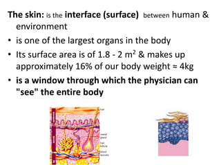
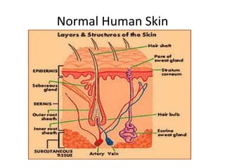
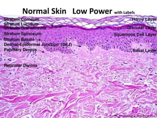
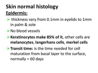
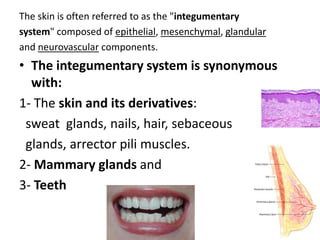
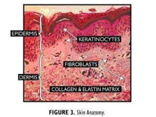
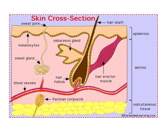
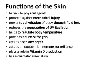
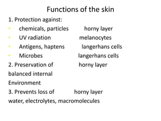
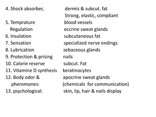
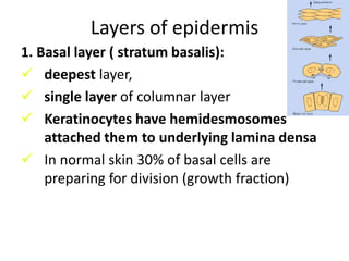
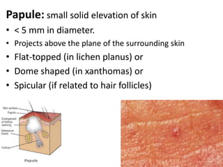
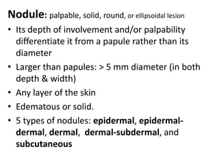
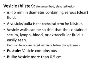
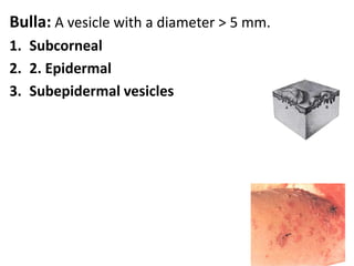
Anatomy, Organization, Histology, Function of Human Skin and DiagnosisBy:Dr. Kazhan Ali Taufiq KadirOctober 2010
The skin: is theinterface (surface)between human & environmentis one of the largest organs in the body  Its surface area is of 1.8 - 2 m2 & makes up approximately 16% of our body weight ≈ 4kgis a window through which the physician can "see" the entire body
Normal Human Skin
Normal Skin   Low Power with Labels
Skin normal histologyEpidermis: thickness vary from 0.1mm in eyelids to 1mm in palm & sole
No blood vessels
Keratinocytes make 85% of it, other cells are melanocytes, langerhans cells, merkel cells
Transit time: is the time needed for cell maturation from basal layer to the surface, normally = 60 daysThe skin is often referred to as the "integumentarysystem" composed of epithelial, mesenchymal, glandularand neurovascular components. The integumentary system is synonymous with: 1- The skin and its derivatives:   sweat  glands, nails, hair, sebaceous    glands, arrectorpili muscles.  2- Mammary glands and 3- Teeth
Functions of the Skinbarrier to physical agentsprotects against mechanical injuryprevents dehydration of body through fluid lossreduces the penetration of UV Radiationhelps to regulate body temperatureprovides a surface for gripacts as a sensory organacts as an outpost for immune surveillanceplays a role in Vitamin D productionhas a cosmetic association
Functions of the skin1. Protection against:chemicals, particles           horny layerUV radiation                    melanocytesAntigens, haptens            langerhans cellsMicrobes                         langerhans cells2. Preservation of                  horny layerbalanced internal Environment3. Prevents loss of              horny layerwater, electrolytes, macromolecules
4. Shock absorber,                  dermis & subcut. fat                                               Strong, elastic, compliant5. Tempratureblood vessels    Regulation                   eccrine sweat glands                                        6. Insulation                    subcutaneous fat7. Sensation                    specialized nerve endings                                        8. Lubrication                  sebaceous glands9. Protection & prisingnails10. Calorie reserve             subcut. Fat11. Vitamine D synthesis  keratinocytes12. Body odor &             apocrinesweat glands     pheromones:                    (chemicals  for communication)13. psychological:            skin, lip, hair & nails display
Based on the thickness of the epidermis, skin can also be classified as thick or thin:1) Thick skin   - covers palms and soles,    - has sweat glands, but lacks hair follicles, arrectorpili muscles, and sebaceous glands2) Thin skin    - covers most of the rest of the body   - contains sweat glands & hair follicles, arrectorpili muscles, and sebaceous glands.   
Layers of epidermis1. Basal layer ( stratum basalis): deepest layer,
single layer of columnar layer
Keratinocytes have hemidesmosomes attached them to underlying lamina densa
In normal skin 30% of basal cells are preparing for division (growth fraction)2. Spinous (prickle) cell layer:keratinocytes larger than basal layer
 7-9 layers of cells
Keratinocytes firmly attached by cytoplasmic process (desmosomes) & intercellular glycoprotein cement
Tonofilaments are small fibers running from cytoplasm  to desmosomes
Tonofilaments packed into bundles called   tonofibrils, more numerous in this layer
3. Granular layer:2-3 layersCells flatter than spinous layer, No nucleusNo intracytoplasmic organellesMore tonofibrilsContain irregular basophilic granules of keratohyalin.
4.Stratum Luidum: palms & soles5. Stratum corneum: SC:Made of layers of flattened dead cells    (corneocytes), has no nuclei or cytoplasmic organelles.Bricks-stuck together by intercellular lipids
Cytoplasm is picked with keratin filaments, embedded in a matrix, enclosed by an envlope derived from keratohyalin granules give corneocytes toughness allow skin to withstand chemical & mechanical insultsDEJ: Dermo-epidermal junction the basement membrane lies at the interface between epidermis & dermis
 plasma membrane of basal layer has hemidesmosomes .Composed of:Lamina Lucida is electron lucent area lies below basal cell layer.
Lamina Densa     Fine anchoring filaments cross lamina lucida & connect lamina densa to plasma membrane of basal cell layer.
Anchoring fibrils, dermal microfibril bundles & single small collagen extend from papillary dermis to deep part of lamina densa
Function of DEJ:Provide mechanical supportEncouraging adhesion, growth, differentiation & migration of overlying basal cellsAct as semipermeable filter that regulate transfer of nutrients & cells between dermis & epidermis
Dermis:DermalLayers:    1. Papillary layer     2. Reticular layerDermal Fibers:   1. Collagen Fibers   2. Elastic (Elastin) Fibers
Dermis:Lies between epidermis & subcut. fat
Support epidermis structurally & nutritionally
Thickness greatest in palm & soles & least in eyelids & penis
Interdigitate with epidermis, upward projection of dermis is dermal papillaeRete ridges: the downward ridges of epidermis is the rete ridges, which allow adhesion between dermis & epidermis as it increases the surface area between themCells & fibers of dermis Collagen fibers:  make 70-80% of dermis dry weightCompose of thinner fibrils packed in bundlesMade of 3 polypeptide chainsContain proline, hydroxyproline, glycinIts tensile strenth prevent tearing when skin is stretchedReticulin fibers: are fine collagen fibers
 seen in fetal skin & around blood vessels & appendages in adult skinElastic fibers: 2% of dermis dry weight
 elastin is made up of polypeptides rich in glycine, desmosine, valine
Have amorphous elastin core surrounded by elastic tissue micrifibrillar componentsGround substance of dermisComposes of glycosaminoglycan (hyaluronic acid & dermatan sulphate) with smaller amount of heparan sulphate & chondroitin sulphate
Functions:
Ground substance binds water, allowing nutrients, hormones & waste products to pass through dermis
Acts as lubricants between fibers
Provides bulk act as shock absorberMusclesSmooth arrectorpili muscle is vestigial in
1- sebum expression   2- goose pimples from cold,    3- nipple erectionRaising of scrotum is by smooth dartos muscle
Striated fibers (platysma) & some of muscles of facial expression found in dermisBlood vesselsRegulate body temprature2 main horizontal plexus, superficial & deepUnder sympathetic nervous control arteriovenous anastamoses can shunt blood to venous plexus reducing surface heat loss by convection
NervesOne million nerve fibers mostly in the face & extremitiesCell bodies are in dorsal root gangliaBoth mylinated & unmyelinated fibers existItch is follow stimulation of free nerve endings close to dermo-epidermal junction
Skin appendages (Adnexa): SHANS:1. Sweat glands2. Hair3. Arrector pilli muscle4. Nails5. Sebaceous glands
Skin appendages (Adnexa): SHANS:Hair: is human plumageHair follicle form before nine week of fetal life when hair germ grows obliquely down into dermis.It includes the following parts: Infandibulum: from surface to entrance of sebaceous g.
Asthmus: between entrance of seb. g to attachment of arrectorpili muscle
Hair Matrix: is the inferior portion including the hair bulb
Free of hair areas: Lip, glans penis, labia minora, palms, soles
Hair cycle:anagen, catagen & telogen
Typesof hair: 1- Lanugo hair   2- Vellus hair 3- Terminal hair
NailsHard keratin of nail plate  formed in nail matrix which lies in an invagination of epidermis on back of terminal phalanx of digits toesIt provide strength & protection for terminal phalanx & allow handling of fine objectsFinger nail growth is 0.5-1.2 mm per weekSlower in toe nailsFaster growth in summer
Skin Emberiology:Origins of the Integumentary System: (The Skin)The skin basically consists of two layers that are derived from two germ layers: ectoderm and mesoderm.The epidermis is from surface ectoderm.The dermis is derived from mesoderm.  Mesenchyme: is the embryonic meshwork of connective tissue (CT).
Hair: A hair follicle develops as a proliferation of the epidermal stratum germinativum and extends into the underlying mesenchyme.Nails:develop from thickened areas of epidermis at the tips of each digit called nail fields. Later these nail fields migrate onto the dorsal surface surrounded laterally and proximally by folds of epidermis called nail folds
Both the sebaceous glands and sweat glands (+ mammary glands) are derived from the epidermis and grow downwards into the dermis.  The majority of sebaceous glands   originate as buds from developing    hair follicle root sheath.Sebaceous glands in the glans penis and labia minora (independent of hair follicles) develop similarly, but as buds from the epidermis.    
Sweat glands: - Eccrine sweat glandslocated throughout the body originate as downgrowths from the epidermis into the underlying mesenchyme.  As a result, these eccrine ducts open onto the surface of the skin.- Apocrine sweat glandsare located in the axilla, pubic region, perineal region, and areolae of the nipples.  originate instead as downgrowths from the epidermal stratum germinativum (which also gives rise to hair follicles).  As a result, these apocrine ducts open into the upper part of hair follicles above sebaceous gland openings.
The Physiology of SkinThe skin is a metabolically active organ with vital functions such as protection and homeostasis.  When discussing the physiology of the skin and its derivatives, the following topics are important:
Keratinocyte Maturation: The aging of basal cells into the corneocytes (dead cells) is crucial.  The stratum corneum is important in preventing all manner of agents from entering the skin, including micro-organisms, water and particulate matter.  It's the epidermis that also prevents loss of vital body fluids. The dividing basal cell on average replicates every 200 to 400 hours, and the resulting cell takes 14 days to differentiate and 14 days to be shed.
Hair Growth Unlike most mammals, humans are considered "nude“ رووت ; thus, hair does not play the same vital role of heat conservation.  However, the hair on the scalp does protect us from UV radiation and minor injury.  Today, hair is of cosmetic value.Hair growth is cyclical and has three phases.  The 3 phases are:
1) Anagen is the growing phase.  Its duration depends on the location of the hair.  For eyebrow hair, this phase only lasts 4 months, but for scalp hair it lasts from 3-7 years.  At any moment, 80-90% of scalp hairs are in anagen; daily, approximately 50-100 scalp follicles switch to the catagen phase. 2) Catagen is the resting phase and lasts 3-4 weeks.  There is a stop to hair protein synthesis as the hair follicle retreats towards the surface.  At any moment, less than 1% of hairs are in the catagen phase.3) Telogen is the shedding phase and is characterized by hairs with a short club root.  Daily, approximately 50-100 scalp hairs are shed.  At any moment, 10-20% of scalp hairs are in telogen.
Dermatological General & Specific Terms:Dermatologists have some universal terms that are used when describing skin conditions:Lesion:Eruption (or rash):2 Types of Skin Lesions:    1- Primary Skin Lesions   2- Secondary Skin Lesions
1- Primary Lesions: are the first to appear and are due to the disease or abnormal state.  Must be distinguished from secondary or induced lesionsMaculePapuleNoduleVesicleBullaPustuleCystPlaqueWheal, Erythema, abscess, Angiodema, Tumor, Petechiae, Purpura, Ecchymosis, Haematoma, Burrow, Comedo, Telangiectasia & Poikiloderma
Macule:small,flat, nonpalpable circumscribed area of change in color or texture of the skin.  are < 0.5 cm in size, more than 0.5 called patches1. Macules (individual) may be the result of (A) hyperpigmentation: e.g. brown as inlentigos) (B) depigmentation (e.g. vitiligo),  (C) vascular dilation (e.g. erythema) 2. Multiple well-defined macules of     various shapes and sizes.     In this case, the macules blanch upon pressure(diascopy) and thus are due to inflammatory vasodilation.
Papule: small solid elevation of skin < 5 mm in diameter.  Projects above the plane of the surrounding skin Flat-topped (in lichen planus) or Dome shaped (in xanthomas) or Spicular (if related to hair follicles)
Nodule:palpable, solid, round, or ellipsoidal lesion  Its depth of involvement and/or palpability differentiate it from a papule rather than its diameter Larger than papules: > 5 mm diameter (in both depth & width)  Any layer of the skin Edematous or solid.  5 types of nodules: epidermal, epidermal-dermal, dermal,  dermal-subdermal, and subcutaneous
Vesicle (blister):circumscribed, elevated lesion is < 5 mm in diameter containing serous (clear) fluid.  A vesicle/bulla is the technical term for blisters Vesicle walls can be so thin that the contained serum, lymph, blood, or extracellular fluid is easily seen.  Fluid can be accumulated within or below the epidermisPustule: Vesicle contains pusBulla: Vesicle more than 0.5 cm
Bulla: A vesicle with a diameter > 5 mm. Subcorneal2. EpidermalSubepidermal vesicles
Pustule:superficial, elevated lesion    that contains pus (pus in a blister).  may vary in size and shape.  The color may appear white, yellow, or greenish-yellow depending on the color of the pus.  Pus is composed of leukocytes with or without cellular debrismay also contain bacteria or may be sterile1. A pustule is basically a papule containing pus2. Superficial, subcorneal pustules ( e.g. pustularpsoriasis) `
Cyst: an epithelial lined cavity containing liquid or semisolid material (fluid, cells, and cell products)  A spherical or oval papule or nodule may be a cyst if, when palpated, is resilient (feels like an eyeball). Plaque: palpable, plateau-like elevation of skin, usually more than 0.5 cm in diameter without substantial growth Often formed by a convergence of papules, as in psoriasis. 1. Plaques occupy a relatively large surface area in comparison with its height above the skin
Wheal: elevated, transitory, compressible papule or plaque produced by dermal edemaThe papule or plaque is usually rounded or flat-toped, and evanescent, disappearing within hours.  The borders of a wheal are sharp, but not stable and can move from involved to adjacent uninvolved areas over hours.  The epidermisis not affected.  can be pale red or white (especially in the center) if edema is sufficient to compress superficial vessels.
Erythema: is redness caused by vascular dilatation
Abscess: localized collection of puss more than 1 cm in diameter
Angiodema: is diffuse swelling caused by subcutaneous oedema
Tumor: is an enlargement of the tissue by normal or pathological material or cell greater than 1 cm in diameter
Petechiae: pinhead sized macules of blood

Dermatology 5th year, 1st lecture (Dr. Kazhan)

  • 1.
    Anatomy, Organization, Histology,Function of Human Skin and DiagnosisBy:Dr. Kazhan Ali Taufiq KadirOctober 2010
  • 2.
    The skin: istheinterface (surface)between human & environmentis one of the largest organs in the body  Its surface area is of 1.8 - 2 m2 & makes up approximately 16% of our body weight ≈ 4kgis a window through which the physician can "see" the entire body
  • 3.
  • 4.
    Normal Skin Low Power with Labels
  • 5.
    Skin normal histologyEpidermis:thickness vary from 0.1mm in eyelids to 1mm in palm & sole
  • 6.
  • 7.
    Keratinocytes make 85%of it, other cells are melanocytes, langerhans cells, merkel cells
  • 8.
    Transit time: isthe time needed for cell maturation from basal layer to the surface, normally = 60 daysThe skin is often referred to as the "integumentarysystem" composed of epithelial, mesenchymal, glandularand neurovascular components. The integumentary system is synonymous with: 1- The skin and its derivatives: sweat glands, nails, hair, sebaceous glands, arrectorpili muscles. 2- Mammary glands and 3- Teeth
  • 11.
    Functions of theSkinbarrier to physical agentsprotects against mechanical injuryprevents dehydration of body through fluid lossreduces the penetration of UV Radiationhelps to regulate body temperatureprovides a surface for gripacts as a sensory organacts as an outpost for immune surveillanceplays a role in Vitamin D productionhas a cosmetic association
  • 12.
    Functions of theskin1. Protection against:chemicals, particles horny layerUV radiation melanocytesAntigens, haptens langerhans cellsMicrobes langerhans cells2. Preservation of horny layerbalanced internal Environment3. Prevents loss of horny layerwater, electrolytes, macromolecules
  • 13.
    4. Shock absorber, dermis & subcut. fat Strong, elastic, compliant5. Tempratureblood vessels Regulation eccrine sweat glands 6. Insulation subcutaneous fat7. Sensation specialized nerve endings 8. Lubrication sebaceous glands9. Protection & prisingnails10. Calorie reserve subcut. Fat11. Vitamine D synthesis keratinocytes12. Body odor & apocrinesweat glands pheromones: (chemicals for communication)13. psychological: skin, lip, hair & nails display
  • 14.
    Based on thethickness of the epidermis, skin can also be classified as thick or thin:1) Thick skin - covers palms and soles, - has sweat glands, but lacks hair follicles, arrectorpili muscles, and sebaceous glands2) Thin skin - covers most of the rest of the body - contains sweat glands & hair follicles, arrectorpili muscles, and sebaceous glands.   
  • 15.
    Layers of epidermis1.Basal layer ( stratum basalis): deepest layer,
  • 16.
    single layer ofcolumnar layer
  • 17.
    Keratinocytes have hemidesmosomesattached them to underlying lamina densa
  • 18.
    In normal skin30% of basal cells are preparing for division (growth fraction)2. Spinous (prickle) cell layer:keratinocytes larger than basal layer
  • 19.
    7-9 layersof cells
  • 20.
    Keratinocytes firmly attachedby cytoplasmic process (desmosomes) & intercellular glycoprotein cement
  • 21.
    Tonofilaments are smallfibers running from cytoplasm to desmosomes
  • 22.
    Tonofilaments packed intobundles called tonofibrils, more numerous in this layer
  • 23.
    3. Granular layer:2-3layersCells flatter than spinous layer, No nucleusNo intracytoplasmic organellesMore tonofibrilsContain irregular basophilic granules of keratohyalin.
  • 24.
    4.Stratum Luidum: palms& soles5. Stratum corneum: SC:Made of layers of flattened dead cells (corneocytes), has no nuclei or cytoplasmic organelles.Bricks-stuck together by intercellular lipids
  • 25.
    Cytoplasm is pickedwith keratin filaments, embedded in a matrix, enclosed by an envlope derived from keratohyalin granules give corneocytes toughness allow skin to withstand chemical & mechanical insultsDEJ: Dermo-epidermal junction the basement membrane lies at the interface between epidermis & dermis
  • 26.
    plasma membraneof basal layer has hemidesmosomes .Composed of:Lamina Lucida is electron lucent area lies below basal cell layer.
  • 27.
    Lamina Densa Fine anchoring filaments cross lamina lucida & connect lamina densa to plasma membrane of basal cell layer.
  • 28.
    Anchoring fibrils, dermalmicrofibril bundles & single small collagen extend from papillary dermis to deep part of lamina densa
  • 29.
    Function of DEJ:Providemechanical supportEncouraging adhesion, growth, differentiation & migration of overlying basal cellsAct as semipermeable filter that regulate transfer of nutrients & cells between dermis & epidermis
  • 30.
    Dermis:DermalLayers: 1. Papillary layer 2. Reticular layerDermal Fibers: 1. Collagen Fibers 2. Elastic (Elastin) Fibers
  • 31.
  • 32.
  • 33.
    Thickness greatest inpalm & soles & least in eyelids & penis
  • 34.
    Interdigitate with epidermis,upward projection of dermis is dermal papillaeRete ridges: the downward ridges of epidermis is the rete ridges, which allow adhesion between dermis & epidermis as it increases the surface area between themCells & fibers of dermis Collagen fibers: make 70-80% of dermis dry weightCompose of thinner fibrils packed in bundlesMade of 3 polypeptide chainsContain proline, hydroxyproline, glycinIts tensile strenth prevent tearing when skin is stretchedReticulin fibers: are fine collagen fibers
  • 35.
    seen infetal skin & around blood vessels & appendages in adult skinElastic fibers: 2% of dermis dry weight
  • 36.
    elastin ismade up of polypeptides rich in glycine, desmosine, valine
  • 37.
    Have amorphous elastincore surrounded by elastic tissue micrifibrillar componentsGround substance of dermisComposes of glycosaminoglycan (hyaluronic acid & dermatan sulphate) with smaller amount of heparan sulphate & chondroitin sulphate
  • 38.
  • 39.
    Ground substance bindswater, allowing nutrients, hormones & waste products to pass through dermis
  • 40.
    Acts as lubricantsbetween fibers
  • 41.
    Provides bulk actas shock absorberMusclesSmooth arrectorpili muscle is vestigial in
  • 42.
    1- sebum expression 2- goose pimples from cold, 3- nipple erectionRaising of scrotum is by smooth dartos muscle
  • 43.
    Striated fibers (platysma)& some of muscles of facial expression found in dermisBlood vesselsRegulate body temprature2 main horizontal plexus, superficial & deepUnder sympathetic nervous control arteriovenous anastamoses can shunt blood to venous plexus reducing surface heat loss by convection
  • 44.
    NervesOne million nervefibers mostly in the face & extremitiesCell bodies are in dorsal root gangliaBoth mylinated & unmyelinated fibers existItch is follow stimulation of free nerve endings close to dermo-epidermal junction
  • 45.
    Skin appendages (Adnexa):SHANS:1. Sweat glands2. Hair3. Arrector pilli muscle4. Nails5. Sebaceous glands
  • 46.
    Skin appendages (Adnexa):SHANS:Hair: is human plumageHair follicle form before nine week of fetal life when hair germ grows obliquely down into dermis.It includes the following parts: Infandibulum: from surface to entrance of sebaceous g.
  • 47.
    Asthmus: between entranceof seb. g to attachment of arrectorpili muscle
  • 48.
    Hair Matrix: isthe inferior portion including the hair bulb
  • 49.
    Free of hairareas: Lip, glans penis, labia minora, palms, soles
  • 50.
  • 51.
    Typesof hair: 1-Lanugo hair 2- Vellus hair 3- Terminal hair
  • 52.
    NailsHard keratin ofnail plate formed in nail matrix which lies in an invagination of epidermis on back of terminal phalanx of digits toesIt provide strength & protection for terminal phalanx & allow handling of fine objectsFinger nail growth is 0.5-1.2 mm per weekSlower in toe nailsFaster growth in summer
  • 53.
    Skin Emberiology:Origins ofthe Integumentary System: (The Skin)The skin basically consists of two layers that are derived from two germ layers: ectoderm and mesoderm.The epidermis is from surface ectoderm.The dermis is derived from mesoderm. Mesenchyme: is the embryonic meshwork of connective tissue (CT).
  • 54.
    Hair: A hairfollicle develops as a proliferation of the epidermal stratum germinativum and extends into the underlying mesenchyme.Nails:develop from thickened areas of epidermis at the tips of each digit called nail fields. Later these nail fields migrate onto the dorsal surface surrounded laterally and proximally by folds of epidermis called nail folds
  • 55.
    Both the sebaceousglands and sweat glands (+ mammary glands) are derived from the epidermis and grow downwards into the dermis. The majority of sebaceous glands originate as buds from developing hair follicle root sheath.Sebaceous glands in the glans penis and labia minora (independent of hair follicles) develop similarly, but as buds from the epidermis.    
  • 56.
    Sweat glands: -Eccrine sweat glandslocated throughout the body originate as downgrowths from the epidermis into the underlying mesenchyme. As a result, these eccrine ducts open onto the surface of the skin.- Apocrine sweat glandsare located in the axilla, pubic region, perineal region, and areolae of the nipples. originate instead as downgrowths from the epidermal stratum germinativum (which also gives rise to hair follicles). As a result, these apocrine ducts open into the upper part of hair follicles above sebaceous gland openings.
  • 57.
    The Physiology ofSkinThe skin is a metabolically active organ with vital functions such as protection and homeostasis.  When discussing the physiology of the skin and its derivatives, the following topics are important:
  • 58.
    Keratinocyte Maturation: Theaging of basal cells into the corneocytes (dead cells) is crucial. The stratum corneum is important in preventing all manner of agents from entering the skin, including micro-organisms, water and particulate matter. It's the epidermis that also prevents loss of vital body fluids. The dividing basal cell on average replicates every 200 to 400 hours, and the resulting cell takes 14 days to differentiate and 14 days to be shed.
  • 59.
    Hair Growth Unlikemost mammals, humans are considered "nude“ رووت ; thus, hair does not play the same vital role of heat conservation. However, the hair on the scalp does protect us from UV radiation and minor injury. Today, hair is of cosmetic value.Hair growth is cyclical and has three phases. The 3 phases are:
  • 61.
    1) Anagen isthe growing phase. Its duration depends on the location of the hair. For eyebrow hair, this phase only lasts 4 months, but for scalp hair it lasts from 3-7 years. At any moment, 80-90% of scalp hairs are in anagen; daily, approximately 50-100 scalp follicles switch to the catagen phase. 2) Catagen is the resting phase and lasts 3-4 weeks. There is a stop to hair protein synthesis as the hair follicle retreats towards the surface. At any moment, less than 1% of hairs are in the catagen phase.3) Telogen is the shedding phase and is characterized by hairs with a short club root. Daily, approximately 50-100 scalp hairs are shed. At any moment, 10-20% of scalp hairs are in telogen.
  • 62.
    Dermatological General &Specific Terms:Dermatologists have some universal terms that are used when describing skin conditions:Lesion:Eruption (or rash):2 Types of Skin Lesions: 1- Primary Skin Lesions 2- Secondary Skin Lesions
  • 63.
    1- Primary Lesions:are the first to appear and are due to the disease or abnormal state.  Must be distinguished from secondary or induced lesionsMaculePapuleNoduleVesicleBullaPustuleCystPlaqueWheal, Erythema, abscess, Angiodema, Tumor, Petechiae, Purpura, Ecchymosis, Haematoma, Burrow, Comedo, Telangiectasia & Poikiloderma
  • 64.
    Macule:small,flat, nonpalpable circumscribedarea of change in color or texture of the skin. are < 0.5 cm in size, more than 0.5 called patches1. Macules (individual) may be the result of (A) hyperpigmentation: e.g. brown as inlentigos) (B) depigmentation (e.g. vitiligo),  (C) vascular dilation (e.g. erythema) 2. Multiple well-defined macules of various shapes and sizes.  In this case, the macules blanch upon pressure(diascopy) and thus are due to inflammatory vasodilation.
  • 65.
    Papule: small solidelevation of skin < 5 mm in diameter. Projects above the plane of the surrounding skin Flat-topped (in lichen planus) or Dome shaped (in xanthomas) or Spicular (if related to hair follicles)
  • 66.
    Nodule:palpable, solid, round,or ellipsoidal lesion Its depth of involvement and/or palpability differentiate it from a papule rather than its diameter Larger than papules: > 5 mm diameter (in both depth & width) Any layer of the skin Edematous or solid. 5 types of nodules: epidermal, epidermal-dermal, dermal,  dermal-subdermal, and subcutaneous
  • 67.
    Vesicle (blister):circumscribed, elevatedlesion is < 5 mm in diameter containing serous (clear) fluid. A vesicle/bulla is the technical term for blisters Vesicle walls can be so thin that the contained serum, lymph, blood, or extracellular fluid is easily seen. Fluid can be accumulated within or below the epidermisPustule: Vesicle contains pusBulla: Vesicle more than 0.5 cm
  • 68.
    Bulla: A vesiclewith a diameter > 5 mm. Subcorneal2. EpidermalSubepidermal vesicles
  • 69.
    Pustule:superficial, elevated lesion that contains pus (pus in a blister). may vary in size and shape. The color may appear white, yellow, or greenish-yellow depending on the color of the pus. Pus is composed of leukocytes with or without cellular debrismay also contain bacteria or may be sterile1. A pustule is basically a papule containing pus2. Superficial, subcorneal pustules ( e.g. pustularpsoriasis) `
  • 70.
    Cyst: an epitheliallined cavity containing liquid or semisolid material (fluid, cells, and cell products) A spherical or oval papule or nodule may be a cyst if, when palpated, is resilient (feels like an eyeball). Plaque: palpable, plateau-like elevation of skin, usually more than 0.5 cm in diameter without substantial growth Often formed by a convergence of papules, as in psoriasis. 1. Plaques occupy a relatively large surface area in comparison with its height above the skin
  • 71.
    Wheal: elevated, transitory,compressible papule or plaque produced by dermal edemaThe papule or plaque is usually rounded or flat-toped, and evanescent, disappearing within hours. The borders of a wheal are sharp, but not stable and can move from involved to adjacent uninvolved areas over hours. The epidermisis not affected. can be pale red or white (especially in the center) if edema is sufficient to compress superficial vessels.
  • 72.
    Erythema: is rednesscaused by vascular dilatation
  • 73.
    Abscess: localized collectionof puss more than 1 cm in diameter
  • 74.
    Angiodema: is diffuseswelling caused by subcutaneous oedema
  • 75.
    Tumor: is anenlargement of the tissue by normal or pathological material or cell greater than 1 cm in diameter
  • 76.
    Petechiae: pinhead sizedmacules of blood