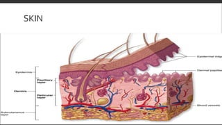
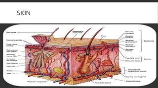
INTEGUMENTARY SYSTEM
By Dr. Nousheen
INTEGUMENTARY SYSTEM
ď‚  Consist of skin and its appendages
ď‚  Appendages include hair, nails, sebaceous glands and sweat glands
FUNCTIONS OF INTEGUMENTARY SYSTEM
1. Protection: prevents the entry of harmful microorganism and foreign
material.
2. Prevents loss of body fluids.
3. Temperature regulation: heat is lost through evaporation of sweat. In
winter heat is preserved by fat and hair.
4. Excretion: through sweating excrete small amount of waste materials. such
as urea excreted through skin.
FUNCTIONS OF INTEGUMENTARY SYSTEM
ď‚  5.Synthens:synthesis of vitamin D under the influence of ultraviolet rays.
ď‚  6.Sensory perception: important sensory organ contain sensory receptor for heat,
cold, touch, pressure and pain
.SKIN
ď‚  largest organ of the body
• At orifices, continuous with mucous membranes
• Mainly two parts
• Epidermis
• dermis
EPIDERMIS
ď‚  Epidermis: outer layer consist of stratified squamous epithelium consisting of
many layers of cells called keratinocytes
ď‚  Histologically, epidermis consists of five(5) layers
ď‚  1.Stratum basale(MERKEL DISC and MELANOCYTES)
ď‚  2.Stratum spinosum
ď‚  3.Stratum granulosum
ď‚  4.Stratum lucidum
ď‚  5.Stratum CORNEUM
STRATUM CORNEUM
• Cover exposed surface of epidermis
• Composed of death cytoplasm of which is packed with filaments composed of a
special protein called keratin
• Keratinized cells of epidermis offer effective resistance both to passage of fluid
through them and to friction
• Most superficial layer of stratum corneum contently shed off and replaced by
cells from deepest layer i.e. stratum basale (also called stratum germinatium )
EPIDERMIS
• epidermis has no blood supply, obtain its nourishment and oxygen by diffusion
by under lying dermis
• Deeper layer of the epidermis also contain melanocytes produce a brownish
pigment called melanin, which help in determination of color of skin
DERMIS
ď‚  Beneath the epidermis, lie dermis or cornium
ď‚  Arranged in two layers:
1. Superficial papillary layer
2. Deep reticular layer
LAYERS OF DERMIS
ď‚  Superficial papillary layer:Form blunt+ conical projections, called dermal papillae, which fit
into reciprocal Depression and the under surface of epidermis
ď‚  COLLAGEN and ELASTIC fibers form loose mesh work
ď‚  FIBROBLASTS,ADIPOCYTES,ABUNDANCEOF SMALL BLOODVESSELS,PHAGOCYTES
andTOUCH RECCEP: (MESSENIERCORPUSCLE)
ď‚  Reticular layer:Consist of inter lying bundles of connective tissues fibers
ď‚  Much thicker
ď‚  Tight meshwork of collagen and elastic fibers
ď‚  Well vascularized
ď‚  Rich sensory and sympathetic nerve supply
DERMIS
ď‚  Contain abundant blood vessels, nerves and nerves endings
ď‚  Lodges hair follicle, sweat glands and sebaceous glands
SKIN
SKIN
CLEAVAGE LINES
ď‚  faint linear clefts can be seen to be present in the skin
ď‚  These clefts are indicative of the direction of collagenous fibers in the dermis
CLEAVAGE LINES
IMPORTANCE OF CLEAVAGE LINES
ď‚  Important for surgoen
• Incision made parallel to cleavage line heals faster and leaves only fine scar
• If an incision is made across the cleavage lines, it results in formative of a
gapping wound that heals slowly and leaves a broad thick scar
APPENDAGES, EPITHELIAL STRUCTURES
DERIVED FROMTHE EPIDERMIS
HAIR(LATIN SYNONYM PILUS)
Thread like structures
composed of dead cornified
cells
HAIR PARTS
1. Shaft: projects above the skin surface
2. Root: embedded in the skin
3. Hair follicle: surround the root of the hair, lies in the dermis and is responsible
for the growth of the hair
4. Hair bulb: deep to hair follicle dilated to form hair bulb
HAIR PARTS
HAIR PAPILLA
Connective tissue of
dermis indents the base
of the hair bulb
ARRECTOR PILI
ď‚  Associated with each hair follicle , bundle of smooth muscle fibres called arrector
pili
• Hairs are constantly shed and replaced by a one . A hair is shed when its growth
is complete
• Scalp hair (3-5 years) eye brows and eye lashes (3-5 months)
ARRECTOR PILI
NAILS
ď‚  Horny plates covering the dorsal surface of the distal segnant fingers and toes
ď‚  Nails consists of three parts:
1. The body
2. Th root
3. The free edge
NAILS
BODY
That part which shows
consist of cornified
dead cells
ROOT
Hidden part that
extends proximally
deep to the fold of
the skin called nail
fold
FREE EDGE
ď‚  Over hangs the tip of the finger or toe. Nails rest on epithelial surface called nail
bed
ď‚  The thicker layer of skin beneath the nail root is the matrix, where new cells are
generated for the growth of the nail
ď‚  Growth rate:
SEBACEOUS GLANDS (OIL GLANDS)
Lie in the dermis
Duct and sebaceous
gland open into neck of
hair follicle
Produce an oily
secretion which
lubricates the skin
surface
SWEAT GLAND:
FOUND OVER MOST OF BODY
ď‚  Secretory portion lies in subcutaneous and is in the form of long tube, which is
coiled to form a ball
ď‚  Duct of glands run through dermis, joint the epidermis and spirals through it to
skin surface it terminate at an opening called sweat pore
SWEAT GLAND
FASCIA
Sheath or band of
connective tissue lies
beneath the skin or
forms an investment for
muscles and various
organs of body
TWOTYPES OF FASCIA
1. Superficial fascia
2. Deep fascia
TWOTYPES OF FASCIA
TWOTYPES OF FASCIA
SUPERFICIAL FASCIA
ď‚  Lies just beneath the skin
ď‚  Covers the whole body but varies in thickness in different regions
ď‚  Consists of two layers
1. Outer fatty layer
2. Inner fibrous layer
OUTER LAYER
Contain fatty tissue
Quantity of fat higher in
women then men
Absent in eyelids, nose,
external ear, penis and scrotum
INNER LAYER
ď‚  Consist of fibrous connective tissue
ď‚  Mostly thin but over anterior abdominal wall it condenses into a membranous
layer rich is elastic fibers
SUPERFICIAL FASCIA
ď‚  Conduct blood vessels nerves and lymphatics of the skin
ď‚  Functions:
1. Storage of energy
2. Insulator
DEEP FASCIA
ď‚  Lies beneath the superficial fascia
ď‚  Tough and inelastic
ď‚  Consist of several layers of fibrous connective tissue
ď‚  Covers almost all the body but ill defined in face and trunk and well defined in
neck and limbs
ď‚  In the limbs,deep fascia form tight sleeves and from its deep surface arise fibrous
sheets which pentrate between the muscles called intermuscular septa
DEEP FASCIA
ď‚  Intermuscular septa divides the limbs into compartments
ď‚  Purpose of compartements:
ď‚  Muscles are divided into functional groups
ď‚  Contraction of muscles within inelastic walls of the osteofacial compartment put
pressure on the deep vein of the limbs and thus aid in return of venous blood
towards the heart.
DEEP FASCIA
ď‚  At wrist and ankle joints,dep fascia is thickened to form transverse band likr
structure called retinacula which is attached to local bony prominences
ď‚  In the limbs,some muscles are attached to internal aspect of deep fascia portion
of which then serve as aponeurosis e.g iliotibial tract
ď‚  In palm and soles,deep fascia is thickened to form palmer and plantar aponeurosis
ILLIOTIBIALTRACT
DEEP FASCIA
LIGAMENTS
ď‚  Band of fibrous connective tissue which serve to strengthen and support the joints
ď‚  Divided into two types:
ď‚  CAPSULAR LIGAMENTS:exist as thickening of articular capsule
ď‚  ACCESSORY LIGAMENTS:not present as a part of capsule.it may be:
ď‚  EXTRACAPSULAR:coracoclavicular ligament of the acromioclavicular joints
ď‚  INTRACAPSULAR :cruciate ligament of the knee joint
LIGAMENTS
LIGAMENTS
ď‚  Tough and inelastic but they are flexible and pliable to permit the normal range of
the movement
ď‚  Due to their non extensible nature,the ligaments prevent the occurrence of
excessive or abnormal movement
ď‚  CLINICAL COORELATIONS:under excessive stress a ligament will tear rather than
stretch such tear is called sprain is associated with severe pain and local swelling
LIGAMENTS

Integumantary system.pptx

  • 1.
  • 2.
    INTEGUMENTARY SYSTEM ď‚  Consistof skin and its appendages ď‚  Appendages include hair, nails, sebaceous glands and sweat glands
  • 3.
    FUNCTIONS OF INTEGUMENTARYSYSTEM 1. Protection: prevents the entry of harmful microorganism and foreign material. 2. Prevents loss of body fluids. 3. Temperature regulation: heat is lost through evaporation of sweat. In winter heat is preserved by fat and hair. 4. Excretion: through sweating excrete small amount of waste materials. such as urea excreted through skin.
  • 4.
    FUNCTIONS OF INTEGUMENTARYSYSTEM ď‚  5.Synthens:synthesis of vitamin D under the influence of ultraviolet rays. ď‚  6.Sensory perception: important sensory organ contain sensory receptor for heat, cold, touch, pressure and pain
  • 5.
    .SKIN  largest organof the body • At orifices, continuous with mucous membranes • Mainly two parts • Epidermis • dermis
  • 6.
    EPIDERMIS ď‚  Epidermis: outerlayer consist of stratified squamous epithelium consisting of many layers of cells called keratinocytes ď‚  Histologically, epidermis consists of five(5) layers ď‚  1.Stratum basale(MERKEL DISC and MELANOCYTES) ď‚  2.Stratum spinosum ď‚  3.Stratum granulosum ď‚  4.Stratum lucidum ď‚  5.Stratum CORNEUM
  • 7.
    STRATUM CORNEUM • Coverexposed surface of epidermis • Composed of death cytoplasm of which is packed with filaments composed of a special protein called keratin • Keratinized cells of epidermis offer effective resistance both to passage of fluid through them and to friction • Most superficial layer of stratum corneum contently shed off and replaced by cells from deepest layer i.e. stratum basale (also called stratum germinatium )
  • 8.
    EPIDERMIS • epidermis hasno blood supply, obtain its nourishment and oxygen by diffusion by under lying dermis • Deeper layer of the epidermis also contain melanocytes produce a brownish pigment called melanin, which help in determination of color of skin
  • 9.
    DERMIS ď‚  Beneath theepidermis, lie dermis or cornium ď‚  Arranged in two layers: 1. Superficial papillary layer 2. Deep reticular layer
  • 10.
    LAYERS OF DERMIS ď‚ Superficial papillary layer:Form blunt+ conical projections, called dermal papillae, which fit into reciprocal Depression and the under surface of epidermis ď‚  COLLAGEN and ELASTIC fibers form loose mesh work ď‚  FIBROBLASTS,ADIPOCYTES,ABUNDANCEOF SMALL BLOODVESSELS,PHAGOCYTES andTOUCH RECCEP: (MESSENIERCORPUSCLE) ď‚  Reticular layer:Consist of inter lying bundles of connective tissues fibers ď‚  Much thicker ď‚  Tight meshwork of collagen and elastic fibers ď‚  Well vascularized ď‚  Rich sensory and sympathetic nerve supply
  • 11.
    DERMIS ď‚  Contain abundantblood vessels, nerves and nerves endings ď‚  Lodges hair follicle, sweat glands and sebaceous glands
  • 12.
  • 13.
  • 14.
    CLEAVAGE LINES ď‚  faintlinear clefts can be seen to be present in the skin ď‚  These clefts are indicative of the direction of collagenous fibers in the dermis
  • 15.
  • 16.
    IMPORTANCE OF CLEAVAGELINES  Important for surgoen • Incision made parallel to cleavage line heals faster and leaves only fine scar • If an incision is made across the cleavage lines, it results in formative of a gapping wound that heals slowly and leaves a broad thick scar
  • 17.
    APPENDAGES, EPITHELIAL STRUCTURES DERIVEDFROMTHE EPIDERMIS HAIR(LATIN SYNONYM PILUS) Thread like structures composed of dead cornified cells
  • 18.
    HAIR PARTS 1. Shaft:projects above the skin surface 2. Root: embedded in the skin 3. Hair follicle: surround the root of the hair, lies in the dermis and is responsible for the growth of the hair 4. Hair bulb: deep to hair follicle dilated to form hair bulb
  • 19.
  • 20.
    HAIR PAPILLA Connective tissueof dermis indents the base of the hair bulb
  • 21.
    ARRECTOR PILI  Associatedwith each hair follicle , bundle of smooth muscle fibres called arrector pili • Hairs are constantly shed and replaced by a one . A hair is shed when its growth is complete • Scalp hair (3-5 years) eye brows and eye lashes (3-5 months)
  • 22.
  • 23.
    NAILS ď‚  Horny platescovering the dorsal surface of the distal segnant fingers and toes ď‚  Nails consists of three parts: 1. The body 2. Th root 3. The free edge
  • 24.
  • 25.
    BODY That part whichshows consist of cornified dead cells
  • 26.
    ROOT Hidden part that extendsproximally deep to the fold of the skin called nail fold
  • 27.
    FREE EDGE ď‚  Overhangs the tip of the finger or toe. Nails rest on epithelial surface called nail bed ď‚  The thicker layer of skin beneath the nail root is the matrix, where new cells are generated for the growth of the nail ď‚  Growth rate:
  • 28.
    SEBACEOUS GLANDS (OILGLANDS) Lie in the dermis Duct and sebaceous gland open into neck of hair follicle Produce an oily secretion which lubricates the skin surface
  • 29.
    SWEAT GLAND: FOUND OVERMOST OF BODY ď‚  Secretory portion lies in subcutaneous and is in the form of long tube, which is coiled to form a ball ď‚  Duct of glands run through dermis, joint the epidermis and spirals through it to skin surface it terminate at an opening called sweat pore
  • 30.
  • 31.
    FASCIA Sheath or bandof connective tissue lies beneath the skin or forms an investment for muscles and various organs of body
  • 32.
    TWOTYPES OF FASCIA 1.Superficial fascia 2. Deep fascia
  • 33.
  • 34.
  • 35.
    SUPERFICIAL FASCIA ď‚  Liesjust beneath the skin ď‚  Covers the whole body but varies in thickness in different regions ď‚  Consists of two layers 1. Outer fatty layer 2. Inner fibrous layer
  • 36.
    OUTER LAYER Contain fattytissue Quantity of fat higher in women then men Absent in eyelids, nose, external ear, penis and scrotum
  • 37.
    INNER LAYER ď‚  Consistof fibrous connective tissue ď‚  Mostly thin but over anterior abdominal wall it condenses into a membranous layer rich is elastic fibers
  • 38.
    SUPERFICIAL FASCIA ď‚  Conductblood vessels nerves and lymphatics of the skin ď‚  Functions: 1. Storage of energy 2. Insulator
  • 39.
    DEEP FASCIA ď‚  Liesbeneath the superficial fascia ď‚  Tough and inelastic ď‚  Consist of several layers of fibrous connective tissue ď‚  Covers almost all the body but ill defined in face and trunk and well defined in neck and limbs ď‚  In the limbs,deep fascia form tight sleeves and from its deep surface arise fibrous sheets which pentrate between the muscles called intermuscular septa
  • 40.
    DEEP FASCIA ď‚  Intermuscularsepta divides the limbs into compartments ď‚  Purpose of compartements: ď‚  Muscles are divided into functional groups ď‚  Contraction of muscles within inelastic walls of the osteofacial compartment put pressure on the deep vein of the limbs and thus aid in return of venous blood towards the heart.
  • 41.
    DEEP FASCIA ď‚  Atwrist and ankle joints,dep fascia is thickened to form transverse band likr structure called retinacula which is attached to local bony prominences ď‚  In the limbs,some muscles are attached to internal aspect of deep fascia portion of which then serve as aponeurosis e.g iliotibial tract ď‚  In palm and soles,deep fascia is thickened to form palmer and plantar aponeurosis
  • 42.
  • 43.
  • 44.
    LIGAMENTS ď‚  Band offibrous connective tissue which serve to strengthen and support the joints ď‚  Divided into two types: ď‚  CAPSULAR LIGAMENTS:exist as thickening of articular capsule ď‚  ACCESSORY LIGAMENTS:not present as a part of capsule.it may be: ď‚  EXTRACAPSULAR:coracoclavicular ligament of the acromioclavicular joints ď‚  INTRACAPSULAR :cruciate ligament of the knee joint
  • 45.
  • 46.
    LIGAMENTS ď‚  Tough andinelastic but they are flexible and pliable to permit the normal range of the movement ď‚  Due to their non extensible nature,the ligaments prevent the occurrence of excessive or abnormal movement ď‚  CLINICAL COORELATIONS:under excessive stress a ligament will tear rather than stretch such tear is called sprain is associated with severe pain and local swelling
  • 47.