SlideShare a Scribd company logo
1 of 56
Unit II
Integumentary system
Skeletal system
Joints
By
Miss. Aishwarya S. Patil
Assistant Professor
Dr. D. Y. Patil College of Pharmacy Akurdi Pune.
Contents:
• Introduction to integumentary system
• Structure of skin
• Functions of skin
• Skeletal system
• Divisions of skeletal system
• Types of bones
• Salient features and function of axial and appendicular skeletal system.
• Organization of skeletal system
• Physiology of muscle contraction.(notes given)
• Neuromuscular junction
• Introduction to joints
• Structural and functional classification of joints.
• Types of joints movement and its articulation.
Introduction to integumentary system
• Integumentary system = Skin (function + structure)
• Skin completely covers body and continuous with linings
• Skin is the largest organ of the body.
• Surface area is about 1.5-2 square meters.
• It contains some accessory structures: Glands, hairs, nails.
• Thickness varies , thickest at palms and feet’s.
• Consist of 2 layers.
• Superficial layers: epidermis.
• Deeper layer: dermis.
• Below dermis the layer is known as subcutaneous composed of areolar
tissue and adipose tissue.
Structure of skin
Epidermis • Most superficial layer of the skin and is composed of stratified keratinised squamous epithelium
• Varies in thickness in different parts of the body.
• No blood vessels or nerve endings in the epidermis, but its deeper layers are bathed in interstitial fluid from
the dermis, which provides oxygen and nutrients, and drains away as lymph.
• There are several layers (strata) of cells in the epidermis which extend from the deepest germinative layer to
the most superficial stratum corneum (a thick horny layer)
• The cells on the surface are flat, thin, non-nucleated, dead cells, or squamous, in which the cytoplasm has
been replaced by the fibrous protein keratin.
• cells are constantly being rubbed off and replaced by cells that originated in the germinative layer and have
undergone gradual change as they progressed towards the surface.
• Continual cell division in the deeper layers with newly formed cells being pushed to the surface.
• Hairs, secretions from sebaceous glands and ducts of sweat glands pass through the epidermis to surface
• The surface of the epidermis is ridged by projections of cells in the dermis called papillae. The pattern of
ridges on the fingertips is unique to every individual and the impression made by them is the ‘fingerprint’.
• Blisters develop when trauma causes separation of the dermis and epidermis and serous fluid collects
between the two layers. Skin colour is affected by various factors.
• Melanin, a dark pigment derived from the amino acid tyrosine and secreted by melanocytes in the deep
germinative layer, is absorbed by surrounding epithelial cells. Colour depend on the amount of melanin
secreted. It protects the skin from the harmful effects of sunlight. Exposure to sunlight promotes synthesis
of melanin.
• Normal saturation of haemoglobin and the amount of blood circulating in the dermis give white skin its pink
colour.
Dermis • Tough and elastic.
• formed from connective tissue and the matrix contains collagen fibres, interlaced with elastic fibres.
• Rupture of elastic fibres occurs when the skin is overstretched, resulting in permanent striae, or stretch marks, that
may be found in pregnancy and obesity.
• Collagen fibres bind water and give the skin its tensile strength, but as this ability declines with age, wrinkles
develop.
• Fibroblasts , macrophages and mast cells are the main cells found in the dermis. Underlying its deepest layer there
is areolar tissue and varying amounts of adipose (fat) tissue.
• The structures in the dermis are: blood vessels, lymph vessels, sensory (somatic) nerve endings, sweat glands and
their ducts, hairs, arrector pili muscles and sebaceous glands.
• Arterioles form a fine network with capillary branches supplying sweat glands, sebaceous glands, hair follicles and
the dermis. Lymph vessels form a network throughout the dermis.
• The skin is an important sensory organ through which individuals receive information about their environment.
Nerve impulses, generated in the sensory receptors in the dermis, are conveyed to the spinal cord by sensory
nerves, then to the sensory area of the cerebrum where the sensations are perceived.
Sensory receptor Stimulus
Meissner’s corpuscle Light pressure
Pacinian corpuscle Deep pressure
Free nerve ending Pain
Sweat glands:
• These are widely distributed throughout the skin and are most numerous in the palms of the
hands, soles of the feet, axillae and groins.
• They are formed from epithelial cells.
• The bodies of the glands lie coiled in the subcutaneous tissue.
• There are two types of sweat gland.
• The commonest type opens onto the skin surface through tiny pores, and the sweat
produced here is a clear, watery fluid important in regulating body temperature.
• The second type opens into hair follicles, and is found, for example, in the axilla. Bacterial
decomposition of these secretions causes an unpleasant odour.
• A specialised example of this type of gland is the ceruminous gland of the outer ear, which
secretes earwax.
• function of sweat, which is secreted by glands, is in the regulation of body temperature.
Excessive sweating may lead to dehydration
Hairs:
• These are formed by a downgrowth of epidermal cells into the dermis
or subcutaneous tissue, called hair follicles.
• At the base of the follicle is a cluster of cells called the papilla or bulb.
• The hair is formed by multiplication of cells of the bulb and as they
are pushed upwards, away from their source of nutrition, the cells die
and become keratinised.
• The part of the hair above the skin is the shaft.
• The colour of the hair is genetically determined and depends on the
amount of melanin present. White hair is the result of the replacement
of melanin by tiny air bubbles.
Arrector pili:
• These are little bundles of smooth muscle fibres attached to the hair follicles.
• Contraction makes the hair stand erect and raises the skin around the hair,
causing ‘goose flesh’.
• The muscles are stimulated by sympathetic nerve fibres in response to fear
and cold.
• Erect hairs trap air, which acts as an insulating layer.
• This is an efficient warming mechanism, especially when accompanied by
shivering, i.e. involuntary contraction of skeletal muscles.
Sebaceous gland
• These consist of secretory epithelial cells derived from the same tissue as the hair follicles.
• They secrete an oily substance, sebum, into the hair follicles and are present in the skin of
all parts of the body except the palms of the hands and the soles of the feet.
• They are most numerous in the skin of the scalp, face, axillae and groins.
• In regions of transition from one type of superficial epithelium to another, such as lips,
eyelids, nipple, labia minora and glans penis, there are sebaceous glands that are
independent of hair follicles, secreting sebum directly onto the surface.
• Sebum keeps the hair soft and pliable and gives it a shiny appearance. On the skin it
provides someSebum keeps the hair soft and pliable and gives it a shiny appearance.
• On the skin it provides some waterproofing and acts as a bactericidal and fungicidal agent,
preventing infection.
• It also prevents drying and cracking of skin, especially on exposure to heat and sunshine.
• The activity of these glands increases at puberty and is less at the extremes of age,
rendering the skin of infants and older adults prone to the effects of excessive moisture
Nails:
• Human nails are equivalent to the claws, horns and hoofs of animals.
• They are derived from the same cells as epidermis and hair and consist
of hard, horny keratin plates.
• They protect the tips of the fingers and toes.
• The root of the nail is embedded in the skin and covered by the cuticle,
which forms the hemispherical pale area called the lunula.
• The nail plate is the exposed part that has grown out from the
germinative zone of the epidermis called the nail bed.
• Finger nails grow more quickly than toe nails and growth is quicker
when the environmental temperature is high.
Functions of skin :
(explain the functions)
• Protection
• Body temperature regulation
• Formation of vitamin D
• Sensation
• Absorption
• Excretion
• Wound healing.
Protection waterproof layer, provided mainly by its keratinised epithelium, which protects the deeper and
more delicate structures. As an important non-specific defence mechanism it acts as a barrier
against: invasion by micro-organisms, chemicals, physical agents, e.g. mild trauma, ultraviolet
light, dehydration.
The epidermis contains specialised immune cells
Body temperature regulation Regulated body temperature through sweating.
Formation of vitamin D
7-dehydrocholesterol is a lipid-based substance in the skin, and ultraviolet rays in sunlight
convert it to vitamin D. This circulates in the blood and is used, with calcium and phosphate, in
the formation and maintenance of bone.
Sensation Sensory receptors are nerve endings in the dermis that are sensitive to touch, pressure,
temperature or pain. Stimulation generates nerve impulses in sensory nerves that are
transmitted to the cerebral cortex .Some areas have more sensory receptors than others causing
them to be especially sensitive, e.g. the lips and fingertips.
Absorption
This property is limited but substances that can be absorbed include:
• some drugs, in transdermal patches, e.g. hormone replacement therapy during the menopause,
nicotine as an aid to stopping smoking
• some toxic chemicals, e.g. mercury.
Excretion
Excretion of waste products such as Nacl and urea.
Skeletal system :
• The skeletal system is made up of your bones.
• Give support structure for the rest of your tissue and organs.
• Gives body shape, supports your muscles, provides movement, and makes
red blood cells, provides protection to delicate organs. Storage for minerals
and lipids,
• Main parts of skeletal system are bones, cartilage, ligaments, and tendons.
• Bone tissues makes up about 18% of the total human body weight.
• 5 main parts of skeletal system: skull, vertebral column, collarbone,
shoulder blades, rib cage, pelvic girdle and the bones of the hands, arms,
feet, and legs.
• Types of skeletal system: Hydrostatic skeleton, exoskeleton and
endoskeleton.
Axial skeleton
• Salient features:
• It consist of skull, vertebral column
(spinal cord) , thoracic cage , sternum
bone.
• The skull, which contains 22 bones,
from which 8 are cranial and 14 are
facial.
• 6 middle ear ossicles (3 in each ear),
• 1 hyoid bone in the neck,
• 26 bones of vertebral column,
• 1 chest bone (sternum), and
• 24 ribs (12 pairs).
Divisions of skeletal system
• Adult human skeleton consists of 206 named bones.
• Bones are divided into two types
• Axial skeleton and appendicular skeleton.
Appendicular
bones:
• Salient Features:
• Appendicular skeleton is
consist of total 126 bones.
• It includes all bones besides
axial skeleton.
• Allow us to move.
• It includes limbs and
supportive girdles
• Pectoral girdle: Upper limbs
• Pelvic girdle: Lower limb.
Functions of axial and appendicular bones:
• The axial skeleton:
• provides support and protection for the brain, spinal cord, and the organs in
the ventral body cavity; it also provides a surface for the attachment of
muscles, directs respiratory movements, and stabilizes portions of the
appendicular skeleton.
• Appendicular skeleton:
• The human appendicular skeleton is composed of the bones of the upper
limbs (which function to grasp and manipulate objects) and the lower limbs
(which permit locomotion). It also includes the pectoral (or shoulder) girdle
and the pelvic girdle, which attach the upper and lower limbs to the body,
respectively.
Organization of skeletal system
Types of bones:
1. According to shape:
• Long bones - Long Bones Are typically longer then they are wide. All of
the limbs, except the wrist and ankle bones. Mostly compact.
• Short bones - Flat Bones are thin, flattened and usually curved. Cube-
shaped and contain mostly spongy bone Found in the skull, sternum,
ribs, and scapula. Have two thin layers of compact bone sandwiching a
layer of spongy bone between them
• Flat bones- The parietal bone of the skull and sternum bone.
• Irregular bone- Irregular Bones Have complex shapes. Do not fit one of
the other categories Vertebrae, Pelvis.
• Sesamoid bones- Sesamoid Bones Special type of short bone
• Form within tendons. Best known example is the patella. Develop inside
tendons near joints of knees, hands, and feet
2. Internal tissues
• Compact -Strong able to bear weight. Femur.
• Spongy – Light, less dense. Ribs, skull, pelvic bones and vertebrae.
3. Bone markings
• Sutures : Sutural Bones Are small, irregular bones are found between
the flat bones of the skull.
Skull :
• Skull consists of 29 bones and divided into 5 bones.
• Cranial bones: 8.
• Facial bones: 14.
• Hyoid bone : 1.
• The skull Consists of the cranium and the bones of the face Sutures
• Lambdoid-between occipital and parietal bones
• Coronal-between parietal and frontal bones
• Sagittal-between parietal bones
• Squamous-between parietal and temporal bones
• Cranial Bones one occipital bone foramen
magnum two temporal bones
• two parietal bones
• one frontal bone
• frontal sinuses
• glabella
• two temporal bones
• auditory ossicles
• one sphenoid
• one ethmoid
Occipital and Parietal Bones
Occipital bone
Foramen magnum
Occipital condyle
External occipital protuberance
Parietal bone
Frontal bone
Supraorbital foramen
Glabella
Temporal bone Squamous suture Zygomatic process Zygomatic arch
Mandibular fossa
External auditory meatus
Styloid process
Temporal bone is divided in regions
Mastoid process
Mastoiditis
Meningitis
Stylomastoid foramen
Passage for cranial nerve VII
Internal acustic meatus
Passage for cranial nerves VII and VIII
Sphenoid bone
Greater wings
Sella turcica
For the pituitary gland
The EthmoidEthmoid bone Crista galli Attachment of the dura mater Cribiform plate
Passage of olfactory nerves
Ethmoid bone Perpendicular plate
Forms the superior part of the nasal septum
Superior and middle nasal conchae (turbinates)
Covered by mucosa
Warms and humidifies the air
• Facial bones Maxillary bones Mandible Inferior nasal
conchae
• Palatine bones
• Nasal bones
• Vomer
• Inferior nasal conchae
• Zygomatic bones
• Lacrimal BONE
• Cranial Foramens to identify
• External view:
• Supraorbital foramen-for blood vessels and nerves
• Infraorbital foramen-blood vessels and nerves
• Mental foramen-blood vessels and nerves
• Stylomastoid foramen- nerve VII
• Carotid canal-for carotid artery
• External auditory meatus-leads to eardrum
• Incisive fossa-for blood vessels and nerves
• Incisive = i as in hit
Maxillae Alveolar margim Palatine process-
anterior hard palate
Incisive fossa- passage for nerves and blood
vessels
Zygomatic bone
articulates with zygomatic process of
temporal bone forming the zygomatic
Mandible Body-
horizontal portion Ramus-vertical portion
Mandibular condyle- articulates with
temporal bone
Coronoid process
Angle
Alveolar margin- with sockets for the teeth
Mandibular foramen – site of Novocain
injection
Vertebral column:
Thoracic cage:
• The thoracic cage is composed of the thoracic vertebrae, ribs and the sternum
• Functions
• Forms a protective cage around the heart, lungs, and great blood vessels
• Supports the shoulder girdles and upper limbs
• Provides attachment for many neck, back, chest, and shoulder muscles
• Sternum (Breastbone)
• A dagger-shaped, flat bone that lies in the anterior midline of the thorax
Ribs
• There are twelve pair of ribs forming the flaring sides of the thoracic cage
• All ribs attach posteriorly to the thoracic vertebrae
• The superior 7 pair (true ribs) attach directly to the sternum via costal cartilages
• Ribs 8-10 (false ribs) attach indirectly to the sternum via costal cartilage
• Ribs (floatingribs) have no anterior attachment
Thoracic cage :
Appendicular skeleton:
Pelvic girdle
Femur, tibia and fibula.
Patella and tarsal bones.
Muscle contraction:
Physiology of muscle contraction/ Sliding
filament theory)
Neuromuscular junction:
Introduction to joints:
• A joint also called as articulation is appoint
of contact.
• Arthrology is the scientific study of joints.
• The site at which any two or more bone
articulated is called joint.
• There are mainly 3 types are as follows:
• Fibrous or fixed joints (immovable joints).
• Cartilaginous or slightly movable joints.
• Synovial or freely moveable.
B pharmacy HAP-1 Sem-1 skin and bones.pptx

More Related Content

Similar to B pharmacy HAP-1 Sem-1 skin and bones.pptx

2015 a&p integumentary handout
2015 a&p integumentary handout2015 a&p integumentary handout
2015 a&p integumentary handoutDINESH sharma
 
Integumentary system
Integumentary systemIntegumentary system
Integumentary systemHimikaRathi
 
Anatomy of skin by dr.sana
Anatomy of skin by dr.sanaAnatomy of skin by dr.sana
Anatomy of skin by dr.sanaRAEESEJAZ
 
Integumentary system
Integumentary systemIntegumentary system
Integumentary systemdrangelosmith
 
Integumentary System.pptx
Integumentary System.pptxIntegumentary System.pptx
Integumentary System.pptxSheetal Patil
 
3. Integumentary system (Skin).pptx
3. Integumentary system (Skin).pptx3. Integumentary system (Skin).pptx
3. Integumentary system (Skin).pptxAbhiDabra
 
Anatomy of skin S.A.pptx
Anatomy of skin S.A.pptxAnatomy of skin S.A.pptx
Anatomy of skin S.A.pptxssuser3ae7cb
 
Integumentary disorders.ppt
Integumentary  disorders.pptIntegumentary  disorders.ppt
Integumentary disorders.pptMohammedAbdela7
 
SKIN AND ITS APPENDAGES.pptx
SKIN AND ITS APPENDAGES.pptxSKIN AND ITS APPENDAGES.pptx
SKIN AND ITS APPENDAGES.pptxlekhakuriakose1
 
Anatomy of skin
Anatomy of skinAnatomy of skin
Anatomy of skinA Y
 
Pdf |Histology| - The skin -
Pdf |Histology| - The skin -Pdf |Histology| - The skin -
Pdf |Histology| - The skin -Ameen ALzaidy
 
Integumentary disorders
Integumentary  disordersIntegumentary  disorders
Integumentary disordersAshagrem
 
The skin (integumentary system)
The skin (integumentary system)The skin (integumentary system)
The skin (integumentary system)Jordan Mwelwa
 
Skin anatomy & physiology for ayush students
Skin anatomy & physiology for ayush studentsSkin anatomy & physiology for ayush students
Skin anatomy & physiology for ayush studentsDr.arun Naragund
 
skin_&_fascia.ppt
skin_&_fascia.pptskin_&_fascia.ppt
skin_&_fascia.pptThuyamani M
 
Cosmetic Science- Hair and Skin
Cosmetic Science- Hair and SkinCosmetic Science- Hair and Skin
Cosmetic Science- Hair and SkinMaruthi.N
 

Similar to B pharmacy HAP-1 Sem-1 skin and bones.pptx (20)

Layers of skin
Layers of skinLayers of skin
Layers of skin
 
2015 a&p integumentary handout
2015 a&p integumentary handout2015 a&p integumentary handout
2015 a&p integumentary handout
 
Integumentary system
Integumentary systemIntegumentary system
Integumentary system
 
Anatomy of skin by dr.sana
Anatomy of skin by dr.sanaAnatomy of skin by dr.sana
Anatomy of skin by dr.sana
 
Integumentary system
Integumentary systemIntegumentary system
Integumentary system
 
Integumentary System.pptx
Integumentary System.pptxIntegumentary System.pptx
Integumentary System.pptx
 
3. Integumentary system (Skin).pptx
3. Integumentary system (Skin).pptx3. Integumentary system (Skin).pptx
3. Integumentary system (Skin).pptx
 
Skin
SkinSkin
Skin
 
Anatomy of skin S.A.pptx
Anatomy of skin S.A.pptxAnatomy of skin S.A.pptx
Anatomy of skin S.A.pptx
 
Integumentary disorders.ppt
Integumentary  disorders.pptIntegumentary  disorders.ppt
Integumentary disorders.ppt
 
SKIN AND ITS APPENDAGES.pptx
SKIN AND ITS APPENDAGES.pptxSKIN AND ITS APPENDAGES.pptx
SKIN AND ITS APPENDAGES.pptx
 
Anatomy of skin
Anatomy of skinAnatomy of skin
Anatomy of skin
 
SemiSolid Dosage Forms
SemiSolid Dosage FormsSemiSolid Dosage Forms
SemiSolid Dosage Forms
 
Pdf |Histology| - The skin -
Pdf |Histology| - The skin -Pdf |Histology| - The skin -
Pdf |Histology| - The skin -
 
Integumentary disorders
Integumentary  disordersIntegumentary  disorders
Integumentary disorders
 
The skin (integumentary system)
The skin (integumentary system)The skin (integumentary system)
The skin (integumentary system)
 
Skin anatomy & physiology for ayush students
Skin anatomy & physiology for ayush studentsSkin anatomy & physiology for ayush students
Skin anatomy & physiology for ayush students
 
skin_&_fascia.ppt
skin_&_fascia.pptskin_&_fascia.ppt
skin_&_fascia.ppt
 
Cosmetic Science- Hair and Skin
Cosmetic Science- Hair and SkinCosmetic Science- Hair and Skin
Cosmetic Science- Hair and Skin
 
skin and appendages Histology
skin and appendages Histologyskin and appendages Histology
skin and appendages Histology
 

Recently uploaded

How to Make a Pirate ship Primary Education.pptx
How to Make a Pirate ship Primary Education.pptxHow to Make a Pirate ship Primary Education.pptx
How to Make a Pirate ship Primary Education.pptxmanuelaromero2013
 
ECONOMIC CONTEXT - LONG FORM TV DRAMA - PPT
ECONOMIC CONTEXT - LONG FORM TV DRAMA - PPTECONOMIC CONTEXT - LONG FORM TV DRAMA - PPT
ECONOMIC CONTEXT - LONG FORM TV DRAMA - PPTiammrhaywood
 
Computed Fields and api Depends in the Odoo 17
Computed Fields and api Depends in the Odoo 17Computed Fields and api Depends in the Odoo 17
Computed Fields and api Depends in the Odoo 17Celine George
 
Presiding Officer Training module 2024 lok sabha elections
Presiding Officer Training module 2024 lok sabha electionsPresiding Officer Training module 2024 lok sabha elections
Presiding Officer Training module 2024 lok sabha electionsanshu789521
 
_Math 4-Q4 Week 5.pptx Steps in Collecting Data
_Math 4-Q4 Week 5.pptx Steps in Collecting Data_Math 4-Q4 Week 5.pptx Steps in Collecting Data
_Math 4-Q4 Week 5.pptx Steps in Collecting DataJhengPantaleon
 
Pharmacognosy Flower 3. Compositae 2023.pdf
Pharmacognosy Flower 3. Compositae 2023.pdfPharmacognosy Flower 3. Compositae 2023.pdf
Pharmacognosy Flower 3. Compositae 2023.pdfMahmoud M. Sallam
 
ENGLISH5 QUARTER4 MODULE1 WEEK1-3 How Visual and Multimedia Elements.pptx
ENGLISH5 QUARTER4 MODULE1 WEEK1-3 How Visual and Multimedia Elements.pptxENGLISH5 QUARTER4 MODULE1 WEEK1-3 How Visual and Multimedia Elements.pptx
ENGLISH5 QUARTER4 MODULE1 WEEK1-3 How Visual and Multimedia Elements.pptxAnaBeatriceAblay2
 
Alper Gobel In Media Res Media Component
Alper Gobel In Media Res Media ComponentAlper Gobel In Media Res Media Component
Alper Gobel In Media Res Media ComponentInMediaRes1
 
Organic Name Reactions for the students and aspirants of Chemistry12th.pptx
Organic Name Reactions  for the students and aspirants of Chemistry12th.pptxOrganic Name Reactions  for the students and aspirants of Chemistry12th.pptx
Organic Name Reactions for the students and aspirants of Chemistry12th.pptxVS Mahajan Coaching Centre
 
How to Configure Email Server in Odoo 17
How to Configure Email Server in Odoo 17How to Configure Email Server in Odoo 17
How to Configure Email Server in Odoo 17Celine George
 
CARE OF CHILD IN INCUBATOR..........pptx
CARE OF CHILD IN INCUBATOR..........pptxCARE OF CHILD IN INCUBATOR..........pptx
CARE OF CHILD IN INCUBATOR..........pptxGaneshChakor2
 
Mastering the Unannounced Regulatory Inspection
Mastering the Unannounced Regulatory InspectionMastering the Unannounced Regulatory Inspection
Mastering the Unannounced Regulatory InspectionSafetyChain Software
 
18-04-UA_REPORT_MEDIALITERAСY_INDEX-DM_23-1-final-eng.pdf
18-04-UA_REPORT_MEDIALITERAСY_INDEX-DM_23-1-final-eng.pdf18-04-UA_REPORT_MEDIALITERAСY_INDEX-DM_23-1-final-eng.pdf
18-04-UA_REPORT_MEDIALITERAСY_INDEX-DM_23-1-final-eng.pdfssuser54595a
 
EPANDING THE CONTENT OF AN OUTLINE using notes.pptx
EPANDING THE CONTENT OF AN OUTLINE using notes.pptxEPANDING THE CONTENT OF AN OUTLINE using notes.pptx
EPANDING THE CONTENT OF AN OUTLINE using notes.pptxRaymartEstabillo3
 
Incoming and Outgoing Shipments in 1 STEP Using Odoo 17
Incoming and Outgoing Shipments in 1 STEP Using Odoo 17Incoming and Outgoing Shipments in 1 STEP Using Odoo 17
Incoming and Outgoing Shipments in 1 STEP Using Odoo 17Celine George
 
Paris 2024 Olympic Geographies - an activity
Paris 2024 Olympic Geographies - an activityParis 2024 Olympic Geographies - an activity
Paris 2024 Olympic Geographies - an activityGeoBlogs
 
Software Engineering Methodologies (overview)
Software Engineering Methodologies (overview)Software Engineering Methodologies (overview)
Software Engineering Methodologies (overview)eniolaolutunde
 
भारत-रोम व्यापार.pptx, Indo-Roman Trade,
भारत-रोम व्यापार.pptx, Indo-Roman Trade,भारत-रोम व्यापार.pptx, Indo-Roman Trade,
भारत-रोम व्यापार.pptx, Indo-Roman Trade,Virag Sontakke
 

Recently uploaded (20)

How to Make a Pirate ship Primary Education.pptx
How to Make a Pirate ship Primary Education.pptxHow to Make a Pirate ship Primary Education.pptx
How to Make a Pirate ship Primary Education.pptx
 
ECONOMIC CONTEXT - LONG FORM TV DRAMA - PPT
ECONOMIC CONTEXT - LONG FORM TV DRAMA - PPTECONOMIC CONTEXT - LONG FORM TV DRAMA - PPT
ECONOMIC CONTEXT - LONG FORM TV DRAMA - PPT
 
Computed Fields and api Depends in the Odoo 17
Computed Fields and api Depends in the Odoo 17Computed Fields and api Depends in the Odoo 17
Computed Fields and api Depends in the Odoo 17
 
Presiding Officer Training module 2024 lok sabha elections
Presiding Officer Training module 2024 lok sabha electionsPresiding Officer Training module 2024 lok sabha elections
Presiding Officer Training module 2024 lok sabha elections
 
_Math 4-Q4 Week 5.pptx Steps in Collecting Data
_Math 4-Q4 Week 5.pptx Steps in Collecting Data_Math 4-Q4 Week 5.pptx Steps in Collecting Data
_Math 4-Q4 Week 5.pptx Steps in Collecting Data
 
Pharmacognosy Flower 3. Compositae 2023.pdf
Pharmacognosy Flower 3. Compositae 2023.pdfPharmacognosy Flower 3. Compositae 2023.pdf
Pharmacognosy Flower 3. Compositae 2023.pdf
 
ENGLISH5 QUARTER4 MODULE1 WEEK1-3 How Visual and Multimedia Elements.pptx
ENGLISH5 QUARTER4 MODULE1 WEEK1-3 How Visual and Multimedia Elements.pptxENGLISH5 QUARTER4 MODULE1 WEEK1-3 How Visual and Multimedia Elements.pptx
ENGLISH5 QUARTER4 MODULE1 WEEK1-3 How Visual and Multimedia Elements.pptx
 
Alper Gobel In Media Res Media Component
Alper Gobel In Media Res Media ComponentAlper Gobel In Media Res Media Component
Alper Gobel In Media Res Media Component
 
Organic Name Reactions for the students and aspirants of Chemistry12th.pptx
Organic Name Reactions  for the students and aspirants of Chemistry12th.pptxOrganic Name Reactions  for the students and aspirants of Chemistry12th.pptx
Organic Name Reactions for the students and aspirants of Chemistry12th.pptx
 
How to Configure Email Server in Odoo 17
How to Configure Email Server in Odoo 17How to Configure Email Server in Odoo 17
How to Configure Email Server in Odoo 17
 
CARE OF CHILD IN INCUBATOR..........pptx
CARE OF CHILD IN INCUBATOR..........pptxCARE OF CHILD IN INCUBATOR..........pptx
CARE OF CHILD IN INCUBATOR..........pptx
 
Mastering the Unannounced Regulatory Inspection
Mastering the Unannounced Regulatory InspectionMastering the Unannounced Regulatory Inspection
Mastering the Unannounced Regulatory Inspection
 
Staff of Color (SOC) Retention Efforts DDSD
Staff of Color (SOC) Retention Efforts DDSDStaff of Color (SOC) Retention Efforts DDSD
Staff of Color (SOC) Retention Efforts DDSD
 
18-04-UA_REPORT_MEDIALITERAСY_INDEX-DM_23-1-final-eng.pdf
18-04-UA_REPORT_MEDIALITERAСY_INDEX-DM_23-1-final-eng.pdf18-04-UA_REPORT_MEDIALITERAСY_INDEX-DM_23-1-final-eng.pdf
18-04-UA_REPORT_MEDIALITERAСY_INDEX-DM_23-1-final-eng.pdf
 
EPANDING THE CONTENT OF AN OUTLINE using notes.pptx
EPANDING THE CONTENT OF AN OUTLINE using notes.pptxEPANDING THE CONTENT OF AN OUTLINE using notes.pptx
EPANDING THE CONTENT OF AN OUTLINE using notes.pptx
 
Incoming and Outgoing Shipments in 1 STEP Using Odoo 17
Incoming and Outgoing Shipments in 1 STEP Using Odoo 17Incoming and Outgoing Shipments in 1 STEP Using Odoo 17
Incoming and Outgoing Shipments in 1 STEP Using Odoo 17
 
Paris 2024 Olympic Geographies - an activity
Paris 2024 Olympic Geographies - an activityParis 2024 Olympic Geographies - an activity
Paris 2024 Olympic Geographies - an activity
 
Software Engineering Methodologies (overview)
Software Engineering Methodologies (overview)Software Engineering Methodologies (overview)
Software Engineering Methodologies (overview)
 
भारत-रोम व्यापार.pptx, Indo-Roman Trade,
भारत-रोम व्यापार.pptx, Indo-Roman Trade,भारत-रोम व्यापार.pptx, Indo-Roman Trade,
भारत-रोम व्यापार.pptx, Indo-Roman Trade,
 
Model Call Girl in Bikash Puri Delhi reach out to us at 🔝9953056974🔝
Model Call Girl in Bikash Puri  Delhi reach out to us at 🔝9953056974🔝Model Call Girl in Bikash Puri  Delhi reach out to us at 🔝9953056974🔝
Model Call Girl in Bikash Puri Delhi reach out to us at 🔝9953056974🔝
 

B pharmacy HAP-1 Sem-1 skin and bones.pptx

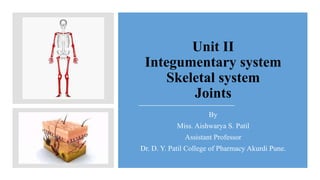
  • 1. Unit II Integumentary system Skeletal system Joints By Miss. Aishwarya S. Patil Assistant Professor Dr. D. Y. Patil College of Pharmacy Akurdi Pune.
  • 2. Contents: • Introduction to integumentary system • Structure of skin • Functions of skin • Skeletal system • Divisions of skeletal system • Types of bones • Salient features and function of axial and appendicular skeletal system. • Organization of skeletal system • Physiology of muscle contraction.(notes given) • Neuromuscular junction • Introduction to joints • Structural and functional classification of joints. • Types of joints movement and its articulation.
  • 3. Introduction to integumentary system • Integumentary system = Skin (function + structure) • Skin completely covers body and continuous with linings • Skin is the largest organ of the body. • Surface area is about 1.5-2 square meters. • It contains some accessory structures: Glands, hairs, nails. • Thickness varies , thickest at palms and feet’s. • Consist of 2 layers. • Superficial layers: epidermis. • Deeper layer: dermis. • Below dermis the layer is known as subcutaneous composed of areolar tissue and adipose tissue.
  • 5. Epidermis • Most superficial layer of the skin and is composed of stratified keratinised squamous epithelium • Varies in thickness in different parts of the body. • No blood vessels or nerve endings in the epidermis, but its deeper layers are bathed in interstitial fluid from the dermis, which provides oxygen and nutrients, and drains away as lymph. • There are several layers (strata) of cells in the epidermis which extend from the deepest germinative layer to the most superficial stratum corneum (a thick horny layer) • The cells on the surface are flat, thin, non-nucleated, dead cells, or squamous, in which the cytoplasm has been replaced by the fibrous protein keratin. • cells are constantly being rubbed off and replaced by cells that originated in the germinative layer and have undergone gradual change as they progressed towards the surface. • Continual cell division in the deeper layers with newly formed cells being pushed to the surface. • Hairs, secretions from sebaceous glands and ducts of sweat glands pass through the epidermis to surface • The surface of the epidermis is ridged by projections of cells in the dermis called papillae. The pattern of ridges on the fingertips is unique to every individual and the impression made by them is the ‘fingerprint’. • Blisters develop when trauma causes separation of the dermis and epidermis and serous fluid collects between the two layers. Skin colour is affected by various factors. • Melanin, a dark pigment derived from the amino acid tyrosine and secreted by melanocytes in the deep germinative layer, is absorbed by surrounding epithelial cells. Colour depend on the amount of melanin secreted. It protects the skin from the harmful effects of sunlight. Exposure to sunlight promotes synthesis of melanin. • Normal saturation of haemoglobin and the amount of blood circulating in the dermis give white skin its pink colour.
  • 6. Dermis • Tough and elastic. • formed from connective tissue and the matrix contains collagen fibres, interlaced with elastic fibres. • Rupture of elastic fibres occurs when the skin is overstretched, resulting in permanent striae, or stretch marks, that may be found in pregnancy and obesity. • Collagen fibres bind water and give the skin its tensile strength, but as this ability declines with age, wrinkles develop. • Fibroblasts , macrophages and mast cells are the main cells found in the dermis. Underlying its deepest layer there is areolar tissue and varying amounts of adipose (fat) tissue. • The structures in the dermis are: blood vessels, lymph vessels, sensory (somatic) nerve endings, sweat glands and their ducts, hairs, arrector pili muscles and sebaceous glands. • Arterioles form a fine network with capillary branches supplying sweat glands, sebaceous glands, hair follicles and the dermis. Lymph vessels form a network throughout the dermis. • The skin is an important sensory organ through which individuals receive information about their environment. Nerve impulses, generated in the sensory receptors in the dermis, are conveyed to the spinal cord by sensory nerves, then to the sensory area of the cerebrum where the sensations are perceived. Sensory receptor Stimulus Meissner’s corpuscle Light pressure Pacinian corpuscle Deep pressure Free nerve ending Pain
  • 7. Sweat glands: • These are widely distributed throughout the skin and are most numerous in the palms of the hands, soles of the feet, axillae and groins. • They are formed from epithelial cells. • The bodies of the glands lie coiled in the subcutaneous tissue. • There are two types of sweat gland. • The commonest type opens onto the skin surface through tiny pores, and the sweat produced here is a clear, watery fluid important in regulating body temperature. • The second type opens into hair follicles, and is found, for example, in the axilla. Bacterial decomposition of these secretions causes an unpleasant odour. • A specialised example of this type of gland is the ceruminous gland of the outer ear, which secretes earwax. • function of sweat, which is secreted by glands, is in the regulation of body temperature. Excessive sweating may lead to dehydration
  • 8. Hairs: • These are formed by a downgrowth of epidermal cells into the dermis or subcutaneous tissue, called hair follicles. • At the base of the follicle is a cluster of cells called the papilla or bulb. • The hair is formed by multiplication of cells of the bulb and as they are pushed upwards, away from their source of nutrition, the cells die and become keratinised. • The part of the hair above the skin is the shaft. • The colour of the hair is genetically determined and depends on the amount of melanin present. White hair is the result of the replacement of melanin by tiny air bubbles.
  • 9. Arrector pili: • These are little bundles of smooth muscle fibres attached to the hair follicles. • Contraction makes the hair stand erect and raises the skin around the hair, causing ‘goose flesh’. • The muscles are stimulated by sympathetic nerve fibres in response to fear and cold. • Erect hairs trap air, which acts as an insulating layer. • This is an efficient warming mechanism, especially when accompanied by shivering, i.e. involuntary contraction of skeletal muscles.
  • 10. Sebaceous gland • These consist of secretory epithelial cells derived from the same tissue as the hair follicles. • They secrete an oily substance, sebum, into the hair follicles and are present in the skin of all parts of the body except the palms of the hands and the soles of the feet. • They are most numerous in the skin of the scalp, face, axillae and groins. • In regions of transition from one type of superficial epithelium to another, such as lips, eyelids, nipple, labia minora and glans penis, there are sebaceous glands that are independent of hair follicles, secreting sebum directly onto the surface. • Sebum keeps the hair soft and pliable and gives it a shiny appearance. On the skin it provides someSebum keeps the hair soft and pliable and gives it a shiny appearance. • On the skin it provides some waterproofing and acts as a bactericidal and fungicidal agent, preventing infection. • It also prevents drying and cracking of skin, especially on exposure to heat and sunshine. • The activity of these glands increases at puberty and is less at the extremes of age, rendering the skin of infants and older adults prone to the effects of excessive moisture
  • 11. Nails: • Human nails are equivalent to the claws, horns and hoofs of animals. • They are derived from the same cells as epidermis and hair and consist of hard, horny keratin plates. • They protect the tips of the fingers and toes. • The root of the nail is embedded in the skin and covered by the cuticle, which forms the hemispherical pale area called the lunula. • The nail plate is the exposed part that has grown out from the germinative zone of the epidermis called the nail bed. • Finger nails grow more quickly than toe nails and growth is quicker when the environmental temperature is high.
  • 12. Functions of skin : (explain the functions) • Protection • Body temperature regulation • Formation of vitamin D • Sensation • Absorption • Excretion • Wound healing.
  • 13. Protection waterproof layer, provided mainly by its keratinised epithelium, which protects the deeper and more delicate structures. As an important non-specific defence mechanism it acts as a barrier against: invasion by micro-organisms, chemicals, physical agents, e.g. mild trauma, ultraviolet light, dehydration. The epidermis contains specialised immune cells Body temperature regulation Regulated body temperature through sweating. Formation of vitamin D 7-dehydrocholesterol is a lipid-based substance in the skin, and ultraviolet rays in sunlight convert it to vitamin D. This circulates in the blood and is used, with calcium and phosphate, in the formation and maintenance of bone. Sensation Sensory receptors are nerve endings in the dermis that are sensitive to touch, pressure, temperature or pain. Stimulation generates nerve impulses in sensory nerves that are transmitted to the cerebral cortex .Some areas have more sensory receptors than others causing them to be especially sensitive, e.g. the lips and fingertips. Absorption This property is limited but substances that can be absorbed include: • some drugs, in transdermal patches, e.g. hormone replacement therapy during the menopause, nicotine as an aid to stopping smoking • some toxic chemicals, e.g. mercury. Excretion Excretion of waste products such as Nacl and urea.
  • 14. Skeletal system : • The skeletal system is made up of your bones. • Give support structure for the rest of your tissue and organs. • Gives body shape, supports your muscles, provides movement, and makes red blood cells, provides protection to delicate organs. Storage for minerals and lipids, • Main parts of skeletal system are bones, cartilage, ligaments, and tendons. • Bone tissues makes up about 18% of the total human body weight. • 5 main parts of skeletal system: skull, vertebral column, collarbone, shoulder blades, rib cage, pelvic girdle and the bones of the hands, arms, feet, and legs. • Types of skeletal system: Hydrostatic skeleton, exoskeleton and endoskeleton.
  • 15. Axial skeleton • Salient features: • It consist of skull, vertebral column (spinal cord) , thoracic cage , sternum bone. • The skull, which contains 22 bones, from which 8 are cranial and 14 are facial. • 6 middle ear ossicles (3 in each ear), • 1 hyoid bone in the neck, • 26 bones of vertebral column, • 1 chest bone (sternum), and • 24 ribs (12 pairs).
  • 16. Divisions of skeletal system • Adult human skeleton consists of 206 named bones. • Bones are divided into two types • Axial skeleton and appendicular skeleton.
  • 17. Appendicular bones: • Salient Features: • Appendicular skeleton is consist of total 126 bones. • It includes all bones besides axial skeleton. • Allow us to move. • It includes limbs and supportive girdles • Pectoral girdle: Upper limbs • Pelvic girdle: Lower limb.
  • 18. Functions of axial and appendicular bones: • The axial skeleton: • provides support and protection for the brain, spinal cord, and the organs in the ventral body cavity; it also provides a surface for the attachment of muscles, directs respiratory movements, and stabilizes portions of the appendicular skeleton. • Appendicular skeleton: • The human appendicular skeleton is composed of the bones of the upper limbs (which function to grasp and manipulate objects) and the lower limbs (which permit locomotion). It also includes the pectoral (or shoulder) girdle and the pelvic girdle, which attach the upper and lower limbs to the body, respectively.
  • 20. Types of bones: 1. According to shape: • Long bones - Long Bones Are typically longer then they are wide. All of the limbs, except the wrist and ankle bones. Mostly compact. • Short bones - Flat Bones are thin, flattened and usually curved. Cube- shaped and contain mostly spongy bone Found in the skull, sternum, ribs, and scapula. Have two thin layers of compact bone sandwiching a layer of spongy bone between them • Flat bones- The parietal bone of the skull and sternum bone. • Irregular bone- Irregular Bones Have complex shapes. Do not fit one of the other categories Vertebrae, Pelvis. • Sesamoid bones- Sesamoid Bones Special type of short bone • Form within tendons. Best known example is the patella. Develop inside tendons near joints of knees, hands, and feet 2. Internal tissues • Compact -Strong able to bear weight. Femur. • Spongy – Light, less dense. Ribs, skull, pelvic bones and vertebrae. 3. Bone markings • Sutures : Sutural Bones Are small, irregular bones are found between the flat bones of the skull.
  • 22. • Skull consists of 29 bones and divided into 5 bones. • Cranial bones: 8. • Facial bones: 14. • Hyoid bone : 1. • The skull Consists of the cranium and the bones of the face Sutures • Lambdoid-between occipital and parietal bones • Coronal-between parietal and frontal bones • Sagittal-between parietal bones • Squamous-between parietal and temporal bones
  • 23. • Cranial Bones one occipital bone foramen magnum two temporal bones • two parietal bones • one frontal bone • frontal sinuses • glabella • two temporal bones • auditory ossicles • one sphenoid • one ethmoid
  • 24. Occipital and Parietal Bones Occipital bone Foramen magnum Occipital condyle External occipital protuberance Parietal bone Frontal bone Supraorbital foramen Glabella Temporal bone Squamous suture Zygomatic process Zygomatic arch Mandibular fossa External auditory meatus Styloid process Temporal bone is divided in regions Mastoid process Mastoiditis Meningitis Stylomastoid foramen Passage for cranial nerve VII Internal acustic meatus Passage for cranial nerves VII and VIII Sphenoid bone Greater wings Sella turcica For the pituitary gland The EthmoidEthmoid bone Crista galli Attachment of the dura mater Cribiform plate Passage of olfactory nerves Ethmoid bone Perpendicular plate Forms the superior part of the nasal septum Superior and middle nasal conchae (turbinates) Covered by mucosa Warms and humidifies the air
  • 25. • Facial bones Maxillary bones Mandible Inferior nasal conchae • Palatine bones • Nasal bones • Vomer • Inferior nasal conchae • Zygomatic bones • Lacrimal BONE • Cranial Foramens to identify • External view: • Supraorbital foramen-for blood vessels and nerves • Infraorbital foramen-blood vessels and nerves • Mental foramen-blood vessels and nerves • Stylomastoid foramen- nerve VII • Carotid canal-for carotid artery • External auditory meatus-leads to eardrum • Incisive fossa-for blood vessels and nerves • Incisive = i as in hit Maxillae Alveolar margim Palatine process- anterior hard palate Incisive fossa- passage for nerves and blood vessels Zygomatic bone articulates with zygomatic process of temporal bone forming the zygomatic Mandible Body- horizontal portion Ramus-vertical portion Mandibular condyle- articulates with temporal bone Coronoid process Angle Alveolar margin- with sockets for the teeth Mandibular foramen – site of Novocain injection
  • 26.
  • 28.
  • 29.
  • 30.
  • 31.
  • 32. Thoracic cage: • The thoracic cage is composed of the thoracic vertebrae, ribs and the sternum • Functions • Forms a protective cage around the heart, lungs, and great blood vessels • Supports the shoulder girdles and upper limbs • Provides attachment for many neck, back, chest, and shoulder muscles • Sternum (Breastbone) • A dagger-shaped, flat bone that lies in the anterior midline of the thorax Ribs • There are twelve pair of ribs forming the flaring sides of the thoracic cage • All ribs attach posteriorly to the thoracic vertebrae • The superior 7 pair (true ribs) attach directly to the sternum via costal cartilages • Ribs 8-10 (false ribs) attach indirectly to the sternum via costal cartilage • Ribs (floatingribs) have no anterior attachment
  • 34.
  • 36.
  • 37.
  • 38.
  • 39.
  • 41.
  • 42. Femur, tibia and fibula.
  • 43.
  • 45.
  • 47. Physiology of muscle contraction/ Sliding filament theory)
  • 49. Introduction to joints: • A joint also called as articulation is appoint of contact. • Arthrology is the scientific study of joints. • The site at which any two or more bone articulated is called joint. • There are mainly 3 types are as follows: • Fibrous or fixed joints (immovable joints). • Cartilaginous or slightly movable joints. • Synovial or freely moveable.