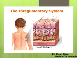
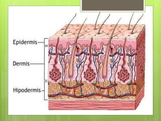
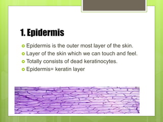
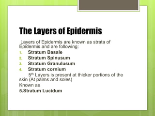
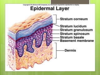
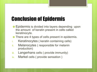
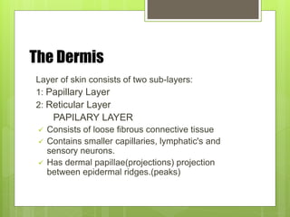
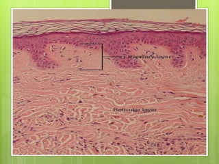
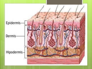
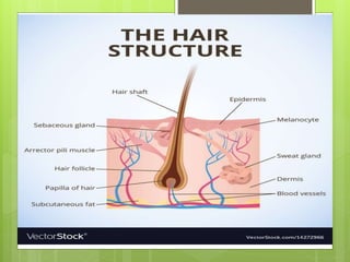
n
By: Umair Khan
Medical Technologist
Introduction
 The Integumentary System is one of the
system of the body which act as integument (
The covering) to the body.
 So, covering will act as protective barrier
between body and environment .
Division of Integumentary
System
 This system consists of following parts
1. Skin
2. Hairs
3. Nails
4. Exocrine glands
1.Skin
 Skin is most important part of integumentary
System.
 It consists of 3 Layers
1. Epidermis
2. Dermis
3. Hypodermis
1. Epidermis
 Epidermis is the outer most layer of the skin.
 Layer of the skin which we can touch and feel.
 Totally consists of dead keratinocytes.
 Epidermis= keratin layer
The Layers of Epidermis
Layers of Epidermis are known as strata of
Epidermis and are following:
1. Stratum Basale
2. Stratum Spinusum
3. Stratum Granulusum
4. Stratum cornium
5th Layers is present at thicker portions of the
skin (At palms and soles)
Known as
5.Stratum Lucidum
1. Stratum basale is act as basement of epidermis. Here
Living cells are present known as keratinocytes.
2. Stratum Spinosum also consists of keratinocytes
containing keratin within.
3. Stratum granulosum contains more keratin than
previous layer.
4. Stratum Cornium contains abundant amount of keratin
which is outer most layer of epidermis.
Conclusion of Epidermis
 Epidermis is divided into layers depending upon
the amount of keratin present in cells called
keratinocyte.
 There are 4 types of cells present in epidermis.
i. Keratinocytes ( keratin containing cells)
ii. Melanocytes ( responsible for melanin
production)
iii. Langerhans cells ( provide immunity)
iv. Markel cells ( provide sensation )
The Dermis
Layer of skin consists of two sub-layers:
1: Papillary Layer
2: Reticular Layer
PAPILARY LAYER
 Consists of loose fibrous connective tissue
 Contains smaller capillaries, lymphatic's and
sensory neurons.
 Has dermal papillae(projections) projection
between epidermal ridges.(peaks)
RETICULAR LAYERS
 It consists of dense irregular connective
tissues.
 It contains larger vessels, lymphatic's and
sensory neurons.
 It contains collagen and elastic fibers.
3. The Hypodermis Layer
 It consists of loose elastic connective tissues
and adipose tissues.
 Connected with Reticular layer via connective
tissue fibers.
 It Stabilizes the skin.
Nails:
 Structure of Nails
 Body of Nail:
The visible portion of the skin.
covers the nail bed.
Lunula:
The crescent like shape at the base
of the nail is called lunula.
Free edge of Nail:
Free edges of nails is the portion of
nails that we can cut without any sensation of pain.
 Sides of Nails:
These sides lie in lateral nail groves .
These sides covered by lateral nail folds.
Function of the Nails:
 Nails protects fingers and toes.
 Helping in griping and holding the things.
 Nails are made up of dead cells filled with
keratin that’s why very hard structures.
 Metabolic disorder can change the Nails
structures, that’s why important in physical
examination of the patient with some
metabolic disorder.
Fun Facts About Nails:
 Nails grow about 0.1 mm a day, so they grow
about 3-4 mm in a month.
 Finger Nails grow faster then the toe nails.
 Finger nails grow faster in days and during
summers.
 Man’s nails grows faster than female.
 Nail of middle finger grow faster than others.
 The little finger nail grows with slowest speed.
 If you loose a nail by injury it will take 6 months to
grow again.
Hairs:
 Introduction:
 One of the appendages of skin.
 Hair follicles are the organs which make the
hairs.
 Hair follicles are present deep in dermis.
 Hairs are non-living.
 They are wrapped in dense connective tissue
sheath.
Structure of the Hair:
 There are 2 main parts of a hair:
1: Hair Root
2: hair Shaft
Hair Root
Lower part of the hair
Originate from the hair follicle
Hair Shaft
Upper visible part of the hair
Thin and long portion of the hair.
Functions:
 Hairs of the Head:
provide UV light protection
Cushion from trauma
Nostrils, ears and eyelashes Hairs:
Prevent entry of foreign material
Hairs of body
Sensory detection
Root of every hair:
Have sensory nerves at the base of hair follicles that
detects slight movement of hairs.
Fun Facts:
 On an Average person have 01 to ½ Lakh of Hair
strands.
 On an average we shed around 50 to 150 hairs
strands per day.
 Hair is 2nd fastest growing tissue in body after
bone marrow.
 A healthy strand of hair can stretch about 30%
when it is wet.
 A new hair start to grow as soon as it is plucked
from hair follicle.
Fun facts:
 A single hair has the lifespan of about 05 years.
 A strand of hair is stronger then a copper wire
with the same diameter.
 When we are scared or cold Goosebumps
appeared when muscle for each hair contracts.
 Hair is made up of keratin which is the same
substance making up the horse hooves.
Glands found on Skin
 There are 3 main types of glands found on human skin:
1) • Eccrine glands - secrete sweat through pores found
in the palms of
hands, soles of feet, and forehead
2) • Sebaceous glands - secrete oily sebum and are
found on the chest,
back, scalp, face, and forehead
3) • Apocrine glands - secrete sweat via canals along
hair follicles in the
underarms, pubic area, anus, and nipple area
Integumentry system

Integumentry system

  • 1.
  • 2.
    Introduction  The IntegumentarySystem is one of the system of the body which act as integument ( The covering) to the body.  So, covering will act as protective barrier between body and environment .
  • 3.
    Division of Integumentary System This system consists of following parts 1. Skin 2. Hairs 3. Nails 4. Exocrine glands
  • 4.
    1.Skin  Skin ismost important part of integumentary System.  It consists of 3 Layers 1. Epidermis 2. Dermis 3. Hypodermis
  • 6.
    1. Epidermis  Epidermisis the outer most layer of the skin.  Layer of the skin which we can touch and feel.  Totally consists of dead keratinocytes.  Epidermis= keratin layer
  • 7.
    The Layers ofEpidermis Layers of Epidermis are known as strata of Epidermis and are following: 1. Stratum Basale 2. Stratum Spinusum 3. Stratum Granulusum 4. Stratum cornium 5th Layers is present at thicker portions of the skin (At palms and soles) Known as 5.Stratum Lucidum
  • 9.
    1. Stratum basaleis act as basement of epidermis. Here Living cells are present known as keratinocytes. 2. Stratum Spinosum also consists of keratinocytes containing keratin within. 3. Stratum granulosum contains more keratin than previous layer. 4. Stratum Cornium contains abundant amount of keratin which is outer most layer of epidermis.
  • 10.
    Conclusion of Epidermis Epidermis is divided into layers depending upon the amount of keratin present in cells called keratinocyte.  There are 4 types of cells present in epidermis. i. Keratinocytes ( keratin containing cells) ii. Melanocytes ( responsible for melanin production) iii. Langerhans cells ( provide immunity) iv. Markel cells ( provide sensation )
  • 11.
    The Dermis Layer ofskin consists of two sub-layers: 1: Papillary Layer 2: Reticular Layer PAPILARY LAYER  Consists of loose fibrous connective tissue  Contains smaller capillaries, lymphatic's and sensory neurons.  Has dermal papillae(projections) projection between epidermal ridges.(peaks)
  • 12.
    RETICULAR LAYERS  Itconsists of dense irregular connective tissues.  It contains larger vessels, lymphatic's and sensory neurons.  It contains collagen and elastic fibers.
  • 14.
    3. The HypodermisLayer  It consists of loose elastic connective tissues and adipose tissues.  Connected with Reticular layer via connective tissue fibers.  It Stabilizes the skin.
  • 16.
    Nails:  Structure ofNails  Body of Nail: The visible portion of the skin. covers the nail bed. Lunula: The crescent like shape at the base of the nail is called lunula. Free edge of Nail: Free edges of nails is the portion of nails that we can cut without any sensation of pain.
  • 17.
     Sides ofNails: These sides lie in lateral nail groves . These sides covered by lateral nail folds.
  • 18.
    Function of theNails:  Nails protects fingers and toes.  Helping in griping and holding the things.  Nails are made up of dead cells filled with keratin that’s why very hard structures.  Metabolic disorder can change the Nails structures, that’s why important in physical examination of the patient with some metabolic disorder.
  • 20.
    Fun Facts AboutNails:  Nails grow about 0.1 mm a day, so they grow about 3-4 mm in a month.  Finger Nails grow faster then the toe nails.  Finger nails grow faster in days and during summers.  Man’s nails grows faster than female.  Nail of middle finger grow faster than others.  The little finger nail grows with slowest speed.  If you loose a nail by injury it will take 6 months to grow again.
  • 21.
    Hairs:  Introduction:  Oneof the appendages of skin.  Hair follicles are the organs which make the hairs.  Hair follicles are present deep in dermis.  Hairs are non-living.  They are wrapped in dense connective tissue sheath.
  • 22.
    Structure of theHair:  There are 2 main parts of a hair: 1: Hair Root 2: hair Shaft Hair Root Lower part of the hair Originate from the hair follicle Hair Shaft Upper visible part of the hair Thin and long portion of the hair.
  • 24.
    Functions:  Hairs ofthe Head: provide UV light protection Cushion from trauma Nostrils, ears and eyelashes Hairs: Prevent entry of foreign material Hairs of body Sensory detection Root of every hair: Have sensory nerves at the base of hair follicles that detects slight movement of hairs.
  • 25.
    Fun Facts:  Onan Average person have 01 to ½ Lakh of Hair strands.  On an average we shed around 50 to 150 hairs strands per day.  Hair is 2nd fastest growing tissue in body after bone marrow.  A healthy strand of hair can stretch about 30% when it is wet.  A new hair start to grow as soon as it is plucked from hair follicle.
  • 26.
    Fun facts:  Asingle hair has the lifespan of about 05 years.  A strand of hair is stronger then a copper wire with the same diameter.  When we are scared or cold Goosebumps appeared when muscle for each hair contracts.  Hair is made up of keratin which is the same substance making up the horse hooves.
  • 27.
    Glands found onSkin  There are 3 main types of glands found on human skin: 1) • Eccrine glands - secrete sweat through pores found in the palms of hands, soles of feet, and forehead 2) • Sebaceous glands - secrete oily sebum and are found on the chest, back, scalp, face, and forehead 3) • Apocrine glands - secrete sweat via canals along hair follicles in the underarms, pubic area, anus, and nipple area