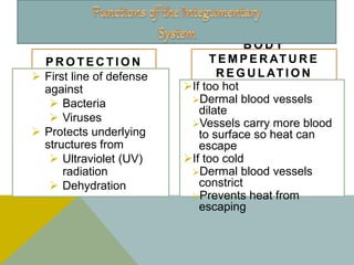
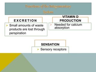
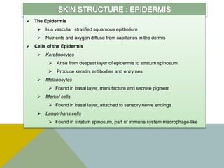
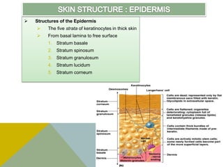
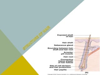
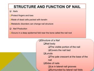
Is the largest system of the body
 16% of body weight,
 1.5 to 2m2
in area,
 The integument is made up of two parts:
1. Cutaneous membrane
a. Epidermis– Superficial epithelium
b. Dermis – underlying CT with blood
supply
2. Accessory structures
a. Hair
b. Nails
c. Exocrine Glands
P R O T E C T I O N
 First line of defense
against
 Bacteria
 Viruses
 Protects underlying
structures from
 Ultraviolet (UV)
radiation
 Dehydration
B O D Y
T E M P E R AT U R E
R E G U L AT I O N
If too hot
Dermal blood vessels
dilate
Vessels carry more blood
to surface so heat can
escape
If too cold
Dermal blood vessels
constrict
Prevents heat from
escaping
E X C R E T I O N
 Small amounts of waste
products are lost through
perspiration
VITAMIN D
PRODUCTION
 Needed for calcium
absorption
SENSATION
 Sensory receptors
Epidermis
Dermis
Hypodermis
or
subcutaneous
layer
 The Epidermis
 Is a vascular stratified squamous epithelium
 Nutrients and oxygen diffuse from capillaries in the dermis
 Cells of the Epidermis
 Keratinocytes
 Arise from deepest layer of epidermis to stratum spinosum
 Produce keratin, antibodies and enzymes
 Melanocytes
 Found in basal layer, manufacture and secrete pigment
 Merkel cells
 Found in basal layer, attached to sensory nerve endings
 Langerhans cells
 Found in stratum spinosum, part of immune system macrophage-like
 Structures of the Epidermis
 The five strata of keratinocytes in thick skin
 From basal lamina to free surface
1. Stratum basale
2. Stratum spinosum
3. Stratum granulosum
4. Stratum lucidum
5. Stratum corneum
Hair
Papillary
layer
Reticular
layer
Cutaneous
plexus
Papillary
plexus
Epidermal
ridges
Dermal
papillae
Capillary loop
of papillary
plexus
 Second major layer of the skin
 Provides mechanical strength, flexibility, and protection for underlying tissues
 Highly vascular and contains a variety of sensory receptors that provide information
about the external environment
 Has two layers
 The papillary layer
 The reticular layer
 Flexure lines - creases on palms
THE PAPILLARRY LAYER
• Underlies epidermis
• Named for dermal papillae
• Aerolar connective tissue
• Supports, nourishes epidermis
• Provides sensory nerves, lymphatics,
and capillaries
THE RETICULAR LAYER
• Tough, dense, fibrous layer
• Dense irregular connective tissue
• Collagen fibers - limit stretch
• Elastic fibers - provide flexibility
• Blends into papillary layer (above)
• Blends into subcutaneous layer
(below)
 The Hypodermis (Subcutaneous Layer)
Lies below the integument
Stabilizes the skin
Allows separate movement
Made of elastic areolar and adipose tissues
Connected to the reticular layer of integument by connective tissue
fibers
 Deposits of Subcutaneous Fat
Distribution patterns determined by hormones
Reduced by cosmetic liposuction (lipoplasty)
T H E H A I R
F O L L I C L E
Hair follicles are the organs that form
the hairs.
Located deep in dermis.
Produces nonliving hairs.
Wrapped in a dense connective
tissue sheath.
Base is surrounded by sensory
nerves (root hair plexus).
Control bacteria
A C C E S S O R Y
S T R U C T U R E S O F H A I R
Arrector pili
Involuntary smooth muscle
Causes hairs to stand up
Produces “goose bumps”
Sebaceous glands
Lubricate the hair
Exposed
shaft
of hair
Sebaceous
gland
Arrector
pili
muscle
Connective
tissue sheath
Root hair
plexus
R E G I O N S O F H A I R
Hair root
Lower part of the hair
Attached to the
integument
Hair shaft
Upper part of the hair
Not attached to the
integument
Boundary
between
hair shaft
and
hair root
Hair shaft
Sebaceous
gland
Arrector
pili muscle
Hair root
Connective
tissue sheat
Hair bulb
Hair matrix
Hair papilla
HAIR
FUNCTION
Head:
UV protection
Cushion from trauma
Insulation
Nostrils, Ear canals, Eyelashes:
Prevent entry of foreign material
Body Hair:
Sensory detection
Root hair plexus:
Sensory nerves at base of hair follicle that detect slight movement of
hair
Arrector pili muscle:
Attached to every hair follicle
Contract to stand hair perpendicular to skin surface
Free edge of
Nail
Body of Nail
Laternal Nail
fold
Lunula
Eponychium
(cuticle)
STRUCTURE AND FUNCTION OF NAIL
 Nails
Protect fingers and toes
Made of dead cells packed with keratin
Metabolic disorders can change nail structure
 Nail Production
Occurs in a deep epidermal fold near the bone called the nail root
Structure of a Nail
Nail body
The visible portion of the nail
Covers the nail bed
Lunula
The pale crescent at the base of the
nail
Sides of nails
Lie in lateral nail grooves
Surrounded by lateral nail folds
A longitudinal section
Eponychium
Proximal nail fold
Nail root
Lunula Nail body
Epidermis Dermis Phalanx Hyponychium
STRUCTURE AND FUNCTION OF NAIL

Integumentary system ppt

  • 2.
    Is the largestsystem of the body  16% of body weight,  1.5 to 2m2 in area,  The integument is made up of two parts: 1. Cutaneous membrane a. Epidermis– Superficial epithelium b. Dermis – underlying CT with blood supply 2. Accessory structures a. Hair b. Nails c. Exocrine Glands
  • 3.
    P R OT E C T I O N  First line of defense against  Bacteria  Viruses  Protects underlying structures from  Ultraviolet (UV) radiation  Dehydration B O D Y T E M P E R AT U R E R E G U L AT I O N If too hot Dermal blood vessels dilate Vessels carry more blood to surface so heat can escape If too cold Dermal blood vessels constrict Prevents heat from escaping
  • 4.
    E X CR E T I O N  Small amounts of waste products are lost through perspiration VITAMIN D PRODUCTION  Needed for calcium absorption SENSATION  Sensory receptors
  • 5.
  • 6.
     The Epidermis Is a vascular stratified squamous epithelium  Nutrients and oxygen diffuse from capillaries in the dermis  Cells of the Epidermis  Keratinocytes  Arise from deepest layer of epidermis to stratum spinosum  Produce keratin, antibodies and enzymes  Melanocytes  Found in basal layer, manufacture and secrete pigment  Merkel cells  Found in basal layer, attached to sensory nerve endings  Langerhans cells  Found in stratum spinosum, part of immune system macrophage-like
  • 7.
     Structures ofthe Epidermis  The five strata of keratinocytes in thick skin  From basal lamina to free surface 1. Stratum basale 2. Stratum spinosum 3. Stratum granulosum 4. Stratum lucidum 5. Stratum corneum
  • 8.
  • 9.
     Second majorlayer of the skin  Provides mechanical strength, flexibility, and protection for underlying tissues  Highly vascular and contains a variety of sensory receptors that provide information about the external environment  Has two layers  The papillary layer  The reticular layer  Flexure lines - creases on palms THE PAPILLARRY LAYER • Underlies epidermis • Named for dermal papillae • Aerolar connective tissue • Supports, nourishes epidermis • Provides sensory nerves, lymphatics, and capillaries THE RETICULAR LAYER • Tough, dense, fibrous layer • Dense irregular connective tissue • Collagen fibers - limit stretch • Elastic fibers - provide flexibility • Blends into papillary layer (above) • Blends into subcutaneous layer (below)
  • 10.
     The Hypodermis(Subcutaneous Layer) Lies below the integument Stabilizes the skin Allows separate movement Made of elastic areolar and adipose tissues Connected to the reticular layer of integument by connective tissue fibers  Deposits of Subcutaneous Fat Distribution patterns determined by hormones Reduced by cosmetic liposuction (lipoplasty)
  • 12.
    T H EH A I R F O L L I C L E Hair follicles are the organs that form the hairs. Located deep in dermis. Produces nonliving hairs. Wrapped in a dense connective tissue sheath. Base is surrounded by sensory nerves (root hair plexus). Control bacteria A C C E S S O R Y S T R U C T U R E S O F H A I R Arrector pili Involuntary smooth muscle Causes hairs to stand up Produces “goose bumps” Sebaceous glands Lubricate the hair Exposed shaft of hair Sebaceous gland Arrector pili muscle Connective tissue sheath Root hair plexus
  • 13.
    R E GI O N S O F H A I R Hair root Lower part of the hair Attached to the integument Hair shaft Upper part of the hair Not attached to the integument Boundary between hair shaft and hair root Hair shaft Sebaceous gland Arrector pili muscle Hair root Connective tissue sheat Hair bulb Hair matrix Hair papilla
  • 14.
    HAIR FUNCTION Head: UV protection Cushion fromtrauma Insulation Nostrils, Ear canals, Eyelashes: Prevent entry of foreign material Body Hair: Sensory detection Root hair plexus: Sensory nerves at base of hair follicle that detect slight movement of hair Arrector pili muscle: Attached to every hair follicle Contract to stand hair perpendicular to skin surface
  • 15.
    Free edge of Nail Bodyof Nail Laternal Nail fold Lunula Eponychium (cuticle)
  • 16.
    STRUCTURE AND FUNCTIONOF NAIL  Nails Protect fingers and toes Made of dead cells packed with keratin Metabolic disorders can change nail structure  Nail Production Occurs in a deep epidermal fold near the bone called the nail root Structure of a Nail Nail body The visible portion of the nail Covers the nail bed Lunula The pale crescent at the base of the nail Sides of nails Lie in lateral nail grooves Surrounded by lateral nail folds
  • 17.
    A longitudinal section Eponychium Proximalnail fold Nail root Lunula Nail body Epidermis Dermis Phalanx Hyponychium STRUCTURE AND FUNCTION OF NAIL