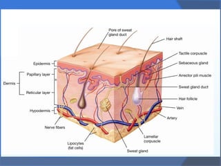
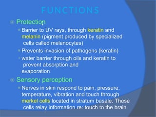
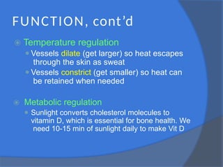
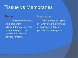
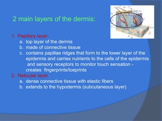
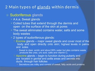
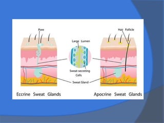
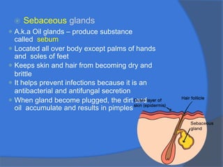
INTEGUMENTARY
SYSTEM
⦿ The term integumentary comes from the
latin word integumentum which means
“covering”
⦿ The integumentary System has been called
both a system, because it covers the body,
and an organ, because it contains several
different types of tissues and organs that
work together to perform important functions
⦿ Most anatomy courses refer to it as a
system because it has organs and other
parts working together to perform specific
functions
FUNCTIONS
:
⦿ Protection
⚫Barrier to UV rays, through keratin and
melanin (pigment produced by specialized
cells called melanocytes)
⚫Prevents invasion of pathogens (keratin)
⚫ water barrier through oils and keratin to
prevent absorption and
evaporation
⦿ Sensory perception
⚫Nerves in skin respond to pain, pressure,
temperature, vibration and touch through
merkel cells located in stratum basale. These
cells relay information re: touch to the brain
FUNCTION, cont’d
⦿ Temperature regulation
⚫Vessels dilate (get larger) so heat escapes
through the skin as sweat
⚫Vessels constrict (get smaller) so heat can
be retained when needed
⦿ Metabolic regulation
⚫ Sunlight converts cholesterol molecules to
vitamin D, which is essential for bone health. We
need 10-15 min of sunlight daily to make Vit D
FUNCTION, cont’d
⦿ Secretion and absorption
⚫Urea and uric acid are eliminated in sweat
⚫Certain substances can be absorbed through
the skin – medications, nicotine, etc. these are
called transdermal medications
⦿ Strength and flexibility
⚫ collagen fibers in skin give it strength to
withstand trauma
⚫ flexibility fibers give it the ability to stretch and
return to normal with movement without tearing
Tissue vs Membranes
Tissue
— ensemble of similar
cells and their
extracellular matrix from
the same origin that
together carry out a
specific function.
Membrane
—thin sheets of tissue
or layer of cells acting as
a boundary, lining, or
partition in an organism
Membranes of
integumentary system
⦿ There are 4 main types of tissues
in the body, 2 of which make up skin:
⚫ Epithelial tissue
⚫ Connective tissue
⦿ Membranes surround and protect body
surfaces, including cavities that open to
the outside world (ie: nasal cavity) and
internal cavities that house organs (ie:
abdominal cavity)
Video link:
https://www.youtube.com/
watch?v=Qj2k8rYrXNM
others;
muscle tissue
nervous tissue
Epithelial Tissue- 3 membranes
⦿Mucous membrane
⚫ Lines body cavities that are open to the outside world,
including all hollow organs of the respiratory, digestive, urinary
and reproductive tracts
⦿Serous membrane
⚫ Line body cavities that are closed to the outside world; pleura
(encloses lungs) & peritoneum (lines abdominal cavity)
⚫ Creates thin, clear fluid called serous fluid that
lubricates between membranes to minimize friction
⦿Cutaneous membrane
⚫ A.k.a. “skin”
⚫ Basic structure of skin is keratinized cells. Keratin is the
tough protein that helps protect the skin against damage
from harmful chemicals, also found in hair and nails that
adds structural strength
Connective T issue - 1 membrane
⦿ Synovial membrane
⚫Line capsules surround synovial joints
(ie: knees and shoulder) and line
tendon .
⚫Serve as cushions and secrete synovial fluid
that provides cushioning and reduces
friction on moving structures
Epithelial tissue
⦿ Provides a lining (covering) for internal and external surfaces
of the body - lungs, digestive system, bronchioles, mouth,
nose, etc.
⦿ Epithelial tissues contain common characteristics
⚫ tightly packed cells: little extra space
⚫ free surface:
○ exposed to environment or body cavities
⚫ deep surface:
○ basement membrane to connect it to the connective tissue
⚫ lack blood supply so they receive nutrients from the surface
and absorb it through the layers
⚫ have rick supply of nerves for sensory perception
⚫ constantly reproduce by mitosis to repair daily damage
Epithelium is classified in 2 ways: shape and layers
SQUAMOUS - thin and flat:
found in air sacs of lungs
CUBOIDAL- cube shaped:
COLUMNAR- taller than wider
with an oval shaped nucleus
PSEUDOSTRATIFIED- various
heights. All connect to basement
membrane and may secrete
mucous.
SHAPES
Epithelium is classified in 2 ways: shape and layers
SIMPLE:
● 1 cell layer thick;
● all layers in contact with
basement membrane
STRATIFIED:
● 2 or more layers of cells.
● only the deepest layer of
cells have contact with the
basement membrane.
● looks like a brick wall to
provide protection
PSEUDOSTRATIFIED
● made up of pseudostratified
cells (various sizes)
LAYERS
when describing, you say simple
squamous, simple cuboidal, etc. to
identify the type and the number
of layers --- or stratified
squamous, statified columnar, etc.
Types of Receptors
⦿ There are several receptors found
throughout the body, these are located in
the skin
⚫Mechanoreceptors
○ Sensory cells that detect muscle contraction and
force
⚫Thermoreceptors
○ Specialized nerve cells that detect difference in
temperature; determine sensitivity of the skin
to changes – heat receptors are closer to
surface, cold receptors are deeper
⚫Chemoreceptors
○ Sensory cells that convert chemical substances
into biological signals that are transmitted to
the brain
Anatomy of the skin
⦿ EPIDERMIS:
⚫ Outermost layer of the skin
⚫ The skin we see
⚫ Made of five small layers(thick skin) 4 layers
everywhere else- but contains no blood
vessels or nerve cells
⚫ Most of cells within
epidermis are
keratinocytes which
produce keratin
⚫ As cells move from
granulosum to corneum, they
become more filled with
Epidermal layers
⚫ Layers (from outside to inside)
⦿ Stratum corneum - outermost layer, , provide
physical barrier for rest of skin layers,constantly
shed and replaced by new cells from the inner
layer – epidermis completely replaces itself
every 25-45 days
⦿ Stratum lucidum - thin layer made of flat,
transparent cells (found ONLY in soles of feet
and palms of hands)
⦿ Stratum granulosum - 2-3 layers of flat cells;
active keratinization; compact & brittle, secretes
lipids (fats) to form a lipid barrier making it
waterproof
⦿ Stratum spinosum – keratinization or cells begin
here; spiny shaped cells
⚫Melanocytes are produced here and connect to
keratinocytes. They are important for
producing melanin.
⚫Also found here is Langerhans cells which
ingest foreign antigens and process them to
activate our immune system
⦿ Stratum germinativum (basale) - innermost layer;
rests on basement membrane. Keratinocytes
are created here and migrate toward the surface
(corneum) making up most of the epidermis.
Merkel cells, or touch receptors, are found here –
these cells relay information to the brain
Layers of
integumentary tissue
⦿ DERMIS:
⚫Considered “true skin”; thickest layer
⚫Found below epidermis
⚫Contains blood vessels, nerves, muscles,
sweat and oil glands and hair follicles
⚫Top of dermis (papillary layer) covered with
papillae – fits into the ridges of the stratum
germinativum of the epidermis
⚫The ridges form lines and make patterns that
become fingerprints and footprints
2 main layers of the dermis:
1. Papillary layer:
a. top layer of the dermis
b. made of connective tissue
c. contains papillae ridges that form to the lower layer of the
epidermis and carries nutrients to the cells of the epidermis
and sensory receptors to monitor touch sensation -
creates fingerprints/toeprints
2. Reticular layer
a. dense connective tissue with elastic fibers
b. extends to the hypodermis (subcutaneous layer)
2 Main types of glands within dermis
⦿ Sudoriferous glands
⚫ A.k.a. Sweat glands
⚫ Coiled tubes that extend through the dermis and
open on the surface of the skin at pores
⚫ The sweat eliminated contains water, salts and some
body wastes
⚫ 2 types of sudoriferous glands:
○ Eccrine glands – major sweat glands and cover most of the
body and open directly onto skin; highest levels in palms
and soles
⚫ Sweat is clear, acidic and about 99% water but also contains waste
products like urea, uric acid, salts and vitamin C;
○ Apocrine glands – begin to function during puberty and
are located in genital and axilla areas and secrete into
ducts through hair follicles
⚫ Secretions are milky and consist of sweat, fatty acids and proteins
⦿ Sebaceous glands
⚫ A.k.a Oil glands – produce substance
called sebum
⚫ Located all over body except palms of hands
and soles of feet
⚫ Keeps skin and hair from becoming dry and
brittle
⚫ It helps prevent infections because it is an
antibacterial and antifungal secretion
⚫ When gland become plugged, the dirt and
oil accumulate and results in pimples
⦿ Arrector pili muscle – small muscles attached to hair
follicles. Contraction of muscle causes hair to “stand
on end” or “goose bumps” as well as force sebum
towards surface of hair.
⦿ Arteries & Veins – transports blood flow through
system
⦿ Nerves – allows transmission of sensation to the brain
⦿ Ruffini endings – mechanoreceptor:
⚫ Responds to sustained touch and pressure to help detect ability
to grip and prevent slipping of objects
⦿ Krause end bulbs: - thermoreceptor:
⚫ Detects cold temps; found in eyes, lips, tongue,
genitals (specialized areas)
⦿ Meissner corpuscle – mechanoreceptor:
⚫ Response to texture changes and slow vibrations; most
sensitive to light touch and is concentrated in fingers and lips
⦿ Pacinian corpuscles – mechanoreceptor:
⚫ Responds to rapid changes and deep pressure
⦿ Merkel disks – mechanoreceptor:
○ Attached to cells in the stratum basale; Responds to slow changes
in position
3rd layer of integumentary
tissue
⦿ Hypodermis – subcutaneous
fascia
⚫Innermost layer
⚫Made of connective tissue and
adipose (fatty) tissue
⚫Connects the skin to muscles
⚫Fat tissue: stores energy,
regulates temperature, provides
cushioning
Lesser known parts:
⦿ Hair – helps protect the body and covers
all body surfaces except for the palms
of the hands and the soles of the feet
⚫Alopecia – baldness, permanent hair loss
⦿ Nails – protect the fingers and toes from
injury, made of dead epidermal epithelial
cells packed closely together to form a
thick, dense surface. Will grow if nail
bed is not damaged.
Pigmentation
⦿ Basic skin color is inherited and determined
by the pigments in the epidermis
⦿ Melanin – blackish-brown pigment leads to
black or brown skin tint; also absorbs UV
light to tan the skin; small concentrations
form freckles
⦿ Carotine – yellow-red pigment
⦿ Albino – person with absence of color
pigments
Abnormal colors
⦿ Erythema – reddish color of skin caused
by burns or congestion of blood
vessels
⦿ Jaundice – yellow discoloration indicates
bile in the blood resulting from liver or
gallbladder disease
⦿ Cyanosis – bluish discoloration of the
skin caused by insufficient oxygen;
associated w/ heart, lung, and
circulatory diseases
Skin Injuries
⦿ Wounds produce different types of damage
to tissue parts
⚫Contusion
○ Bruising of tissue caused by blunt force –
capillaries break and leak blood under the
epidermis
⦿ Incision
⚫Cutting of the skin usually by surgical
knife
⦿ Laceration
⚫A tear or cut in the skin
⦿ Puncture
⚫Hole created in the skin by a sharp,
pointed object
⦿ Abrasion
⚫Superficial scrape to the skin
Fungal Infections
⦿ Athlete’s foot
⦿ Ringworm
Viral Infections
⦿ Herpes varicella
⦿ Herpes zoster
⦿ Herpes simplex virus (HSV-1)
⦿ Human papilloma virus (HPV)
Tinea Pedis (Athlete’s foot)
⦿ Tinea is a fungal infection that occurs in
areas of the body that are moist
⦿ Symptoms include itchy skin, blisters
and cracks into open sores
⦿ Treatment is using antifungal medication
and keeping area dry and clean
Ringworm
⦿ Highly contagious fungal infection
⦿ Symptom Is the formation of flat or
raised circular area with a clear central
area surrounded by an itchy, scaly or
crusty outer ring
⦿ Antifungal medications are used in
treatment
Viral Infections
⦿ Herpes varicella
⦿ Herpes zoster
⦿ Herpes simplex virus (HSV-1)
⦿ Human Papilloma Virus (HPV)
Herpes varicella
⦿ A.k.a. “chickenpox”
⦿ Contagious fluid-filled blisters
⦿ Incubation period 14 days
⦿ Small fever prior to rash outbreak
⦿ Vaccine available
Herpes zoster
⦿ Chickenpox virus – lays
dormant but re-occurs in
adults as “shingles”
⦿ Painful, blistering rash
⦿ Headache, fever and
general achy-ness
⦿ Vaccine recommended
for >55yo.
Herpes simplex virus
(HSV-1)
⦿ Aka – “cold sore”
⦿ Associated with
common colds
⦿ Highly contagious and
can be transmitted to
the mouth or genital
area
⦿ 1 in 6 people have
HSV-1
⦿ Once infected, remains
dormant but can be
reactivated
Human papilloma virus
(HPV)
⦿ A.k.a. “warts”
⦿ Group of 150 viruses
⦿ Raised, painless growths
on skin
⦿ Can spread from one
part of body to another
⦿ Plantar warts grow
inward on soles of foot
causing sever pain
Bacterial Infections
⦿ Acne vulgaris
⦿ cellulitis
⦿ Impetigo
Acne vulgaris
⦿ Inflammation of sebaceous
glands
⦿ Cause unknown, but thought to
be related to hormone
changes
⦿ Symptoms include papules,
pustules and blackheads that
occur when follicles become
blocked
⦿ Treated w/ good skin hygiene;
antibiotic or vitamin A
ointments; avoiding makeup
and heavy creams;
Cellulitis
⦿ Staph infection
⦿ Inflammation and
redness of skin causing
pain
⦿ Can be life threatening if
left untreated
Impetigo
⦿ Highly contagious skin infection caused
by strep or staph organisms
⦿ Symptoms include erythema, oozing
vesicles, formation of yellow crusts and
lesions
⦿ Wash lesions w/ soap and water and
antibiotics
Inflammatory conditions
⦿ Dermatitis
⦿ Eczema
⦿ Psoriasis
Dermatitis
⦿ Inflammation of the skin caused
by irritants
⚫Can lead to cellulitis –
⦿ Possible allergic reactions to
detergents, soaps, cosmetics,
etc.
⦿ Symptoms include dry skin,
erythema, itching, edema,
rashes, etc.
⦿ Treated with anti-inflammatories
and antihistamines and
possible steroids
Eczema
⦿ Noncontagious, inflammatory
skin disorder caused by an
allergen or irritant
⦿ Symptoms include dry skin,
itching, vesicles, crusts
and scaling
⦿ Treatment involves removing
irritant and applying cortisone to
reduce inflammation
Psoriasis
⦿ Chronic, noncontagious,
inherited skin disease
⦿ Symptoms include thick, red
areas covered w/ white or
silver scales
⦿ No cure, treatment methods
include steroid creams, UV
light and scale removal
Other disorders
⦿ Decubitus ulcer
⦿ Skin cancer
⦿ Burns
⚫Decubitus ulcer
○ Bed sores caused by pressure that restricts
blood flow
○ Typically occur with prolonged bed rest when
not turned often enough, usually occurs over
boney areas (hips, tailbone, elbows, etc)
Skin cancer
⦿ Occurs in several forms
⚫ Malignant – cancerous
⚫ Benign – non cancerous
⚫ Basal cell carcinoma, squamous cell carcinoma,
melanomas
Develops from moles, exposure to sun, prolonged
use of tanning beds, chemicals, or radiation
⦿ Treatment involves surgical removal or
radiation
⦿ Concerns (what to look for)
⚫ Asymmetry
⚫ Border irregularity
⚫ Color
⚫ diameter
Burns
⦿ Injuries that come from exposure to
excessive heat, chemicals, electricity, or
UV radiation
⦿ Vary in severity and can cause cell
damage or death
⦿
Rule of 9’s
⦿ when a large area of
skin has been
burned, practitioners
use the “rule of
nine’s” to estimate
the extent of tissue
that has been
burned

Integumentary System Overview Anatomy & Physiology

  • 1.
  • 3.
    ⦿ The termintegumentary comes from the latin word integumentum which means “covering” ⦿ The integumentary System has been called both a system, because it covers the body, and an organ, because it contains several different types of tissues and organs that work together to perform important functions ⦿ Most anatomy courses refer to it as a system because it has organs and other parts working together to perform specific functions
  • 4.
    FUNCTIONS : ⦿ Protection ⚫Barrier toUV rays, through keratin and melanin (pigment produced by specialized cells called melanocytes) ⚫Prevents invasion of pathogens (keratin) ⚫ water barrier through oils and keratin to prevent absorption and evaporation ⦿ Sensory perception ⚫Nerves in skin respond to pain, pressure, temperature, vibration and touch through merkel cells located in stratum basale. These cells relay information re: touch to the brain
  • 5.
    FUNCTION, cont’d ⦿ Temperatureregulation ⚫Vessels dilate (get larger) so heat escapes through the skin as sweat ⚫Vessels constrict (get smaller) so heat can be retained when needed ⦿ Metabolic regulation ⚫ Sunlight converts cholesterol molecules to vitamin D, which is essential for bone health. We need 10-15 min of sunlight daily to make Vit D
  • 6.
    FUNCTION, cont’d ⦿ Secretionand absorption ⚫Urea and uric acid are eliminated in sweat ⚫Certain substances can be absorbed through the skin – medications, nicotine, etc. these are called transdermal medications ⦿ Strength and flexibility ⚫ collagen fibers in skin give it strength to withstand trauma ⚫ flexibility fibers give it the ability to stretch and return to normal with movement without tearing
  • 7.
    Tissue vs Membranes Tissue —ensemble of similar cells and their extracellular matrix from the same origin that together carry out a specific function. Membrane —thin sheets of tissue or layer of cells acting as a boundary, lining, or partition in an organism
  • 8.
    Membranes of integumentary system ⦿There are 4 main types of tissues in the body, 2 of which make up skin: ⚫ Epithelial tissue ⚫ Connective tissue ⦿ Membranes surround and protect body surfaces, including cavities that open to the outside world (ie: nasal cavity) and internal cavities that house organs (ie: abdominal cavity) Video link: https://www.youtube.com/ watch?v=Qj2k8rYrXNM others; muscle tissue nervous tissue
  • 9.
    Epithelial Tissue- 3membranes ⦿Mucous membrane ⚫ Lines body cavities that are open to the outside world, including all hollow organs of the respiratory, digestive, urinary and reproductive tracts ⦿Serous membrane ⚫ Line body cavities that are closed to the outside world; pleura (encloses lungs) & peritoneum (lines abdominal cavity) ⚫ Creates thin, clear fluid called serous fluid that lubricates between membranes to minimize friction ⦿Cutaneous membrane ⚫ A.k.a. “skin” ⚫ Basic structure of skin is keratinized cells. Keratin is the tough protein that helps protect the skin against damage from harmful chemicals, also found in hair and nails that adds structural strength
  • 10.
    Connective T issue- 1 membrane ⦿ Synovial membrane ⚫Line capsules surround synovial joints (ie: knees and shoulder) and line tendon . ⚫Serve as cushions and secrete synovial fluid that provides cushioning and reduces friction on moving structures
  • 11.
    Epithelial tissue ⦿ Providesa lining (covering) for internal and external surfaces of the body - lungs, digestive system, bronchioles, mouth, nose, etc. ⦿ Epithelial tissues contain common characteristics ⚫ tightly packed cells: little extra space ⚫ free surface: ○ exposed to environment or body cavities ⚫ deep surface: ○ basement membrane to connect it to the connective tissue ⚫ lack blood supply so they receive nutrients from the surface and absorb it through the layers ⚫ have rick supply of nerves for sensory perception ⚫ constantly reproduce by mitosis to repair daily damage
  • 12.
    Epithelium is classifiedin 2 ways: shape and layers SQUAMOUS - thin and flat: found in air sacs of lungs CUBOIDAL- cube shaped: COLUMNAR- taller than wider with an oval shaped nucleus PSEUDOSTRATIFIED- various heights. All connect to basement membrane and may secrete mucous. SHAPES
  • 13.
    Epithelium is classifiedin 2 ways: shape and layers SIMPLE: ● 1 cell layer thick; ● all layers in contact with basement membrane STRATIFIED: ● 2 or more layers of cells. ● only the deepest layer of cells have contact with the basement membrane. ● looks like a brick wall to provide protection PSEUDOSTRATIFIED ● made up of pseudostratified cells (various sizes) LAYERS when describing, you say simple squamous, simple cuboidal, etc. to identify the type and the number of layers --- or stratified squamous, statified columnar, etc.
  • 14.
    Types of Receptors ⦿There are several receptors found throughout the body, these are located in the skin ⚫Mechanoreceptors ○ Sensory cells that detect muscle contraction and force ⚫Thermoreceptors ○ Specialized nerve cells that detect difference in temperature; determine sensitivity of the skin to changes – heat receptors are closer to surface, cold receptors are deeper ⚫Chemoreceptors ○ Sensory cells that convert chemical substances into biological signals that are transmitted to the brain
  • 15.
    Anatomy of theskin ⦿ EPIDERMIS: ⚫ Outermost layer of the skin ⚫ The skin we see ⚫ Made of five small layers(thick skin) 4 layers everywhere else- but contains no blood vessels or nerve cells ⚫ Most of cells within epidermis are keratinocytes which produce keratin ⚫ As cells move from granulosum to corneum, they become more filled with
  • 16.
    Epidermal layers ⚫ Layers(from outside to inside) ⦿ Stratum corneum - outermost layer, , provide physical barrier for rest of skin layers,constantly shed and replaced by new cells from the inner layer – epidermis completely replaces itself every 25-45 days ⦿ Stratum lucidum - thin layer made of flat, transparent cells (found ONLY in soles of feet and palms of hands) ⦿ Stratum granulosum - 2-3 layers of flat cells; active keratinization; compact & brittle, secretes lipids (fats) to form a lipid barrier making it waterproof
  • 17.
    ⦿ Stratum spinosum– keratinization or cells begin here; spiny shaped cells ⚫Melanocytes are produced here and connect to keratinocytes. They are important for producing melanin. ⚫Also found here is Langerhans cells which ingest foreign antigens and process them to activate our immune system ⦿ Stratum germinativum (basale) - innermost layer; rests on basement membrane. Keratinocytes are created here and migrate toward the surface (corneum) making up most of the epidermis. Merkel cells, or touch receptors, are found here – these cells relay information to the brain
  • 18.
    Layers of integumentary tissue ⦿DERMIS: ⚫Considered “true skin”; thickest layer ⚫Found below epidermis ⚫Contains blood vessels, nerves, muscles, sweat and oil glands and hair follicles ⚫Top of dermis (papillary layer) covered with papillae – fits into the ridges of the stratum germinativum of the epidermis ⚫The ridges form lines and make patterns that become fingerprints and footprints
  • 19.
    2 main layersof the dermis: 1. Papillary layer: a. top layer of the dermis b. made of connective tissue c. contains papillae ridges that form to the lower layer of the epidermis and carries nutrients to the cells of the epidermis and sensory receptors to monitor touch sensation - creates fingerprints/toeprints 2. Reticular layer a. dense connective tissue with elastic fibers b. extends to the hypodermis (subcutaneous layer)
  • 20.
    2 Main typesof glands within dermis ⦿ Sudoriferous glands ⚫ A.k.a. Sweat glands ⚫ Coiled tubes that extend through the dermis and open on the surface of the skin at pores ⚫ The sweat eliminated contains water, salts and some body wastes ⚫ 2 types of sudoriferous glands: ○ Eccrine glands – major sweat glands and cover most of the body and open directly onto skin; highest levels in palms and soles ⚫ Sweat is clear, acidic and about 99% water but also contains waste products like urea, uric acid, salts and vitamin C; ○ Apocrine glands – begin to function during puberty and are located in genital and axilla areas and secrete into ducts through hair follicles ⚫ Secretions are milky and consist of sweat, fatty acids and proteins
  • 22.
    ⦿ Sebaceous glands ⚫A.k.a Oil glands – produce substance called sebum ⚫ Located all over body except palms of hands and soles of feet ⚫ Keeps skin and hair from becoming dry and brittle ⚫ It helps prevent infections because it is an antibacterial and antifungal secretion ⚫ When gland become plugged, the dirt and oil accumulate and results in pimples
  • 24.
    ⦿ Arrector pilimuscle – small muscles attached to hair follicles. Contraction of muscle causes hair to “stand on end” or “goose bumps” as well as force sebum towards surface of hair. ⦿ Arteries & Veins – transports blood flow through system ⦿ Nerves – allows transmission of sensation to the brain ⦿ Ruffini endings – mechanoreceptor: ⚫ Responds to sustained touch and pressure to help detect ability to grip and prevent slipping of objects ⦿ Krause end bulbs: - thermoreceptor: ⚫ Detects cold temps; found in eyes, lips, tongue, genitals (specialized areas) ⦿ Meissner corpuscle – mechanoreceptor: ⚫ Response to texture changes and slow vibrations; most sensitive to light touch and is concentrated in fingers and lips ⦿ Pacinian corpuscles – mechanoreceptor: ⚫ Responds to rapid changes and deep pressure ⦿ Merkel disks – mechanoreceptor: ○ Attached to cells in the stratum basale; Responds to slow changes in position
  • 25.
    3rd layer ofintegumentary tissue ⦿ Hypodermis – subcutaneous fascia ⚫Innermost layer ⚫Made of connective tissue and adipose (fatty) tissue ⚫Connects the skin to muscles ⚫Fat tissue: stores energy, regulates temperature, provides cushioning
  • 26.
    Lesser known parts: ⦿Hair – helps protect the body and covers all body surfaces except for the palms of the hands and the soles of the feet ⚫Alopecia – baldness, permanent hair loss ⦿ Nails – protect the fingers and toes from injury, made of dead epidermal epithelial cells packed closely together to form a thick, dense surface. Will grow if nail bed is not damaged.
  • 27.
    Pigmentation ⦿ Basic skincolor is inherited and determined by the pigments in the epidermis ⦿ Melanin – blackish-brown pigment leads to black or brown skin tint; also absorbs UV light to tan the skin; small concentrations form freckles ⦿ Carotine – yellow-red pigment ⦿ Albino – person with absence of color pigments
  • 28.
    Abnormal colors ⦿ Erythema– reddish color of skin caused by burns or congestion of blood vessels ⦿ Jaundice – yellow discoloration indicates bile in the blood resulting from liver or gallbladder disease ⦿ Cyanosis – bluish discoloration of the skin caused by insufficient oxygen; associated w/ heart, lung, and circulatory diseases
  • 31.
    Skin Injuries ⦿ Woundsproduce different types of damage to tissue parts ⚫Contusion ○ Bruising of tissue caused by blunt force – capillaries break and leak blood under the epidermis
  • 32.
    ⦿ Incision ⚫Cutting ofthe skin usually by surgical knife ⦿ Laceration ⚫A tear or cut in the skin ⦿ Puncture ⚫Hole created in the skin by a sharp, pointed object ⦿ Abrasion ⚫Superficial scrape to the skin
  • 33.
    Fungal Infections ⦿ Athlete’sfoot ⦿ Ringworm Viral Infections ⦿ Herpes varicella ⦿ Herpes zoster ⦿ Herpes simplex virus (HSV-1) ⦿ Human papilloma virus (HPV)
  • 34.
    Tinea Pedis (Athlete’sfoot) ⦿ Tinea is a fungal infection that occurs in areas of the body that are moist ⦿ Symptoms include itchy skin, blisters and cracks into open sores ⦿ Treatment is using antifungal medication and keeping area dry and clean
  • 35.
    Ringworm ⦿ Highly contagiousfungal infection ⦿ Symptom Is the formation of flat or raised circular area with a clear central area surrounded by an itchy, scaly or crusty outer ring ⦿ Antifungal medications are used in treatment
  • 36.
    Viral Infections ⦿ Herpesvaricella ⦿ Herpes zoster ⦿ Herpes simplex virus (HSV-1) ⦿ Human Papilloma Virus (HPV)
  • 37.
    Herpes varicella ⦿ A.k.a.“chickenpox” ⦿ Contagious fluid-filled blisters ⦿ Incubation period 14 days ⦿ Small fever prior to rash outbreak ⦿ Vaccine available
  • 38.
    Herpes zoster ⦿ Chickenpoxvirus – lays dormant but re-occurs in adults as “shingles” ⦿ Painful, blistering rash ⦿ Headache, fever and general achy-ness ⦿ Vaccine recommended for >55yo.
  • 39.
    Herpes simplex virus (HSV-1) ⦿Aka – “cold sore” ⦿ Associated with common colds ⦿ Highly contagious and can be transmitted to the mouth or genital area ⦿ 1 in 6 people have HSV-1 ⦿ Once infected, remains dormant but can be reactivated
  • 40.
    Human papilloma virus (HPV) ⦿A.k.a. “warts” ⦿ Group of 150 viruses ⦿ Raised, painless growths on skin ⦿ Can spread from one part of body to another ⦿ Plantar warts grow inward on soles of foot causing sever pain
  • 41.
    Bacterial Infections ⦿ Acnevulgaris ⦿ cellulitis ⦿ Impetigo
  • 42.
    Acne vulgaris ⦿ Inflammationof sebaceous glands ⦿ Cause unknown, but thought to be related to hormone changes ⦿ Symptoms include papules, pustules and blackheads that occur when follicles become blocked ⦿ Treated w/ good skin hygiene; antibiotic or vitamin A ointments; avoiding makeup and heavy creams;
  • 43.
    Cellulitis ⦿ Staph infection ⦿Inflammation and redness of skin causing pain ⦿ Can be life threatening if left untreated
  • 44.
    Impetigo ⦿ Highly contagiousskin infection caused by strep or staph organisms ⦿ Symptoms include erythema, oozing vesicles, formation of yellow crusts and lesions ⦿ Wash lesions w/ soap and water and antibiotics
  • 45.
  • 46.
    Dermatitis ⦿ Inflammation ofthe skin caused by irritants ⚫Can lead to cellulitis – ⦿ Possible allergic reactions to detergents, soaps, cosmetics, etc. ⦿ Symptoms include dry skin, erythema, itching, edema, rashes, etc. ⦿ Treated with anti-inflammatories and antihistamines and possible steroids
  • 47.
    Eczema ⦿ Noncontagious, inflammatory skindisorder caused by an allergen or irritant ⦿ Symptoms include dry skin, itching, vesicles, crusts and scaling ⦿ Treatment involves removing irritant and applying cortisone to reduce inflammation
  • 48.
    Psoriasis ⦿ Chronic, noncontagious, inheritedskin disease ⦿ Symptoms include thick, red areas covered w/ white or silver scales ⦿ No cure, treatment methods include steroid creams, UV light and scale removal
  • 49.
    Other disorders ⦿ Decubitusulcer ⦿ Skin cancer ⦿ Burns
  • 50.
    ⚫Decubitus ulcer ○ Bedsores caused by pressure that restricts blood flow ○ Typically occur with prolonged bed rest when not turned often enough, usually occurs over boney areas (hips, tailbone, elbows, etc)
  • 51.
    Skin cancer ⦿ Occursin several forms ⚫ Malignant – cancerous ⚫ Benign – non cancerous ⚫ Basal cell carcinoma, squamous cell carcinoma, melanomas Develops from moles, exposure to sun, prolonged use of tanning beds, chemicals, or radiation ⦿ Treatment involves surgical removal or radiation ⦿ Concerns (what to look for) ⚫ Asymmetry ⚫ Border irregularity ⚫ Color ⚫ diameter
  • 52.
    Burns ⦿ Injuries thatcome from exposure to excessive heat, chemicals, electricity, or UV radiation ⦿ Vary in severity and can cause cell damage or death ⦿
  • 53.
    Rule of 9’s ⦿when a large area of skin has been burned, practitioners use the “rule of nine’s” to estimate the extent of tissue that has been burned